
54-year-old male with a remote history of blunt trauma
CXR Traumatic Diaphragmatic Hernia
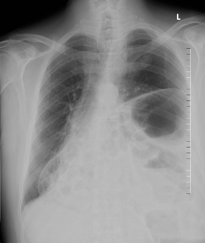
54-year-old male with a remote history of blunt trauma presents for pre-op evaluation. CXR in the frontal projection shows an elevated left hemidiaphragm with herniation of abdominal contents into more than half the left chest cavity. In addition, there is evidence of gas filled bowel to the right of the right heart border as well.
Findings are consistent with a large traumatic diaphragmatic injury with secondary herniation of abdominal contents into the chest cavity
Ashley Davidoff MD TheCommonVein.net 273Lu 136259.8.
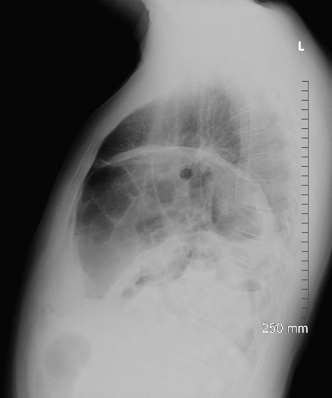
54-year-old male with a remote history of blunt trauma presents for pre-op evaluation. CXR in the lateral projection shows an elevated left hemidiaphragm with herniation of abdominal contents into more than 2/3rds of the left chest cavity.
Findings are consistent with a large traumatic diaphragmatic injury with secondary herniation of abdominal contents into the chest cavity
Ashley Davidoff MD TheCommonVein.net 273Lu 136260.8.
CT Scout
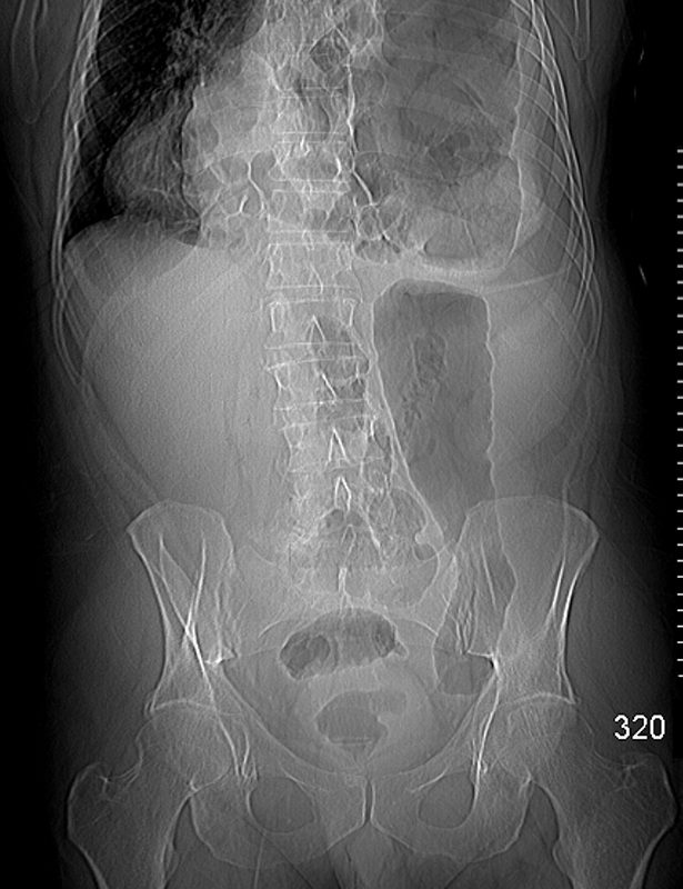
54-year-old male with a remote history of blunt trauma presents for pre-op evaluation. CT scout in the frontal projection shows herniation of abdominal contents into more than 2/3rds of the left chest cavity and a portion of the right thoracic cavity alongside the right heart border. The descending colon in the abdomen is featureless and air-filled. The remaining contents in the abdomen are unremarkable
Findings are consistent with a large traumatic diaphragmatic injury with secondary herniation of abdominal contents into the chest cavity
Ashley Davidoff MD TheCommonVein.net 273Lu 136260.8.
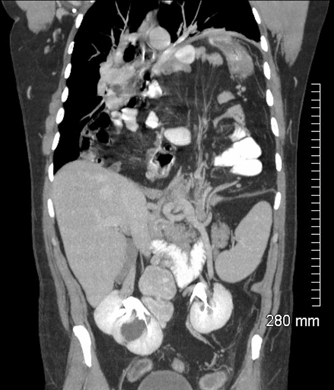
Coronal CT Diaphragmatic Defect and Mesenteric Venous Distension
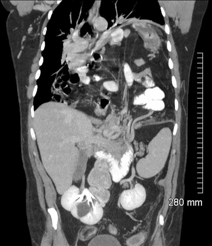
54-year-old male with a remote history of blunt trauma presents for pr-op evaluation. CT scan in the coronal projection shows herniation of abdominal contents into more than 2/3rds of the left chest cavity and a portion of the right thoracic cavity. The hernial orifice is close to 6cms in diameter. There is mild obstruction of venous structures traversing the left side of the hernial orifice. The kidneys have been pulled medially and their hila are oriented toward the vector of the hernia
Findings are consistent with a large traumatic diaphragmatic injury with secondary herniation of abdominal contents into the chest cavity
Ashley Davidoff MD TheCommonVein.net 273Lu 136298.8
54-year-old male with a remote history of blunt trauma presents for pre-op evaluation. CT scan in the coronal projection shows herniation of abdominal contents into more than 2/3rds of the left chest cavity and a portion of the right thoracic cavity. The hernial orifice is close to 6cms in diameter. There is obstruction of mesenteric venous structures traversing the hernial orifice. The kidneys have been pulled medially and their hila are oriented toward the vector of the hernia. The spleen liver and kidneys remain in the abdominal cavity.
Findings are consistent with a large traumatic diaphragmatic injury with secondary herniation of abdominal contents into the chest cavity
Ashley Davidoff MD TheCommonVein.net 273Lu 136308.8.
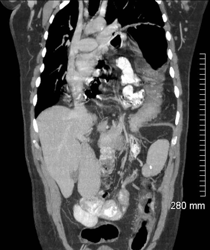
54-year-old male with a remote history of blunt trauma presents for pre-op evaluation. CT scan in the coronal projection shows herniation of abdominal contents into more than 2/3rds of the left chest cavity. Portions of the non-obstructed small bowel and colon are noted in the herniated content
Findings are consistent with a large traumatic diaphragmatic injury with secondary herniation of abdominal contents into the chest cavity
Ashley Davidoff MD TheCommonVein.net 273Lu 136302.8
Superior Extent of the Hernia at the Level of the Aortic Arch
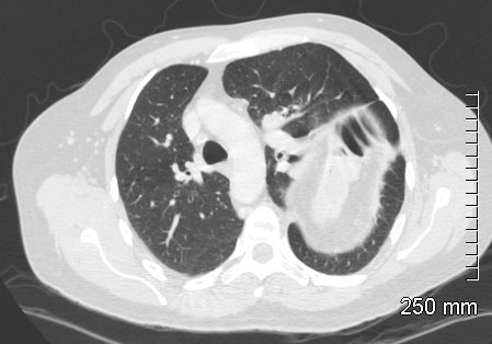
54-year-old male with a remote history of blunt trauma presents for pre-op evaluation. CT in the axial projection shows the superior extent of the herniation of abdominal contents to the level of the aortic arch. The haustra of the colon within the peritoneal fat is apparent at the dome of the herniation.
Findings are consistent with a large traumatic diaphragmatic injury with secondary herniation of abdominal contents into the chest cavity
Ashley Davidoff MD TheCommonVein.net 273Lu 136263
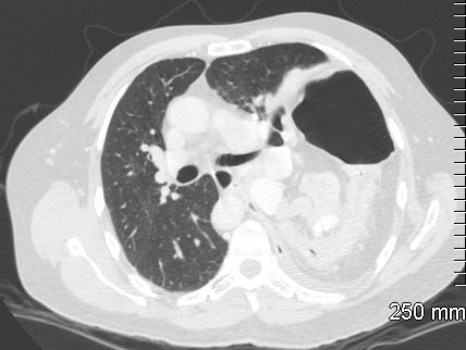
54-year-old male with a remote history of blunt trauma presents for pre-op evaluation. CT in the axial projection shows the herniated abdominal contents to the level of the carina. The anteriorly positioned colon is distended with air and the posteriorly position colon is decompressed. Non distended medially positioned small bowel is present.
Findings are consistent with a large traumatic diaphragmatic injury with secondary herniation of abdominal contents into the chest cavity
Ashley Davidoff MD TheCommonVein.net 273Lu 136265
CT Traumatic Diaphragmatic Hernia Extending to Left and Right Sides of the Thoracic Cavity
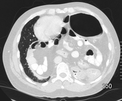
54-year-old male with a remote history of blunt trauma presents for pre-op evaluation. CT in the axial projection shows the herniated abdominal contents at the level of the heart. The anteriorly positioned colon is distended with air. Non distended medially positioned small bowel is present and together with peritoneal fat extends across the midline and pushes the heart toward the right.
Findings are consistent with a large traumatic diaphragmatic injury with secondary herniation of abdominal contents into the chest cavity
Ashley Davidoff MD TheCommonVein.net 273Lu 136275.
Barium Enema Herniation of the Colon into the Thoracic Cavity
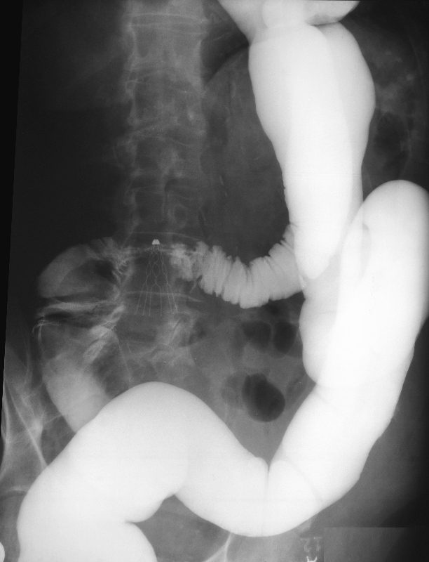
This is a different patient, also with a remote history of blunt trauma single contrast barium enema herniation of the distal portion of transverse colon, splenic flexure, and part of the descending colon into the thoracic cavity. The proximal colon is relatively decompressed but contains contrast suggesting mild obstruction.
Findings are consistent with a large traumatic diaphragmatic injury with secondary herniation of the colon into the chest cavity
Ashley Davidoff MD TheCommonVein.net 273Lu 115261.8