
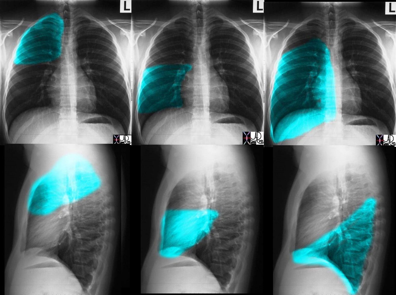
Frontal Examination of the Lungs
Ashley Davidoff MD
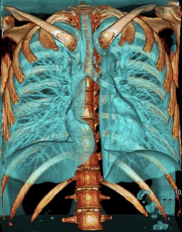
Parts of the Lungs- Basics
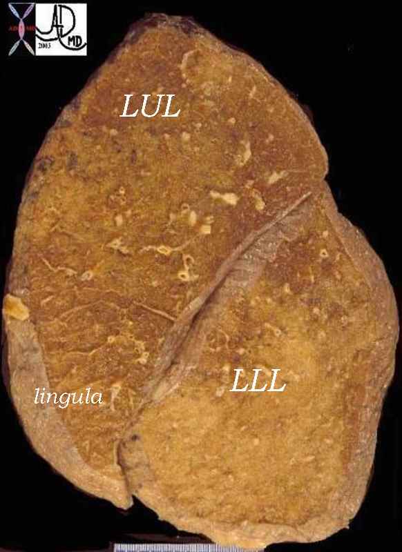
Left Lung – Left Upper Lobe – Frontal Projection
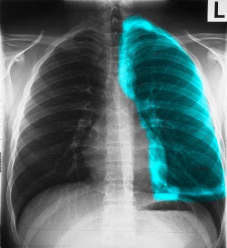
Ashley Davidoff MD
Frontal Projection Left Lower Lobe
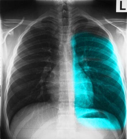
Ashley Davidoff MD
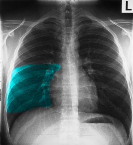
Right Lung – Right Upper Lobe – Frontal Projection
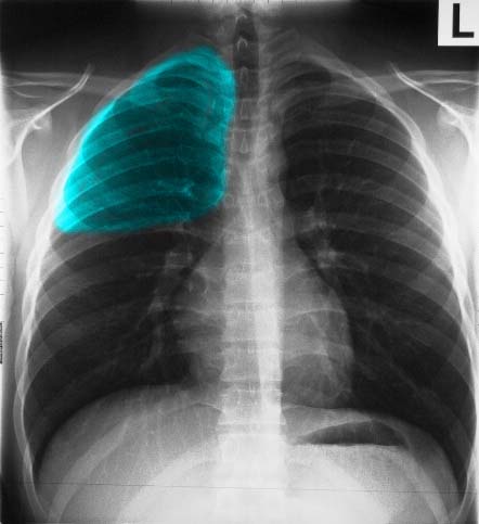
Ashley Davidoff MD
Right Middle Lobe – Frontal Projection
Ashley Davidoff MD
Right Lower Lobe – Frontal Projection
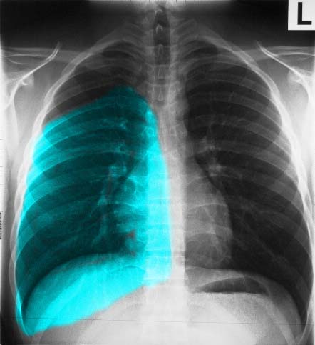
Ashley Davidoff MD
Lateral Examination of the Lungs
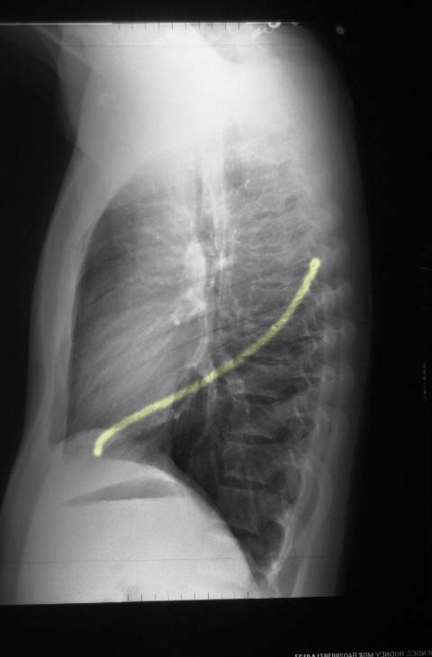
Major Fissure on the Left
Ashley Davidoff MD
Ashley Davidoff MD
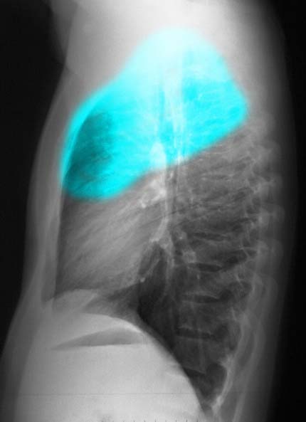
Lateral Projection – Left Upper Lobe
Ashley Davidoff MD
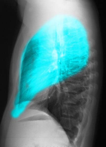
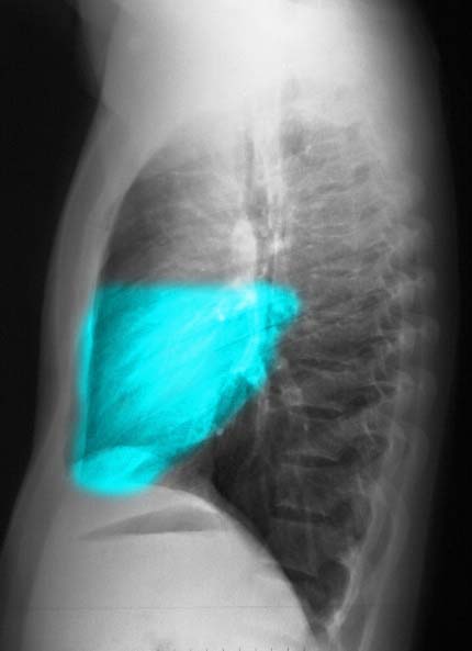
Lateral Projection left Lower Lobe
Ashley Davidoff MD
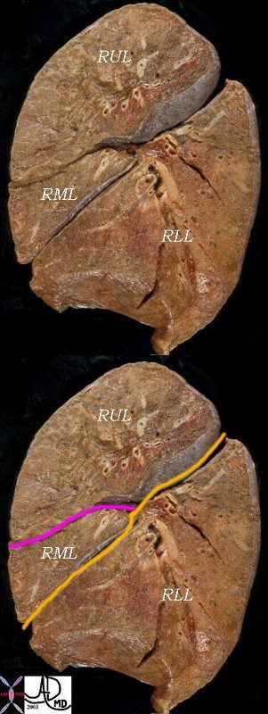
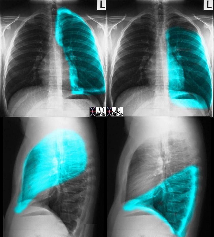
Major and Minor Fissures on the Right
The right lung has a small right upper lobe (RUL) separated from the middle lobe (RML) by the minor fissure (pink,lower image) . Both the RUL and RML are anterior and are separated from the lower lobe by the major fissure (orange line)
Ashley Davidoff MD
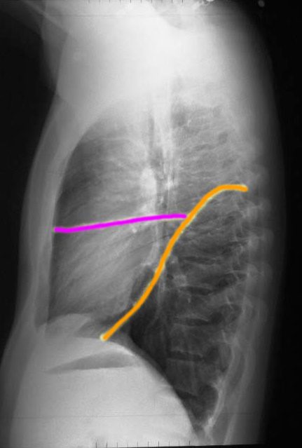
The right lung has a relatively small right upper lobe (RUL) separated from the middle lobe (RML) by the minor fissure (pink,lower image). Both the RUL and RML are anterior and are separated from the lower lobe by the major fissure (orange line)
Ashley Davidoff MD
Right Upper Lobe – Lateral Projection
Ashley Davidoff MD
Right Middle Lobe – Lateral Projection
Ashley Davidoff MD
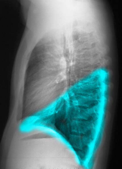
Right Lower Lobe – Lateral Projection
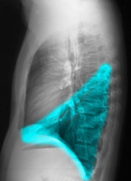
Ashley Davidoff MD
Summary
CXR of LEFT LUNG
Ashley Davidoff MD
Ashley Davidoff MD
Trachea, Main Stem Bronchi, and Carina
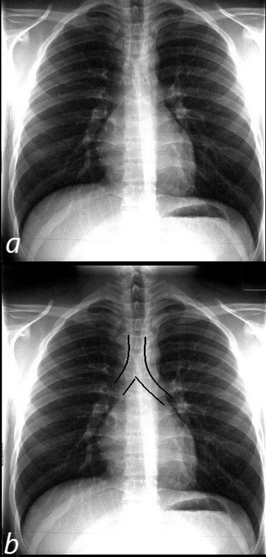
NORMAL FRONTAL CXR NORMAL ASYMMETRIC BRANCHING OF MAINSTEM BRONCHI
The normal CXR shows the characteristic asymmetric branching of the main stem bronchi. The right is short and stout and slightly more vertical while the left is long and thin and slightly more obtuse.
The normal carinal angle is between 40-80 degrees.
Ashley Davidoff MD
CARINAL ANGLE – 40-80 degrees
Ashley Davidoff MD

THE LEFT – TALL THIN AND GRACILE
The carinal angle

http://www.wikiradiography.net/

Courtesy Radiopaedia
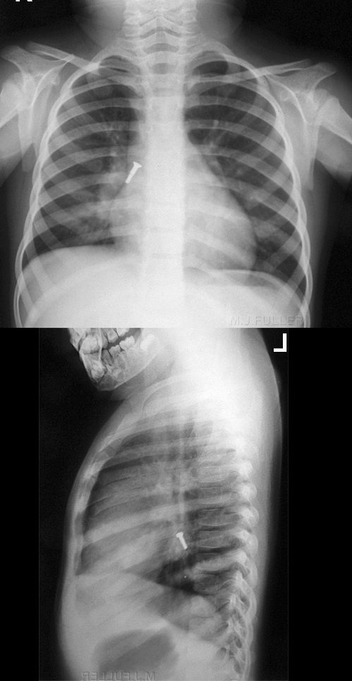
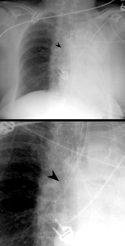
 ET TUBE IN RIGHT MAIN STEM BRONCHUS
Courtesy Radiopaedia
References and Links