
ACTIVE TB – Reactivation PLEEUARAL EFFUSIONS
80 year old Russian woman who intially presented with a cavitating LUL nodule that was biopsied and thought to represent sarcoidosis
In December the nodules in the LUL enlarged with an arborising pattern involving the posterior subsegment of the LUL as well as an unchanged RUL ground glass infiltrate
Susequent diagnosis of TB was made
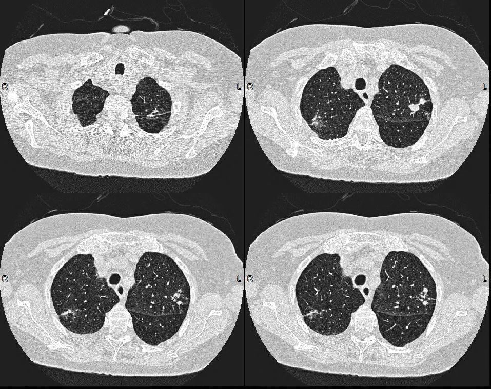
Initially there wasprogressive disease in the LUL and lingula with new cavitation in the lingula infiltrate/nodule and extension of the infiltrate in the LUL with a new calcification. These findings were consistent with reactivation TB .
Repeated sputa were positive for acid fast bacilli
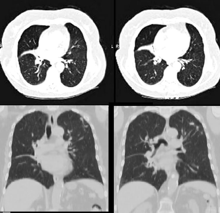
More recently new micronodularity was noted in the right lung .
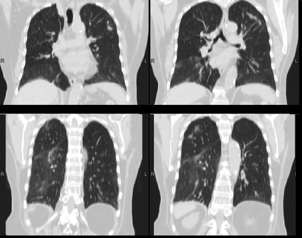
Now 1 month later she presents with a large right pleural effusion and a smaller left effusion
2.5 years prior
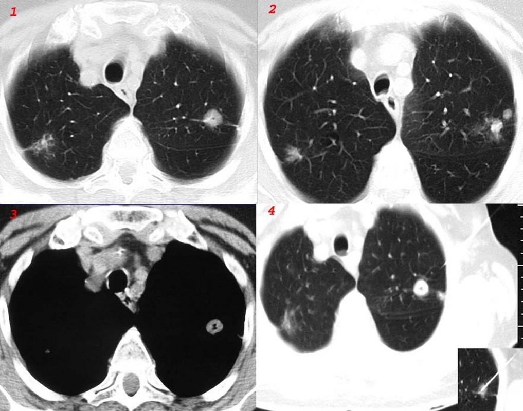
80 year old Russian woman who initially presented with a cavitating LUL nodule that was biopsied and thought to represent sarcoidosis. Subsequently confirmed to be TB
Ashley Davidoff MD TheCommonVein.net
2 Months after the Biopsy
2 months after the biopsy, the nodules in the LUL enlarged with an arborising pattern involving the posterior subsegment of the LUL as well as an unchanged RUL ground glass infiltrate
At this time neither calcification nor cavitation were present and there was no change on this examination when compared to the study 4 months earlier
Ashley Davidoff MD Ashley Davidoff MD TheCommonVein.net
Chronic atelectasis of the middle lobe is likely a sequela of the primaryTB
Ashley Davidoff MD Ashley Davidoff MD TheCommonVein.net
In the next 3 Months
Progressive ground glass opacities and extension of the nodules suggest reactivation
Ashley Davidoff MD Ashley Davidoff MD TheCommonVein.net
Multiple Test ConfirmTB
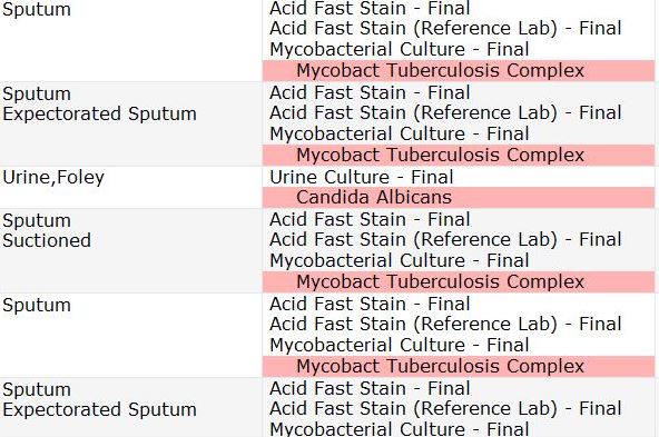
Multiple Tests confirm active TB
Ashley Davidoff MD TheCommonVein.net
3 Months Later
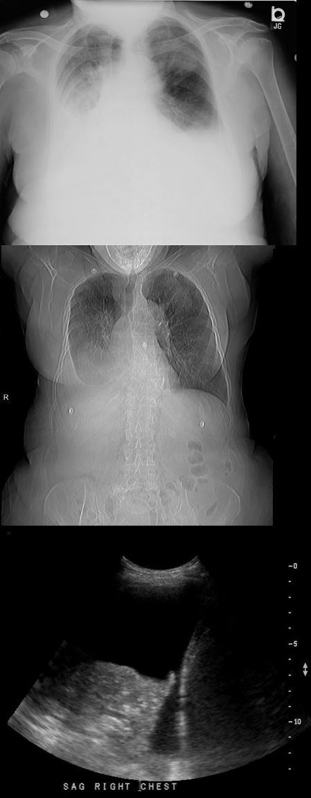
3 months later she presents with a large right pleural effusion and a smaller left effusion noted on the CXR and Ultrasound and the CT confirms the effusions and reveals progressive nodular enlargement and consolidation
Ashley Davidoff MD TheCommonVein.net
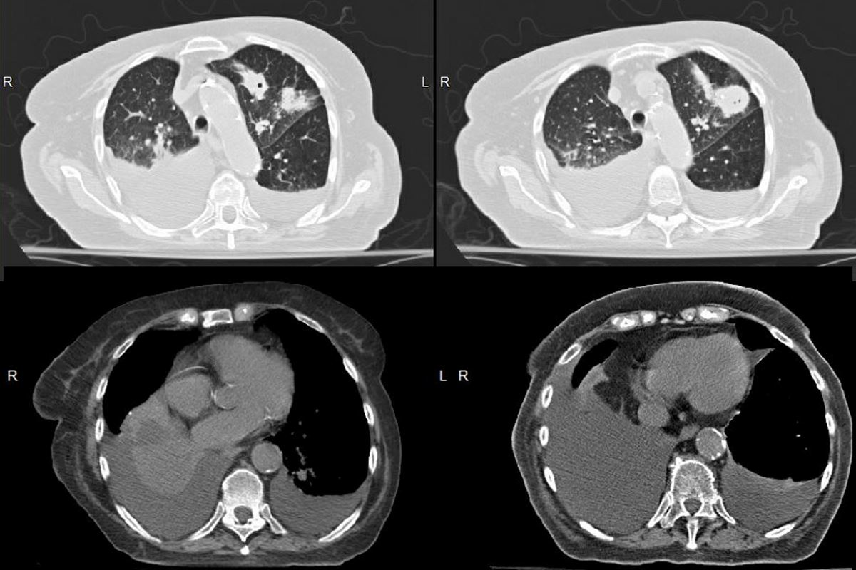
3 months later she presents with a large right pleural effusion and a smaller left effusion noted on the CXR and Ultrasound and the CT confirms the effusions and reveals progressive nodular enlargement and consolidation
Ashley Davidoff MD TheCommonVein.net