
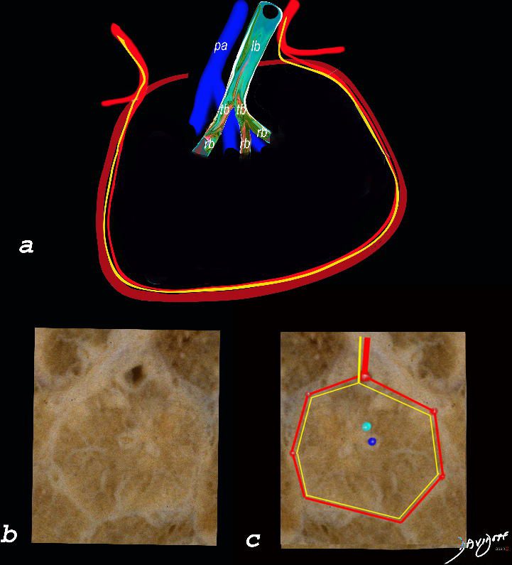
The top image (a) shows an anatomic drawing of a secondary lobule of the lung subtended by a lobular bronchiole (lb) and arteriole (pa). The interlobular septum contains the venule (red) lymphatic (yellow) and septum (maroon)
The anatomical specimen of the lung (b) shows normal intralobular parenchyma while image c shows the centrilobular arteriole (navy blue) and centrilobular bronchiole (teal) and interlobular venule (red) and lymphatics (yellow) The interlobular septum is slightly thickened
Ashley Davidoff TheCommonVein.net
CHF
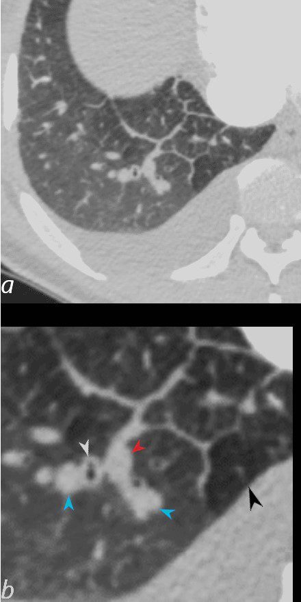
50-year-old female with diabetes, chronic renal failure and congestive heart failure. CT in the axial plane through the right posterior recess, shows thickened interlobular septa at the right base, congested arterioles (light blue arrowheads, b), alongside the bronchioles, peribronchial cuffing (white arrowheads, b), a congested pulmonary venule in the interlobular septum (red arrowhead arrowheads, b), ground glass changes and a secondary lobule demonstrating mosaic attenuation (black arrowhead arrowheads, b). The IVC is dilated and a small complex effusion is present.
Ashley Davidoff MD TheCommonvein.net 135783cL 193Lu
Venous Congestion and Thrombosis S/P PV Ablation
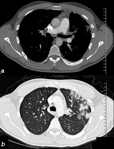
26 year old male who presents with shortness of breath a few months following RFA ablation for atrial fibrillation. Image a shows the left upper lobe thrombosed and the left lower vein contrast filled
Image b (the lung windows) shows thickening of the interlobular septa and secondary lobules distended with material, porobably representing blood.
following male SOB s/p RFA ablation for atrial fibrillation lung pulmonary let upper lobe pulmonary vein thrombosed secondary lobule interlobular septa are thickened secondary lobule destroyed dx pulmonary vein thrombosis secondary to radiofrequency ablation therapy iatrogenic CTscan Courtesy Ashley Davidoff MD Scott Tsai MD key words
ground glass changes pulmonary infarction venous infarction
TheCommonVein.net 75413c02