
Infection Inflammation Malignancy Mechanical/Atelectasis Trauma Metabolic Circulatory- Hemorrhage Immune Infiltrative Idiopathic Iatrogenic Idiopathic
Size
Shape
Position
Superior Segmental Lingular Infiltrate/Atelectasis Obstructing Hamartoma
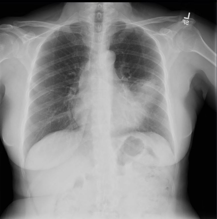
55-year-old female presents with a chronic cough
Frontal CXR shows an infiltrate involving the superior segment of the lingula, with partial silhouetting of the left heart border, and without associated secondary changes of volume loss
Final diagnosis was an obstructing hamartoma of the superior lingula bronchus
Courtesy Ashley Davidoff MD TheCommonVein.net 290 Lu 136563
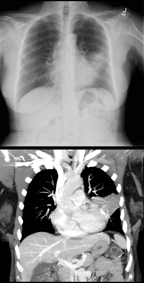
55-year-old female presents with a chronic cough
Frontal CXR and CT in the coronal plane shows an infiltrate involving the superior segment of the lingula, reflecting segmental post obstructive atelectasis and partial silhouetting of the superior aspect of the left heart border.
Final diagnosis was an obstructing hamartoma of the superior lingula bronchus
Courtesy Ashley Davidoff MD TheCommonVein.net 290 Lu 136564b
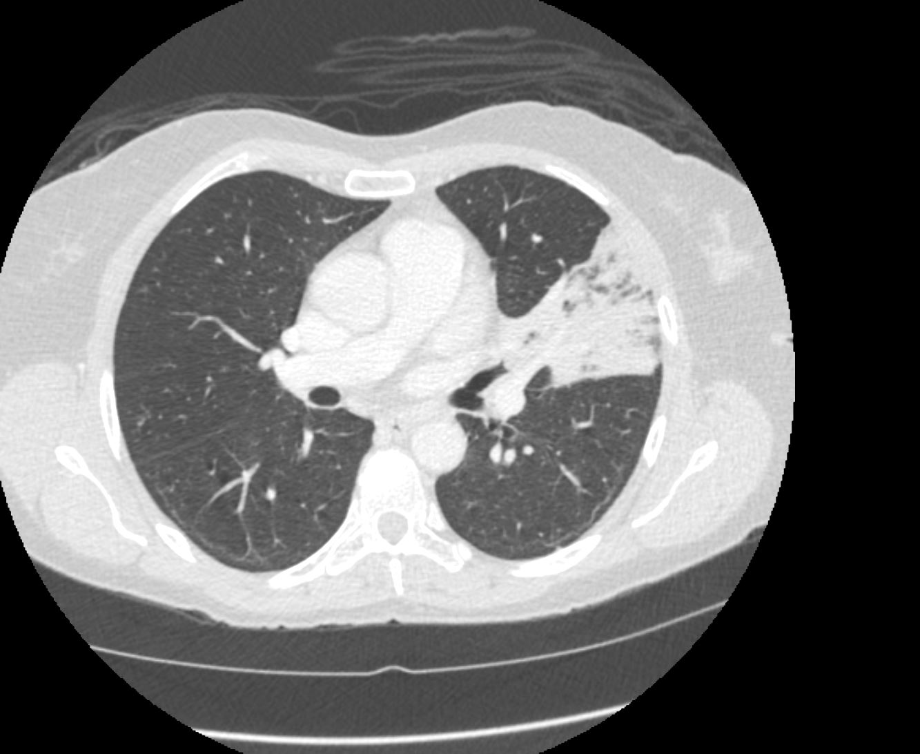
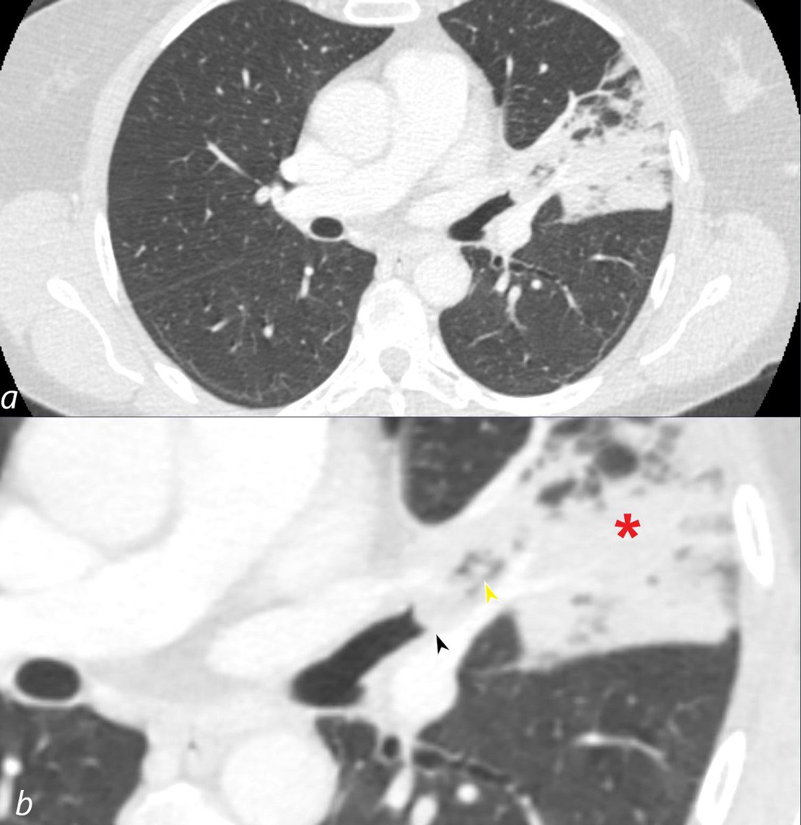
55-year-old female presents with a chronic cough
CT in the axial plane shows an infiltrate involving the superior segment of the lingula, reflecting segmental post obstructive atelectasis. A rounded soft tissue filling defect is noted in the subtending bronchus with downstream mucus accumulation
Final diagnosis was an obstructing hamartoma of the superior lingula bronchus
Courtesy Ashley Davidoff MD TheCommonVein.net 290 Lu 136565
Character
Infection
6.

52 year old male presents with a cough and fever
Frontal CXR shows a lingular infiltrate with a positive silhouette sign. Both the superior and inferior lingular segments appear to be involved
Ashley Davidoff MD TheCommonVein.net
Lingula Infiltrate in an Immunocompromised Individual
46 year old immunocompromised male presents with a fever. CXR shows a cavitating nodule in the left apex and a lingula infiltrate partially silhouetting the left heart border.
Ashley Davidoff TheCommonVein.net
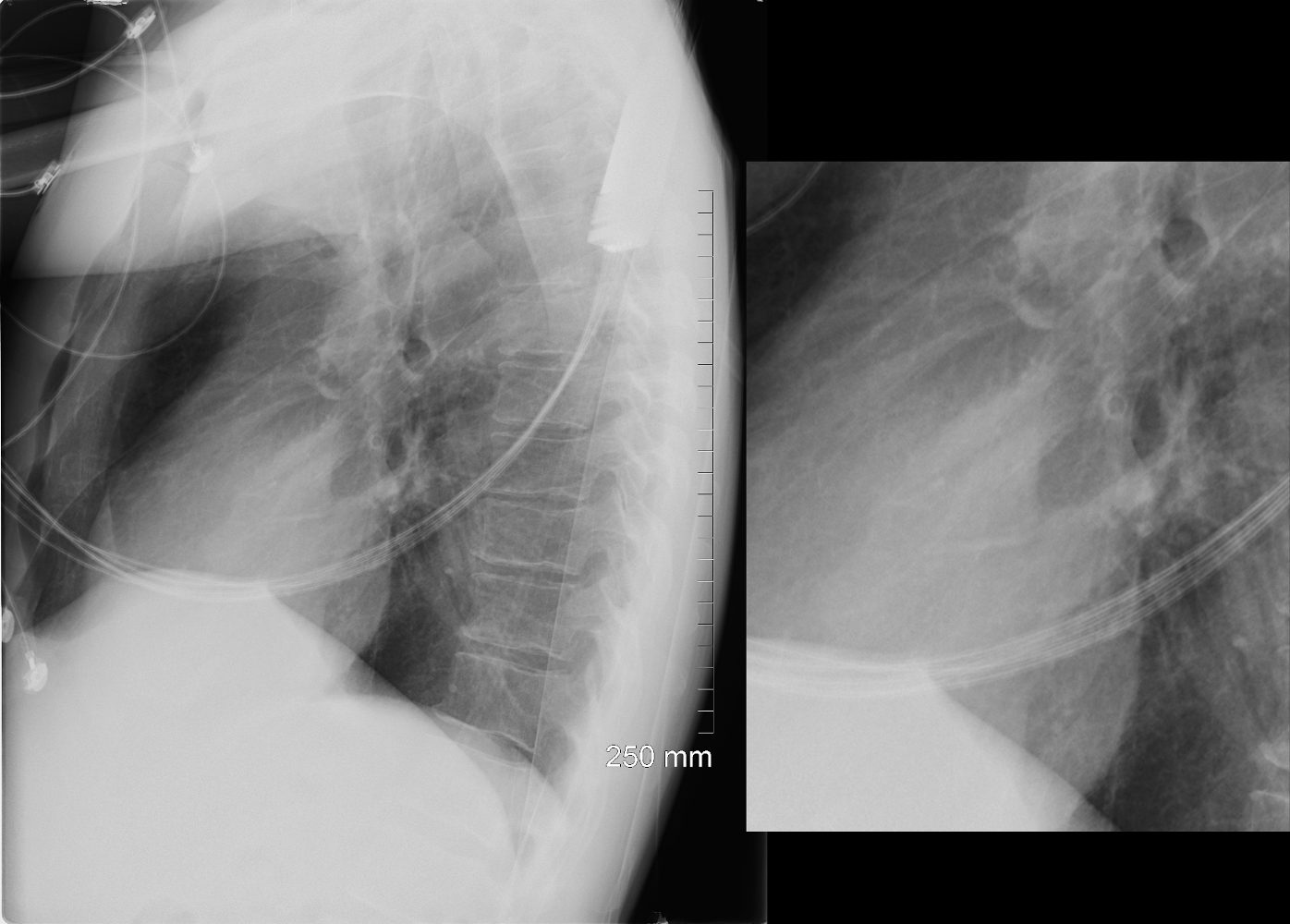
46 year old immunocompromised male presents with a fever. Lateral CXR shows a lingula infiltrate, partially silhouetting the left heart border better seen in the magnified view.
Ashley Davidoff TheCommonVein.net
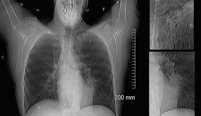
46 year old immunocompromised male presents with a fever. Scout for the CT scan shows a cavitating nodule in the left apex and a lingula infiltrate partially silhouetting the left heart border.
Ashley Davidoff TheCommonVein.net
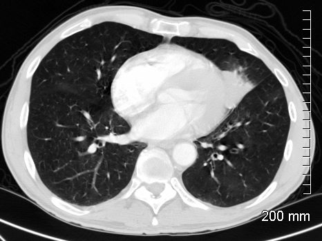
46 year old immunocompromised male presents with a fever. Axial CT shows a subsegmental lingula infiltrate, abutting and silhouetting the left heart border
Ashley Davidoff TheCommonVein.net
Neoplasm
Hamartoma
55-year-old female presents with a chronic cough
Frontal CXR shows an infiltrate involving the superior segment of the lingula, with partial silhouetting of the left heart border, and without associated secondary changes of volume loss
Final diagnosis was an obstructing hamartoma of the superior lingula bronchus
Courtesy Ashley Davidoff MD TheCommonVein.net 290 Lu 136563
55-year-old female presents with a chronic cough
CT in the axial plane shows an infiltrate involving the superior segment of the lingula, reflecting segmental post obstructive atelectasis (b red asterisk). A rounded soft tissue filling defect is noted in the subtending bronchus (b, black arrowhead) with downstream mucus accumulation (b yellow arrowhead)
Final diagnosis was an obstructing hamartoma of the superior lingula bronchus
Courtesy Ashley Davidoff MD TheCommonVein.net 290 Lu 136566cL
Mechanical
Atelectasis Due to ABPA Obstruction of the RLL
RLL Silhouetting the Diaphragm
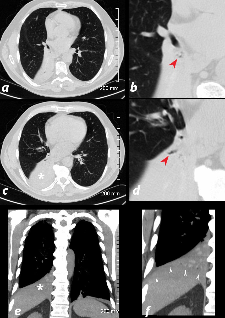
77 year old male presents chest discomfort
CT scan without contrast shows atelectasis of the right lower lobe )asterisk c and r) and also seen axial projection (a) magnified in (b) and in (c) magnified in {d) Red arrowheads in b and d show airways filled with material. Aspergillus was isolated at bronchoscopy. Coronal imaging (e magnified in f) show silhouetting of the right hemidiaphragm by the atelectatic lung (white arrowheads
Ashley Davidoff TheCommonVein.net 117786cL