
Nodules
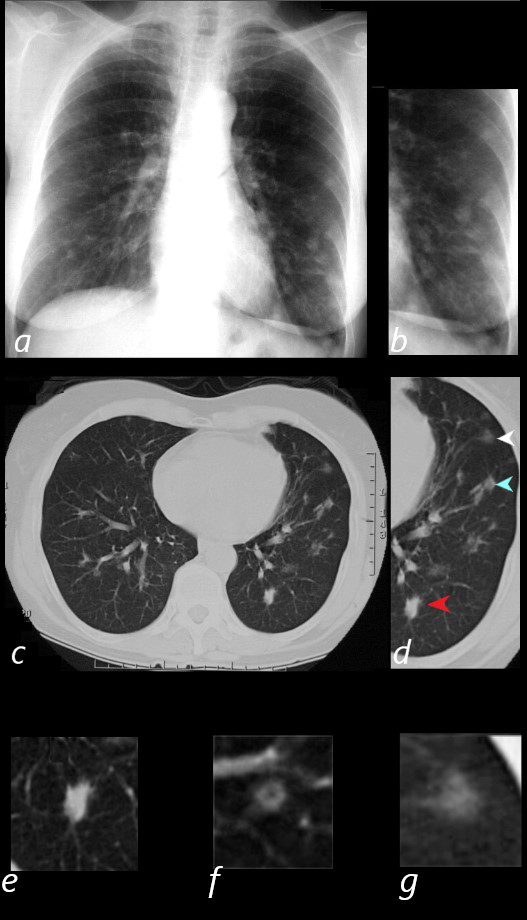
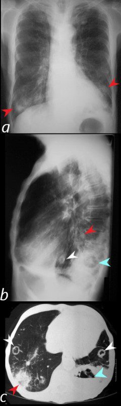
65 year old female presents with epistaxis and with nodular changes on CXR (a) magnified in b.
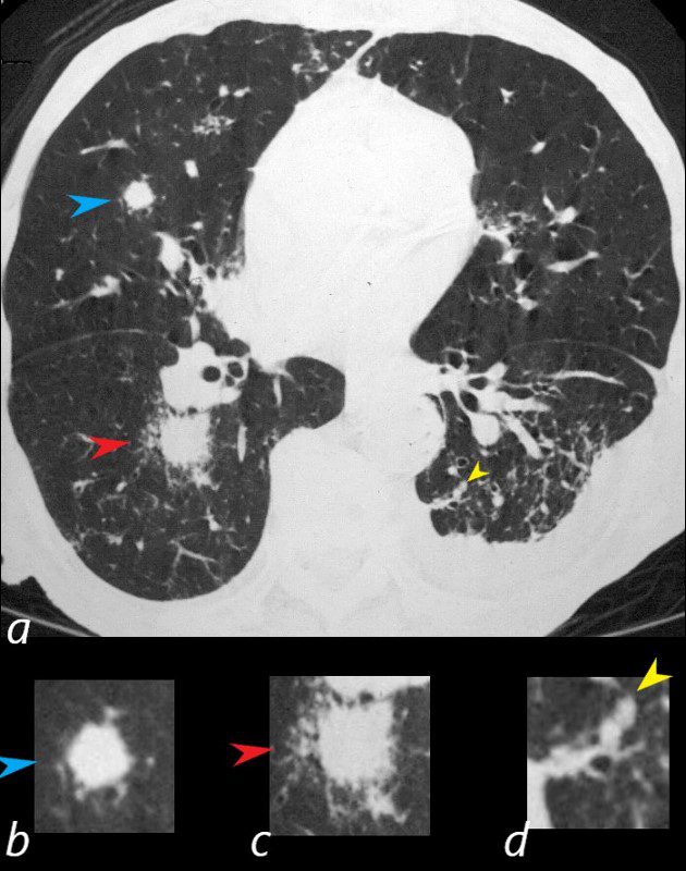
CT scan in axial projection (c) and magnified in d, reveals 3 types of nodules.
A spiculated solid nodule (red arrow head) is magnified in e, a bronchocentric nodule (teal arrowhead) is magnified in e. This may represent a cavitating nodule or hemorrhagic change around a bronchiole (cheerio sign) A ground glass nodule (white arrowhead) is magnified in g.
Ashley Davidoff MD
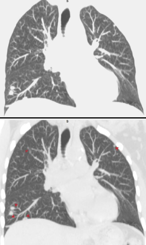
Coronal imaging shows multiple small nodules in the upper and lower lung field with noted subtending vessels
(feeding vessel sign)
Ashley Davidoff MD
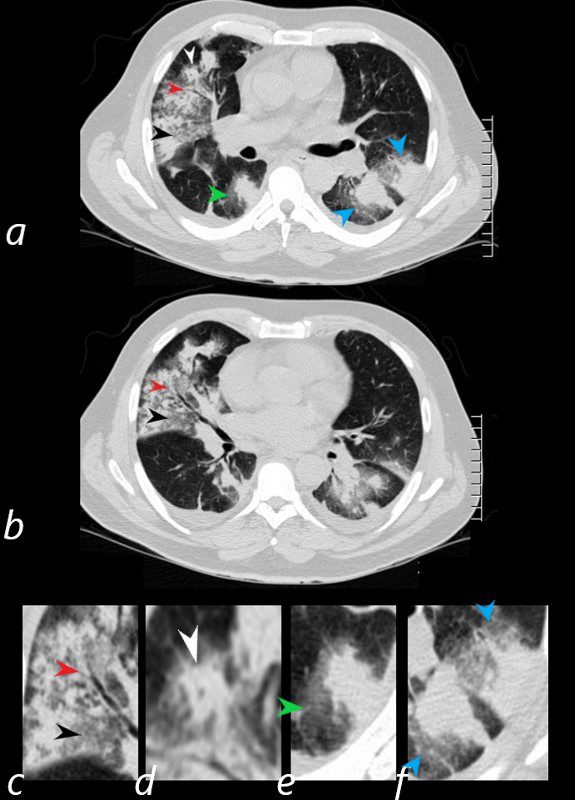
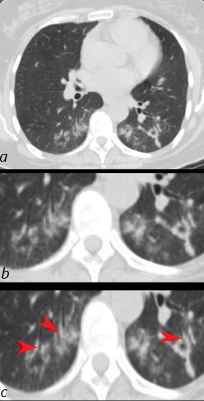
57 year old male presents with a history of hemoptysis and dyspnea.
Axial CT scans show multicentric nodular consolidations with air bronchograms (red arrowheads a ,b, c), ground glass infiltrate (black arrowheads, a,b,c) halo sound around nodules and masses in the RLL (a,e, green arrowheads) and in the LLL 9a,f, blue arrowheads). Lastly there is a cheerio sign (a,d, white arrowheads) either representing granulomatous mass surrounding an airway, or central cavitation of a nodule.
Ashley Davidoff MD
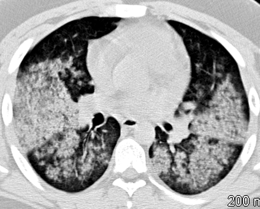
81-year-old male with weight loss, renal failure, and hemoptysis
CT axial view (a) shows a 2 cm solid nodule in the RUL surrounded by A HALO SIGN OF GROUND GLASS CHANGES AND RETICULAR CHANGES (a,b, red arrowheads), indicating surrounding hemorrhage, and subtle air bronchograms (a,b,c, teal arrowheads) best appreciated in c with narrowed windows.
Priscilla Slanetz MPH MD
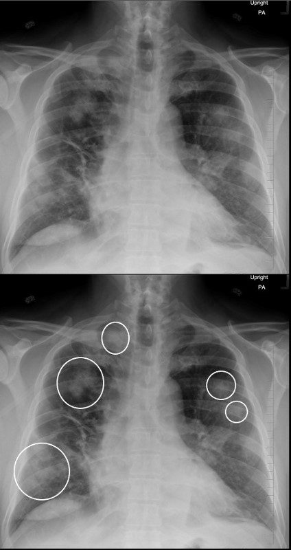
57 year old male presents with a history of hemoptysis and dyspnea. Frontal CXR shows nodular infiltrates dominantly involving the upper lung zones but also the lower lung zones (circled in white) The patient was subsequently shown to have ANCA positive vasculitis and further characterized as Wegener’s granulomatosis
Ashley Davidoff MD
Nodules and Masses
57 year old male presents with a history of hemoptysis and dyspnea.
Axial CT scans show multicentric nodular consolidations with air bronchograms (red arrowheads a ,b, c), ground glass infiltrate (black arrowheads, a,b,c) halo sound around nodules and masses in the RLL (a,e, green arrowheads) and in the LLL 9a,f, blue arrowheads). Lastly there is a cheerio sign (a,d, white arrowheads) either representing granulomatous mass surrounding an airway, or central cavitation of a nodule.
Ashley Davidoff MD.
57 year old male presents with a history of hemoptysis and dyspnea.
Axial CT scans show multicentric nodular consolidations with air bronchograms (red arrowheads a ,b, c), ground glass infiltrate (black arrowheads, a,b,c) halo sound around nodules and masses in the RLL (a,e, green arrowheads) and in the LLL 9a,f, blue arrowheads). Lastly there is a cheerio sign (a,d, white arrowheads) either representing granulomatous mass surrounding an airway, or central cavitation of a nodule.
Ashley Davidoff MD
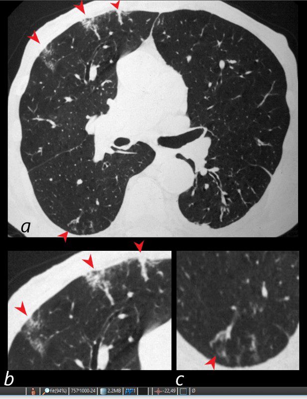
81-year-old male with weight loss, renal failure, and hemoptysis
CT axial view (a) shows a 1 cm solid nodule in the RUL surrounded by small vessels (a,b, blue arrowheads), a 1.8cm nodule in the superior segment of the right lower lobe with a halo sign indicating surrounding hemorrhage(a,c, red arrowheads), and a 3mm nodule in the superior segment of the left lower lobe (a,d, yellow arrowheads).
Priscilla Slanetz MPH MD
Masses
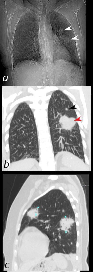
54 year old female presented with painless persistent dry cough, loss of appetite, weight loss, and worsening renal function. Urinary sediment showed white cells suggestive of glomerulonephritis. ANCA test and ANA were negative, ANA negative. Vasculitis was suspected and she was started on solumedrol and cyclophosphamide which improved her symptoms.
She had an uneventful renal biopsy.
The scout film (a) shows two mass like lesions in the left mid and upper lung zones,(white arrowheads) with coronal (b) imaging showing a nodule (black arrowhead) and a mass (red arrowhead) also in the mid and upper left lung. Image c in sagittal projection shows a large mass in the superior segment of the LUL and a second in the anterior segment of the LUL both both with air bronchograms(teal arrowheads). For the large mass like lesions, subacute hemorrhage is a radiological consideration and for the smaller nodules granulomatous nodules of Wgeners seems to be more likely.
Ashley Davidoff MD
81-year-old male with weight loss, renal failure, and hemoptysis
CT axial view (a) shows a 2 cm solid nodule in the RUL surrounded by A HALO SIGN OF GROUND GLASS CHANGES AND RETICULAR CHANGES (a,b, red arrowheads), indicating surrounding hemorrhage, and subtle air bronchograms (a,b,c, teal arrowheads) best appreciated in c with narrowed windows.
Priscilla Slanetz MPH MD
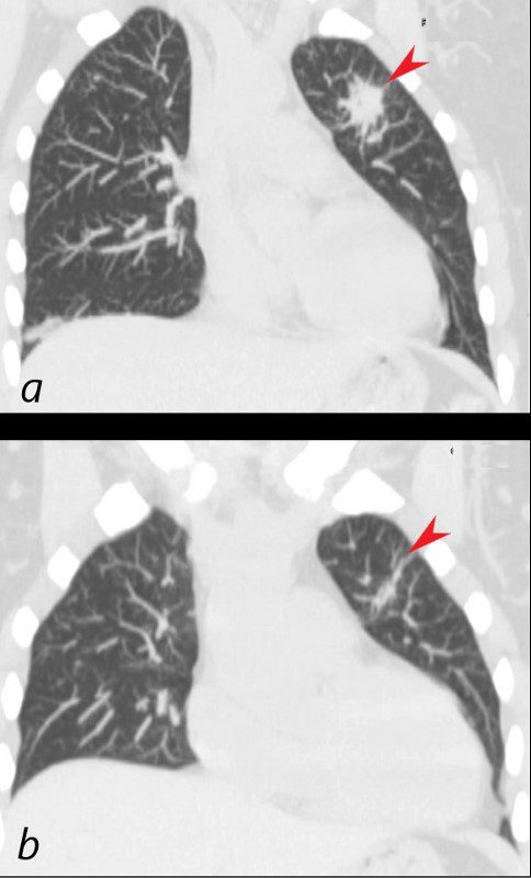
Initial mass like consolidation (a, arrowhead) has almost resolved (b, arrowhead) after 3 months of treatment with steroids and cyclophosphamide
Ashley Davidoff MD
Cavitating Nodules and Masses
81-year-old male with weight loss, renal failure, and hemoptysis
CXR frontal view (a) shows lower lobe nodules (red arrowheads). The lateral examination (b) shows a small solid nodule (red arrowhead), cavitating thick walled nodule (white arrowhead), and an air pocket posteriorly within a mass like abnormality (teal arrowhead)
Priscilla Slanetz MPH MD
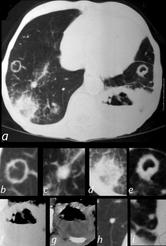
81-year-old male with weight loss, renal failure, and hemoptysis
CT axial view (a) shows cavitating masses in the upper lobes bilaterally magnified (b and e). In addition there is a large necrotic mass with an air fluid level in the left lower lobe (a), magnified ( f and g). A mass with a halo sign noted in the RLL is magnified in d, and scattered bilateral smaller nodules are magnified (c and h) in the RLL and in (i) in the LLL .
Priscilla Slanetz MPH MD
Hemorrhage
Acute Pulmonary Hemorrhage
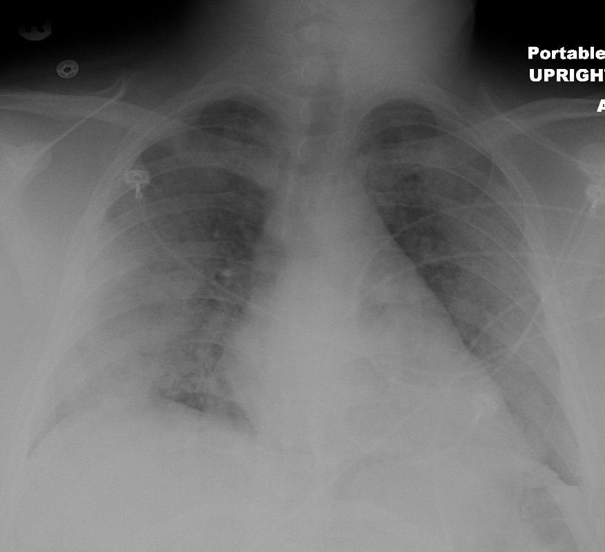
19 year old male previously well with history of hemoptysis, sweating, fevers, myalgias, arthritis over 3 weeks.
The chest x-ray shows diffuse bilateral lobar infiltrates sparing the hilar regions.
Lab shows ANCA positivity, acute renal failure (creatinine 6) and renal biopsy showing crescentic glomerulonephritis.
Treated with cyclophosphamide
Ashley Davidoff MD
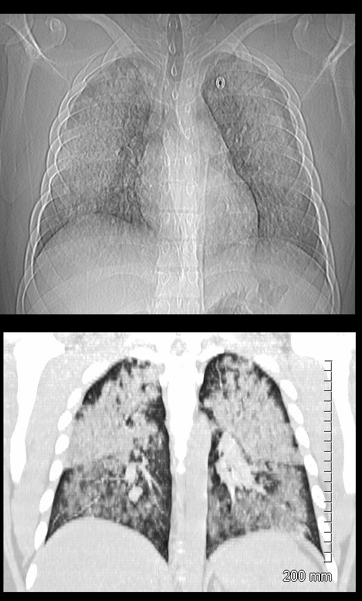
19 year old male previously well with history of hemoptysis, sweating, fevers, myalgias, arthritis over 3 weeks.
CT scan scout (above ) shows diffuse bilateral lobar infiltrates sparing the hilar regions.
Coronal CT shows bilateral symmetrical lobar infiltrates involving the upper and lower lobes with acinar pattern. The upper lobes are more affected than the lower lobes.
Lab shows ANCA positivity, acute renal failure (creatinine 6) and renal biopsy showing crescentic glomerulonephritis.
Treated with cyclophosphamide
Ashley Davidoff MD
57 year old male presents with a history of hemoptysis and dyspnea.
Axial CT scans show multicentric nodular consolidations with air bronchograms (red arrowheads a ,b, c), ground glass infiltrate (black arrowheads, a,b,c) halo sound around nodules and masses in the RLL (a,e, green arrowheads) and in the LLL 9a,f, blue arrowheads). Lastly there is a cheerio sign (a,d, white arrowheads) either representing granulomatous mass surrounding an airway, or central cavitation of a nodule.
Ashley Davidoff MD
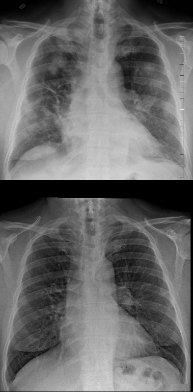
Frontal CXR (above) shows nodular infiltrates dominantly involving the upper lung zones but also the lower lung zones The patient was subsequently shown to have ANCA positive vasculitis and further characterized as Wegener’s granulomatosis
He was treated and the lower frontal X Ray of the chest (CXR) shows resolution of the infiltrates
Ashley Davidoff MD
Air Bronchograms
54 year old female presented with painless persistent dry cough, loss of appetite, weight loss, and worsening renal function. Urinary sediment showed white cells suggestive of glomerulonephritis. ANCA test and ANA were negative, ANA negative. Vasculitis was suspected and she was started on solumedrol and cyclophosphamide which improved her symptoms.
She had an uneventful renal biopsy.
The scout film (a) shows two mass like lesions in the left mid and upper lung zones,(white arrowheads) with coronal (b) imaging showing a nodule (black arrowhead) and a mass (red arrowhead) also in the mid and upper left lung. Image c in sagittal projection shows a large mass in the superior segment of the LUL and a second in the anterior segment of the LUL both both with air bronchograms(teal arrowheads). For the large mass like lesions, subacute hemorrhage is a radiological consideration and for the smaller nodules granulomatous nodules of Wgeners seems to be more likely.
Ashley Davidoff MD
6 days after presentation and early treatment there are resolving consolidations (a, magnified in b ) with red arrowheads in c showing the air bronchograms.
Ashley Davidoff MD
57 year old male presents with a history of hemoptysis and dyspnea.
Axial CT scans show multicentric nodular consolidations with air bronchograms (red arrowheads a ,b, c), ground glass infiltrate (black arrowheads, a,b,c) halo sound around nodules and masses in the RLL (a,e, green arrowheads) and in the LLL 9a,f, blue arrowheads). Lastly there is a cheerio sign (a,d, white arrowheads) either representing granulomatous mass surrounding an airway, or central cavitation of a nodule.
Ashley Davidoff MD.
57 year old male presents with a history of hemoptysis and dyspnea.
Axial CT scans show multicentric nodular consolidations with air bronchograms (red arrowheads a ,b, c), ground glass infiltrate (black arrowheads, a,b,c) halo sound around nodules and masses in the RLL (a,e, green arrowheads) and in the LLL 9a,f, blue arrowheads). Lastly there is a cheerio sign (a,d, white arrowheads) either representing granulomatous mass surrounding an airway, or central cavitation of a nodule.
Ashley Davidoff MD
Vasculitis and Pulmonary Infarction
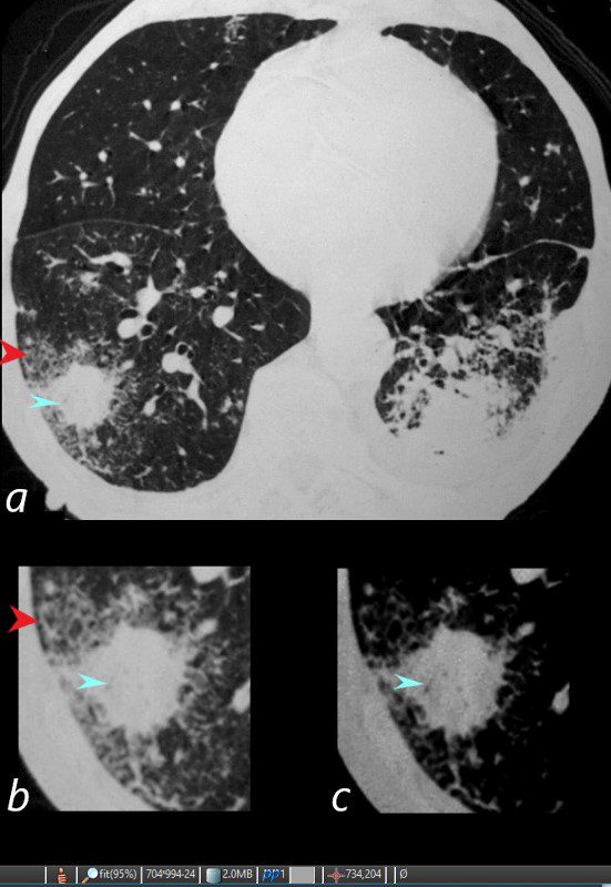
81-year-old male with weight loss, renal failure, and hemoptysis
CT axial view (a) shows multiple peripheral wedge shaped ground glass densities subtended by distended feeding vessels (a,b,c, red arrowheads) reflecting areas of microinfarction due to vasculitis that affects both the arterioles and venules.
Priscilla Slanetz MPH MD
Airways
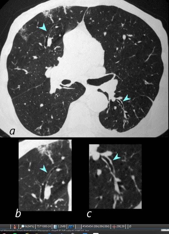
81-year-old male with weight loss, renal failure, and hemoptysis
CT axial view (a) shows focal nodular thickening of the small airways (a,b,c, teal arrowheads) likely reflecting areas of granulomatous changes.
Priscilla Slanetz MPH MD
Treatment – Then and Now
Frontal CXR (above) shows nodular infiltrates dominantly involving the upper lung zones but also the lower lung zones The patient was subsequently shown to have ANCA positive vasculitis and further characterized as Wegener’s granulomatosis
He was treated and the lower frontal X Ray of the chest (CXR) shows resolution of the infiltrates
Ashley Davidoff MD
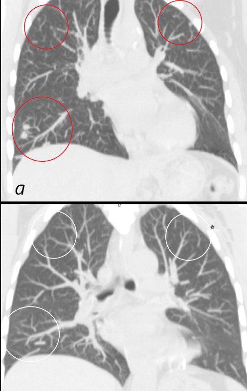
Multiple nodules noted on initial presentation (a, red circles) have almost resolved (b, white circles) after 3 months of treatment with steroids and cyclophosphamide.
Ashley Davidoff MD
Initial mass like consolidation (a, arrowhead) has almost resolved (b, arrowhead) after 3 months of treatment with steroids and cyclophosphamide
Ashley Davidoff MD
is an ANCA-associated vasculitis, (anti-neutrophil cytoplasm antibodies (ANCA) ) which includes 3 diseases
-
- Wegener’s granulomatosis now known as granulomatosis with polyangiitis (GPA)
- Churg-Strauss syndrome now known as eosinophilic granulomatosis with polyangiitis (EGPA)
- Microscopic polyangiitis (MPA)
References and Links
“Granulomatosis with polyangiitis (Wegener granulomatosis), is a multi-system systemic necrotizing non-caseating granulomatous vasculitis affecting small to medium-sized arteries, capillaries and veins 1, and the lungs are the most frequently involved organ, seen in 95% of cases.