
86year old patient who presented with long standing history of a productive cough
6 Months Prior
Prominence of the
Basilar Broncho-vacsclar Bundles
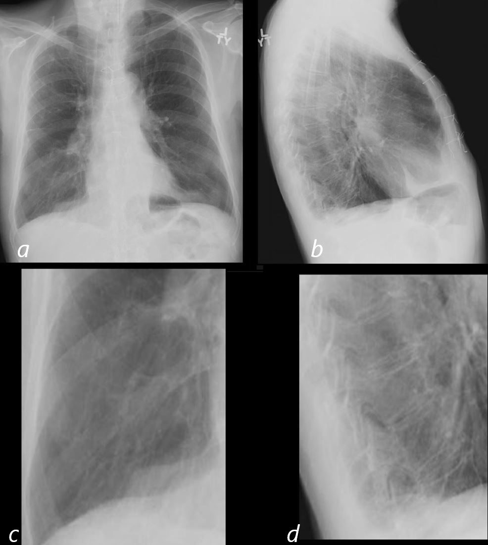
86year old patient with known emphysema and chronic bronchitis CXR 6 months prior to admission shows evidence of hyperinflation with flattened hemidiaphragms and increase in the retrosternal air space (b) On the PA view (a magnified in c) there is a suggestion of bronchovascular thickening. On the lateral view (b and magnified in d) the expected progressive lucency of the vertebral bodies is lost and there is outlining of a bronchus just anterior to the vertebral bodies confirming the bronchovascular thickening.
Ashley Davidoff MD TheCommonVein.net 30602d03cL
Acute on Chronic Cough and Fever
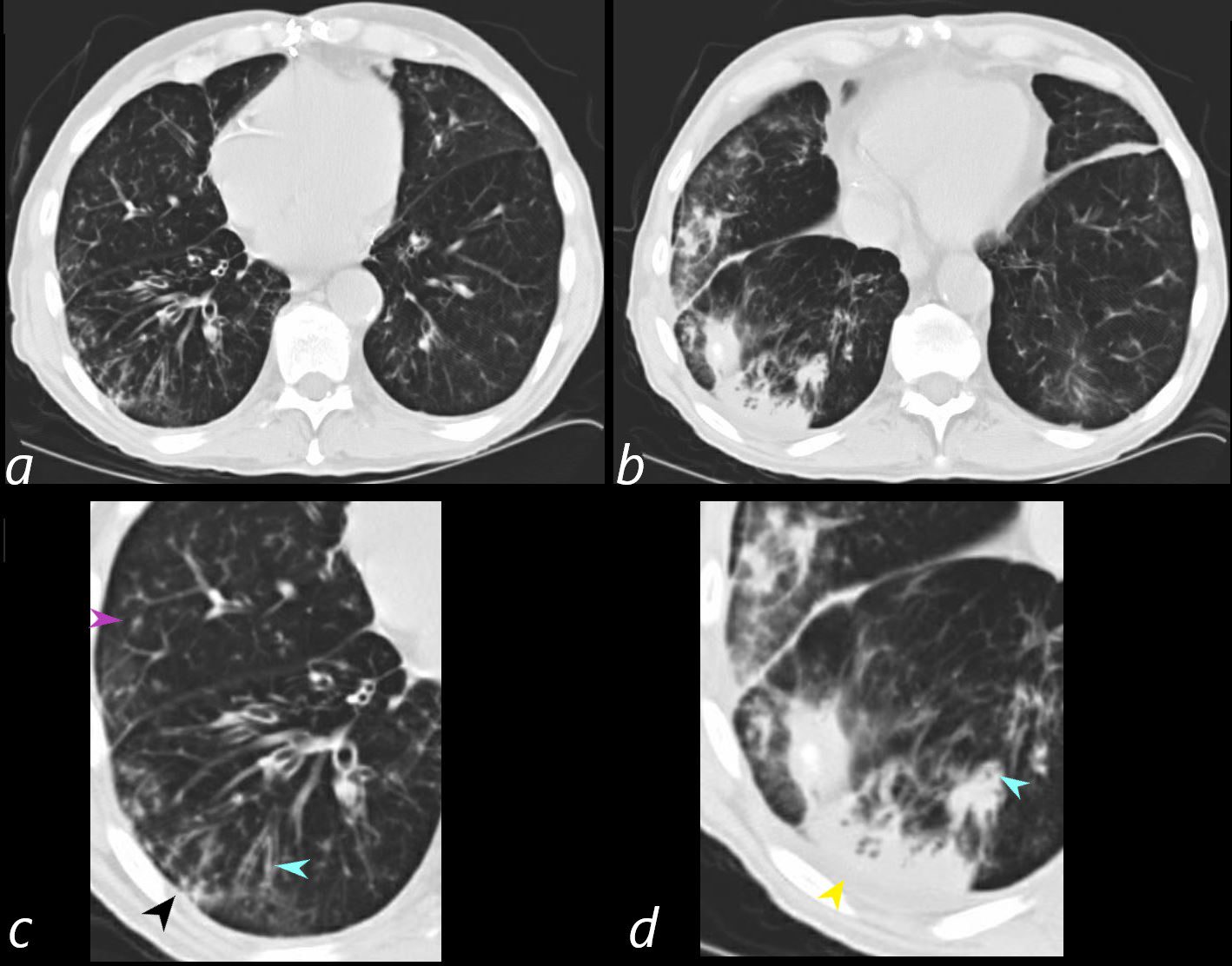
86year old patient with known emphysema and chronic bronchitis presents with fever and an acute on chronic productive cough The CT scan of the chest through the lung bases shows chronic changes at the left lung base (a and magnified in c) with thickening and partial impaction of the subsegmental airways (blue arrowhead c and d) centrilobular nodule (c purple arrowhead) and tree in bud nodules posteriorly (c, black arrowhead). There is an acute segmental consolidation in the right lower lobe (b and d yellow arrowhead) and a subsegmental infiltrate in the lateral segment of the middle lobe. These findings are consistent with an acute on chronic aspiration pneumonia
Ashley Davidoff MD TheCommonVein.net 30602b01L
Chronic Changes Segmental Subsegmental and
Small Airway Disease
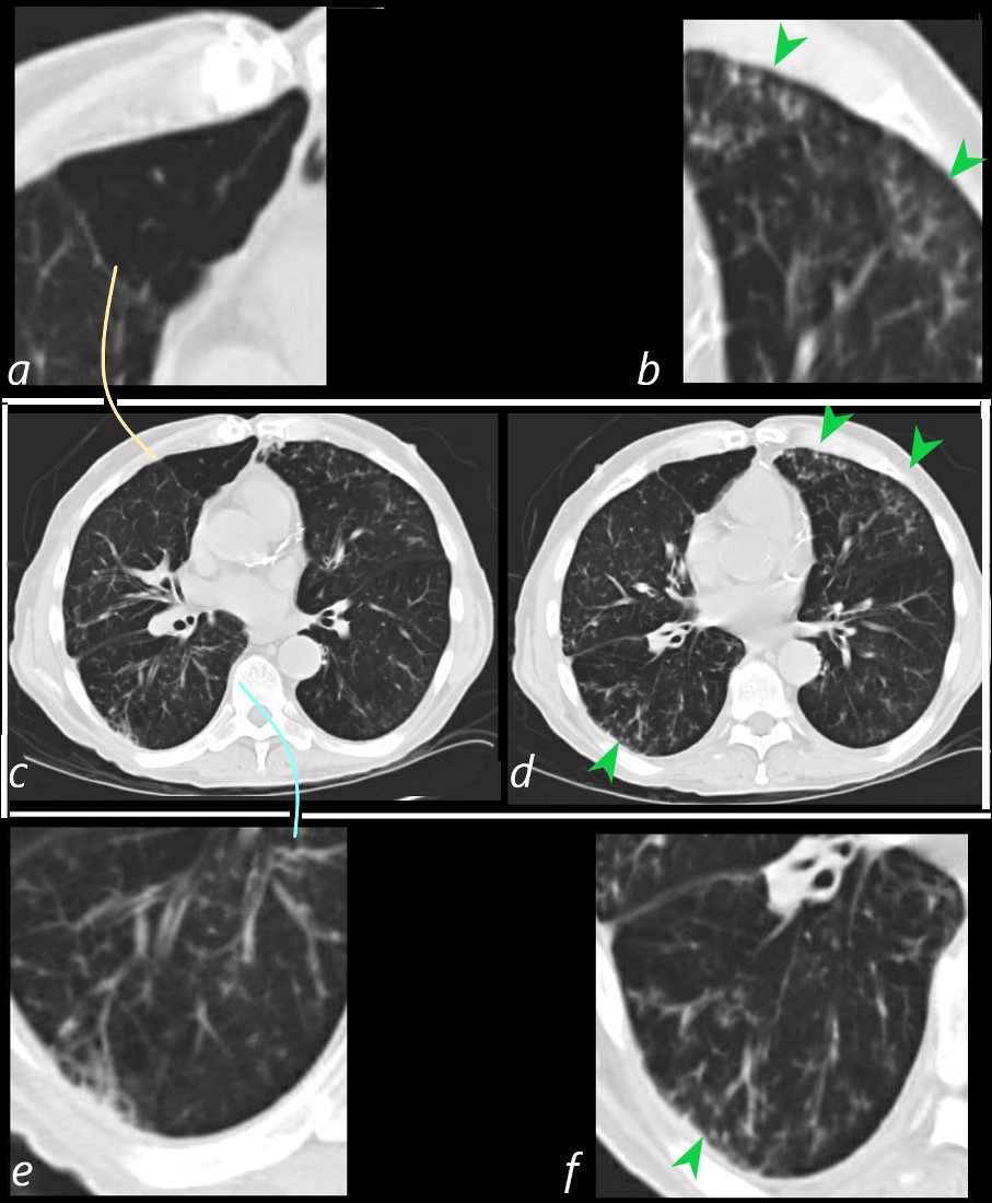
86year old patient with known emphysema and chronic bronchitis presents with a fever and an acute on chronic productive cough The CT scan of the chest through the mid lung fields shows a subsegmental region of air trapping in the medial segment of the middle lobe (c and magnified in a – orange arc). There is evidence of segmental and subsegmental airway thickening (c magnified in e – teal arc) with evidence of small airway disease characterised by tree in bud changes (green arrowhead d magnified in b and f). These findings are non-specific, but in the current context represent manifestations of aspiration.
Ashley Davidoff MD TheCommonVein.net 30602bL
Barium Swallow Aspiration
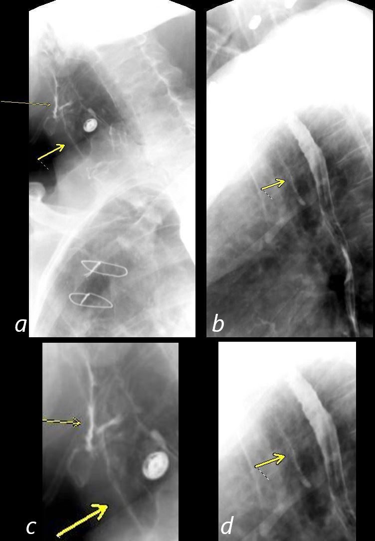
86year old patient with known emphysema and chronic bronchitis presents with fever and an acute on chronic productive cough. Barium swallow confirms the presence of aspiration into the vestibule and onto the cords (non-solid arrow a and c) and into the proximal trachea (solid yellow arrow) . Images b and magnified in d show contrast in the distal trachea – solid yellow arrow.
Ashley Davidoff MD TheCommonVein.net 30602d04L
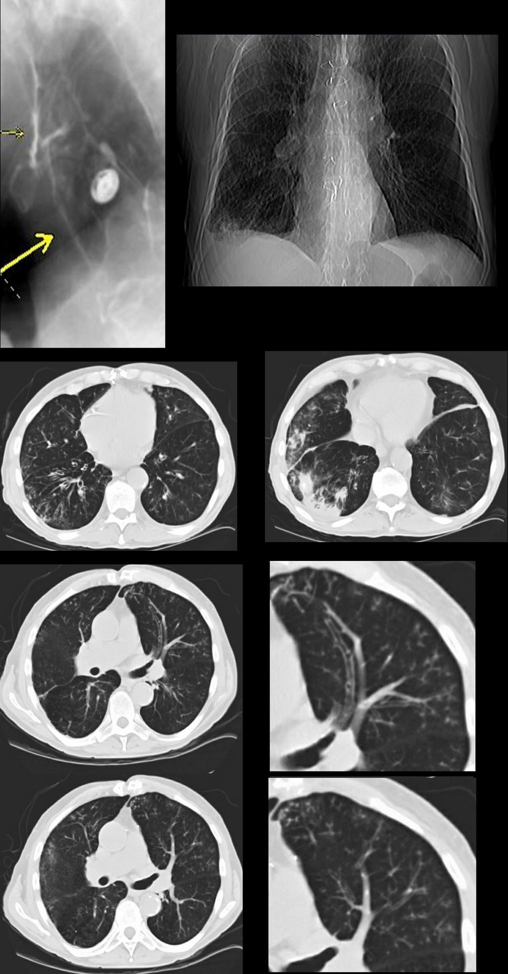
87 year old male with history of cough and suspicion of aspiration shows barium aspiration into the proximal trachea (upper right) The scout view ( upper right) shows an infiltrate at the right base, Thickened airways in the right lower lobe (2nd row left ) is associated with a pneumonic infiltrate in the right lower lobe (lower right) consistent with aspiration. There are thickened airways to the lingula (3rd and 4th row) with magnified view showing tree in bud changes (right sided images 3rd and 4th row)
All these finding likely relate to spiration though lingula involvement is not usual
Ashley Davidoff MD Ashley Davidoff MD TheCommonVein.net
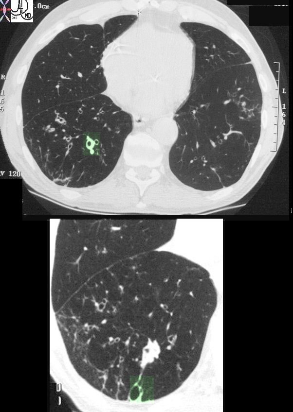
This image represents the CT scan of the chest with a focus on the right lower lobe in an 86year old patient who presented with long standing history of a productive cough. The segmental airways in the upper image are thickened (green overlay) as are the subsegmental airways in the lower panel. Additionally an small airways is abnormally visualised within 1cms of the pleura, indicating traction bronchiolectasis. Aspiration is a radiological consideration
Ashley Davidoff MD TheCommonVein.net 30602c