
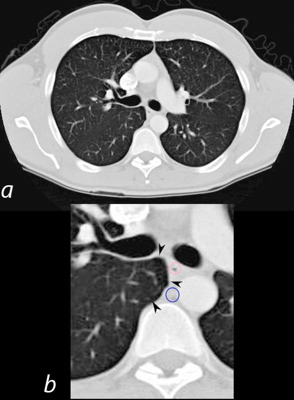
CT in the axial plain shows the azygoesophageal recess, (black arrowheads, bounded medially by the esophagus (pink ring ) and the azygous vein (blue ring
Ashley Davidoff MD TheCommonVein.net 38803c
Fleischner Society
azygoesophageal recess
Anatomy.—The azygoesophageal recess is a right posterior mediastinal recess into which the edge of the right lower lobe extends. It is limited superiorly by the azygos arch, posteriorly by the azygos vein and pleura anterior to the vertebral column, and medially by the esophagus and adjacent structures.
Radiographs and CT scans.—On a frontal chest radiograph, the recess is seen as a vertically oriented interface between the right lower lobe and the adjacent mediastinum (the medial limit of the recess). Superiorly, the interface is seen as a smooth arc with convexity to the left. Disappearance or distortion of part of the interface suggests disease (eg, subcarinal lymphadenopathy). On CT scans, the recess (,Fig 9) merits attention because small lesions located in the recess will often be invisible on chest radiographs (,23).