
Size
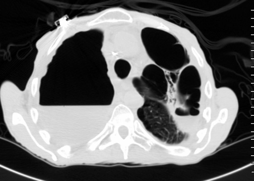
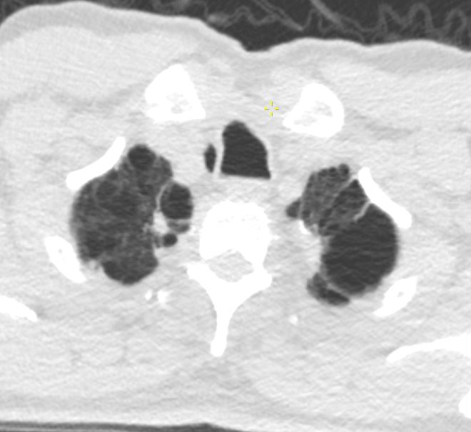
65-year-old male with emphysema of the lungs presents with a cough, fever and leukocytosis. CT in the axial plane shows extensive apical bullous lung disease. There is a large right upper lobe bulla with an air fluid level and a smaller left upper lobe bulla with an air fluid level.
Ashley Davidoff MD TheCommonVein.net 259Lu 117471
Shape
Position
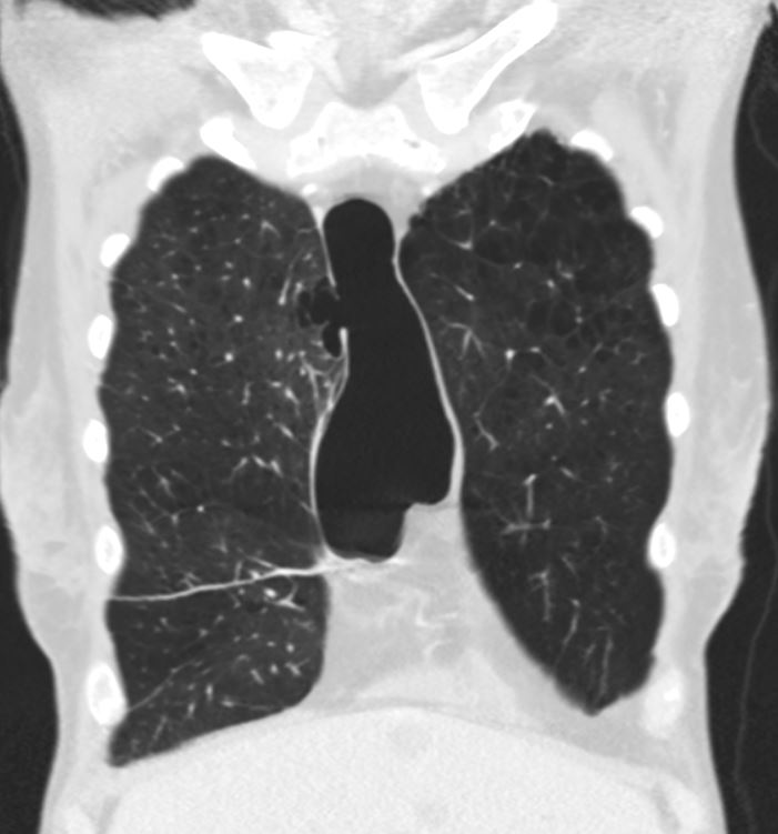
CT Scan Bilateral Apical Bulla Centrilobular Emphysema
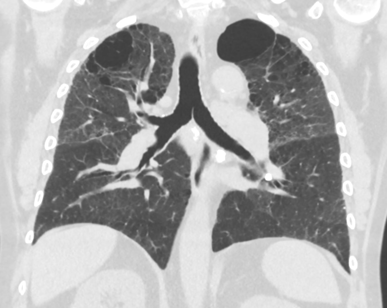
CT scan in the coronal plane of a 64- year-old man with emphysema shows bilateral apical bullous lung disease
Ashley Davidoff MD TheCommonVein.net 136439
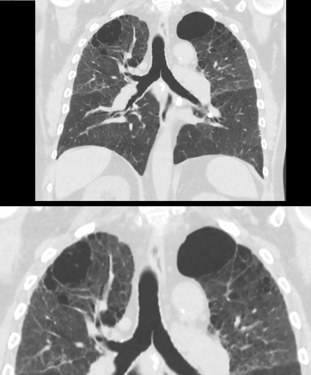
CT scan in the coronal plane of a 64- year-old man with emphysema shows bilateral apical bullous lung disease, magnified in the lower image
Ashley Davidoff MD TheCommonVein.Net 136439c
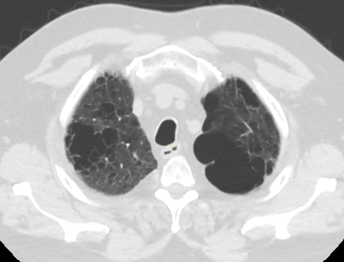
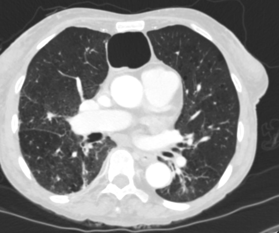
CT scan in the axial plane of a 64- year-old man with emphysema shows bilateral apical bullous lung disease,
Ashley Davidoff MD TheCommonVein.net 136440
71-year old male with a history COPD showing biapical bullous lung disease with a nodule noted in the right apex
Ashley Davidoff MD TheCommonVein.net 136522
Anterior Middle Chest
Ashley Davidoff MD The CommonVein.net
Ashley Davidoff MD The CommonVein.net
Ashley Davidoff MD The CommonVein.net
Ashley Davidoff MD The CommonVein.net
Character
Infection
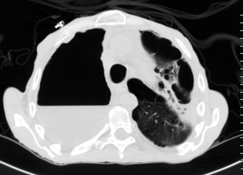
65-year-old male with emphysema of the lungs presents with a cough, fever and leukocytosis. CT in the axial plane shows extensive apical bullous lung disease. There is a large right upper lobe bulla with an air fluid level, and a smaller left upper lobe bulla with an air fluid level. The bulla in the left upper lobe, cause compressive atelectasis of a segment of the left upper lobe.
Ashley Davidoff MD TheCommonVein.net 259Lu 117474
Inflammation
Malignancy
Mechanical
Atelectasis
Trauma
Metabolic
Circulatory-
Hemorrhage
Immune Infiltrative Idiopathic Iatrogenic