
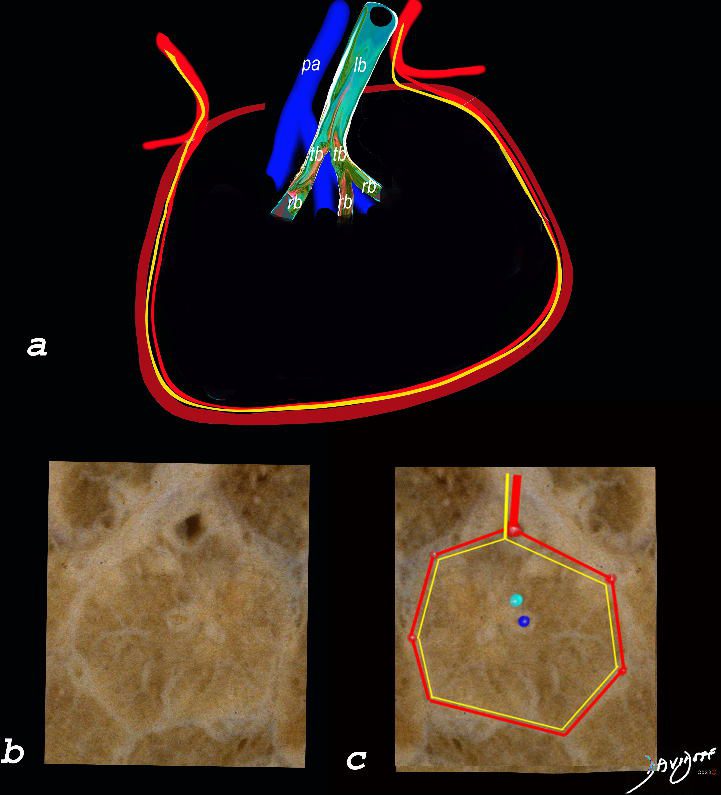
The top image (a) shows an anatomic drawing of a secondary lobule of the lung subtended by a lobular bronchiole (lb) and arteriole (pa). The interlobular septum contains the venule (red) lymphatic (yellow) and septum (maroon)
The anatomical specimen of the lung (b) shows normal intralobular parenchyma while image c shows the centrilobular arteriole (navy blue) and centrilobular bronchiole (teal) and interlobular venule (red) and lymphatics (yellow) The interlobular septum is slightly thickened
Ashley Davidoff TheCommonVein.net
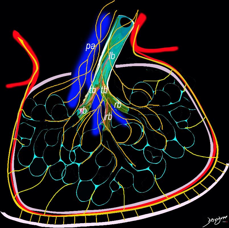
The diagram shows the 2 systems of lymphatic drainage at the level of the secondary lobule. The superficial system drains some of the interstitium of the secondary lobule, runs in the interlobular septa and drains all the pleura. Thee pathway to the lymph nodes in the mediastinum is via the pulmonary veins. The deeper system drains the interstitium in the interalveolar septa, and then they travel along the bronchovascular bundle accompanying the bronchi and pulmonary artery and into the lymph nodes of the hila and mediastinum
Ashley Davidoff MD TheCommonVein.net lungs-0768
The diagram shows the 2 systems of lymphatic drainage at the level of the secondary lobule. The superficial system drains some of the interstitium of the secondary lobule, runs in the interlobular septa and drains all the pleura. Thee pathway to the lymph nodes in the mediastinum is via the pulmonary veins. The deeper system drains the interstitium in the interalveolar septa, and then they travel along the bronchovascular bundle accompanying the bronchi and pulmonary artery and into the lymph nodes of the hila and mediastinum
Ashley Davidoff MD TheCommonVein.net lungs-0767
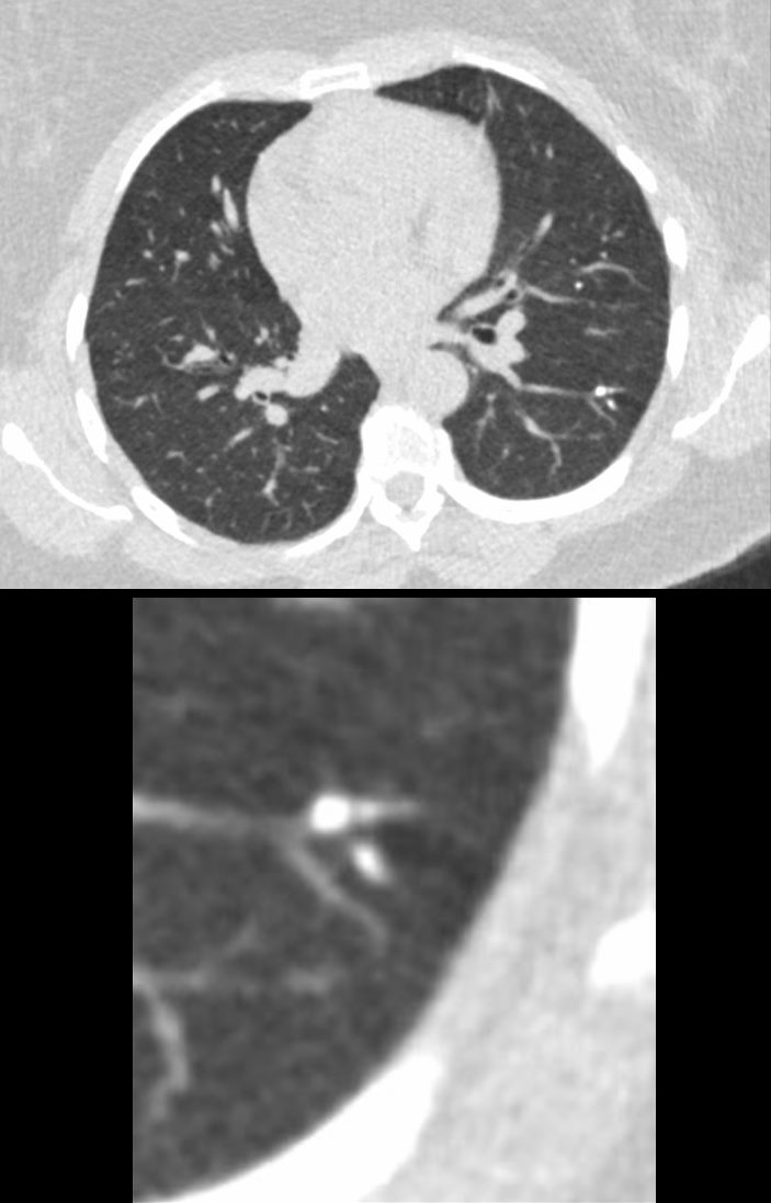
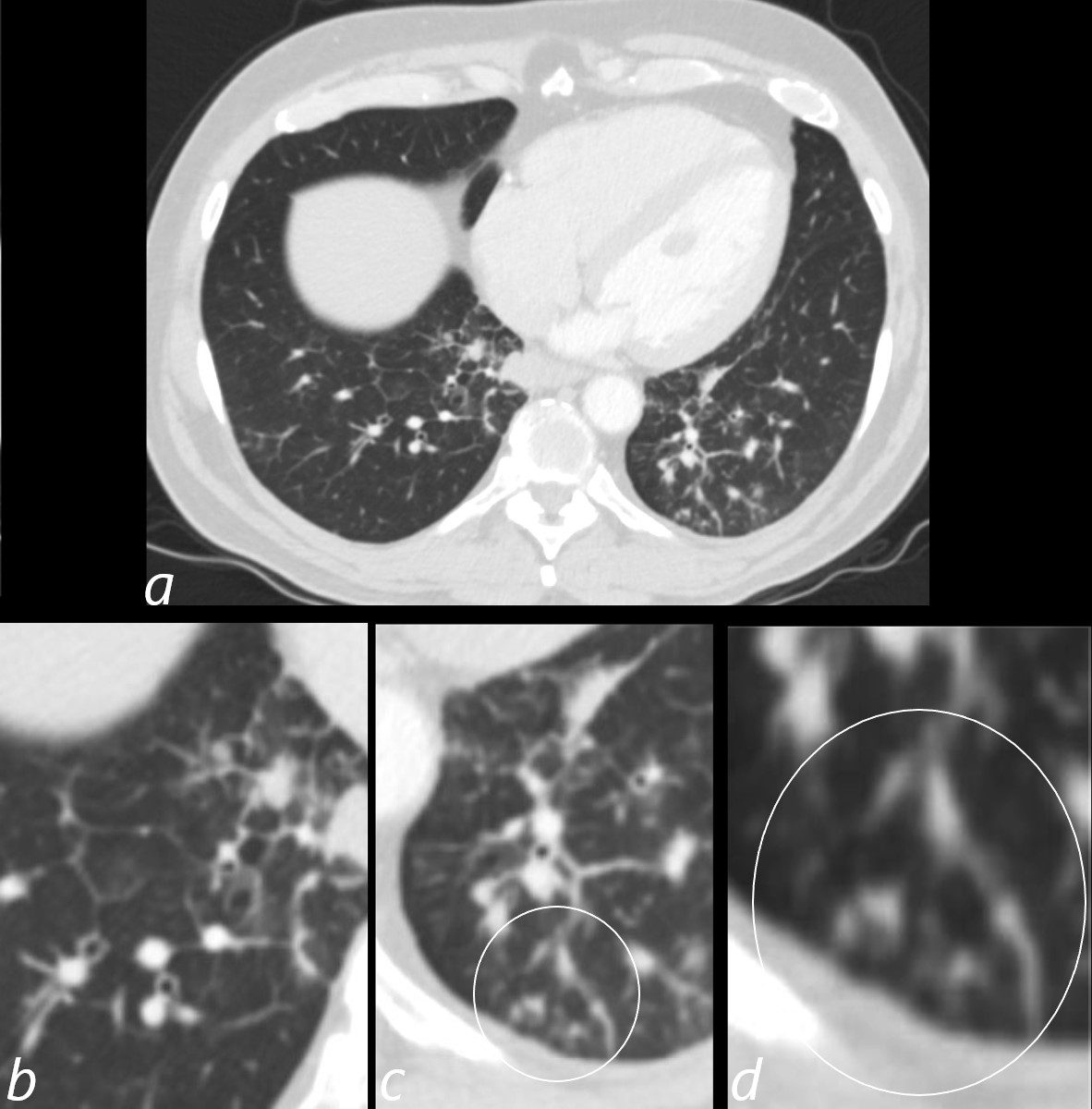
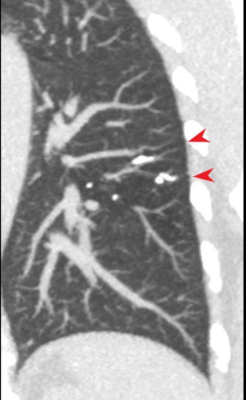
Thickening of the Interlobular Septum -Right Upper Lobe
Active TB
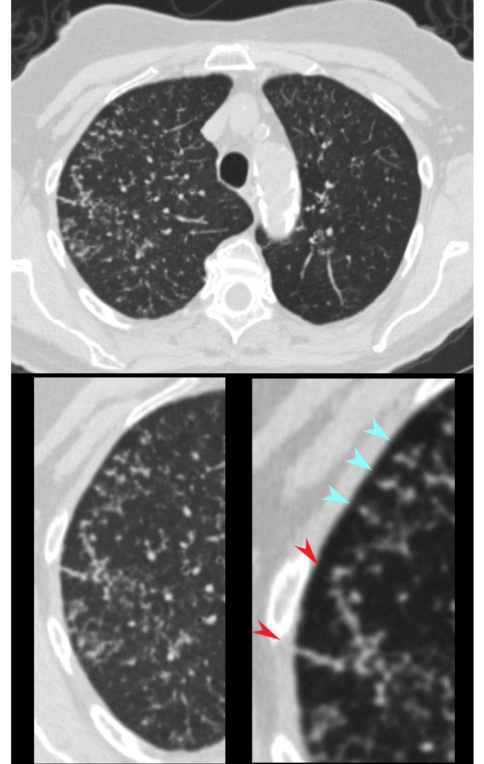
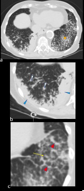
CT in the axial plane shows extensive small airway disease dominant in the right upper lobe characterized by innumerable, ground glass micronodules, centrilobular nodules (teal blue arrowheads) and nodular thickening of the interlobular septa likely reflecting lymphatic involvement (red arrowheads)
Ashley Davidoff MD TheCommonVein.net 135825cL 192Lu
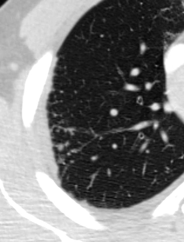
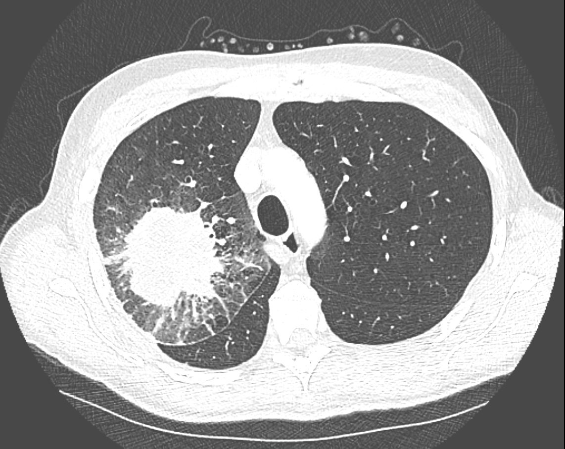
Calcified Septal Granulomas and Calcified Centrilobular Nodule
48-year-old male with history of TB
48-year-old male with history of TB
Axial CT through the mid chest shows calcified granulomas along an interlobular septum of a secondary lobule. There is also a centrilobular calcified granuloma These calcifications are likely centered around the lymphatics of the bronchovascular bundle and in the interlobular septum
Ashley Davidoff MD TheCommonvein.net 132604c
Inflammation
Sarcoidosis
Has both Intralobular and Septal Nodularity
SARCOIDOSIS, ACTIVE – ALVEOLAR FORM
Ashley Davidoff MD
SARCOIDOSIS, ACTIVE – ALVEOLAR FORM
Ashley Davidoff MD
Lymphangitis Carcinomatosis
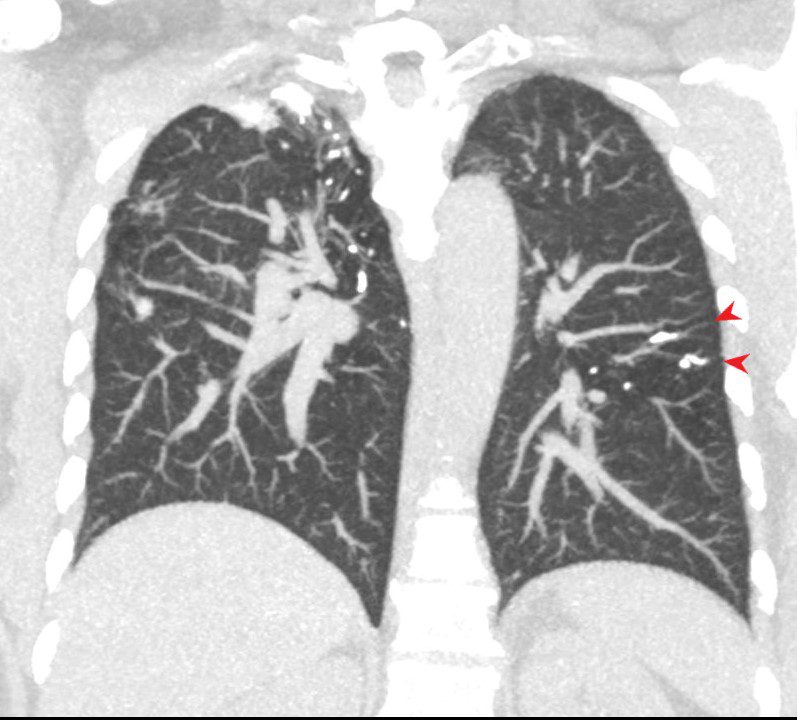
67-year-old man with prior history of bladder cancer. CT scan in the axial plane inferior to the lung mass shows extensive irregular thickening of the interlobular septa magnified in the right lower panel. These findings reflect lymphangitis carcinomatosis. There is background evidence of centrilobular emphysema, dominant in both upper lobes. A magnified view in the right lower panel offers an internal comparison of the abnormal vs the normal size and shape of the interlobular septa
Ashley Davidoff MD TheCommonVein.net 32287c 243Lu
Known Head and Neck Cancer with Metastases – Lymphangitis Carcinomatosa
3 months later the patient presented with chest pain and a cough. CT of the chest in the axial plane shows new bilateral lower lobar regions of irregular interlobular septal thickening noted in the right lower lobe a, magnified in b). Ringed in image c and d are 2 side by side secondary lobules with irregular septal thickening centrilobular nodules and other intralobular nodules likely reflecting lymphatic involvement.
Given the changes in the right upper lobe these findings likely reflect lymphangitis carcinomatosa
Ashley Davidoff MD TheCommonVein.net 013Lu 136062cL
CT in the axial plane demonstrates a large, spiculated mass in the right upper lobe with surrounding halo likely reflecting hemorrhage or lymphatic edema around the mass. In addition, there is evidence of irregular interlobular septal thickening likely reflecting lymphatic invasion and indicating lymphangitis carcinomatosa. There is irregular thickening of the major fissure suggesting involvement.
Ashley Davidoff MD TheCommonVein.net 135865
Adenocarcinoma of Left Lung with
Coarsened Septal Thickening LUL
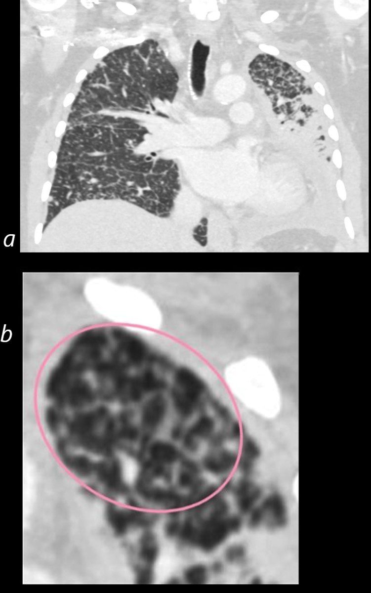
50 year old female with primary adenocarcinoma with the primary lesion presenting as pneumonic consolidation of the left lower lobe, and diffuse reticulonodular changes bilaterally
Image b is a magnified view of the left upper lobe and shows nodular thickening of the interlobular septa representing lymphatic spread along the lymphovascular bundles (pink oval)
The right lung shows interlobular septal thickening centrilobular nodules, and nodular thickening of the minor fissure
These findings are consistent with the diagnosis of lymphangitis carcinomatosa
Ashley Davidoff MD TheCommonVein.net 158Lu 131023c01L
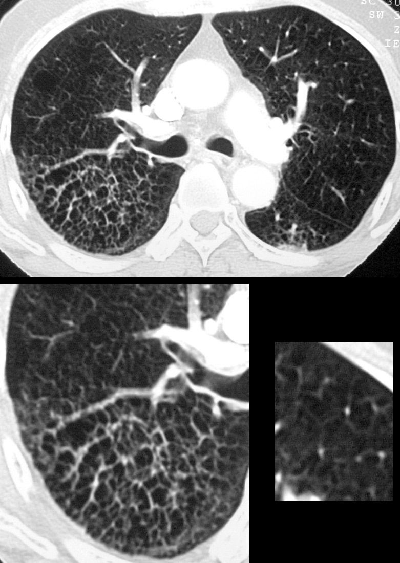
CHF
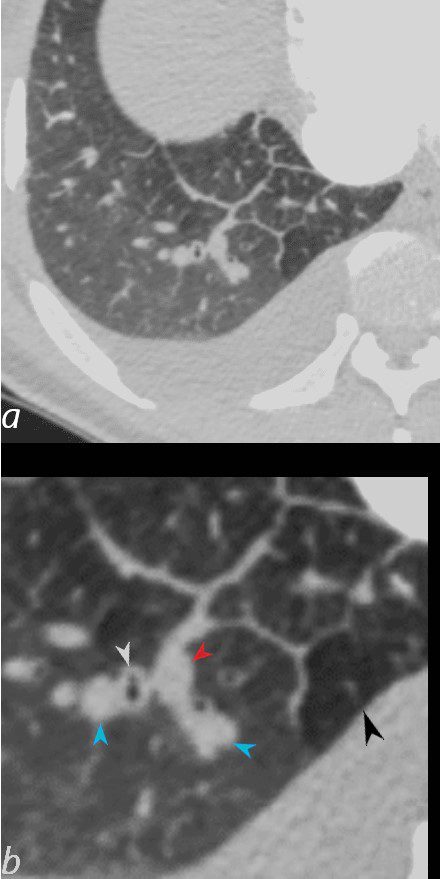
50-year-old female with diabetes, chronic renal failure and congestive heart failure. CT in the axial plane through the right posterior recess, shows thickened interlobular septa at the right base, congested arterioles (light blue arrowheads, b), alongside the bronchioles, peribronchial cuffing (white arrowheads, b), a congested pulmonary venule in the interlobular septum (red arrowhead arrowheads, b), ground glass changes and a secondary lobule demonstrating mosaic attenuation (black arrowhead arrowheads, b). The IVC is dilated and a small complex effusion is present.
Ashley Davidoff MD TheCommonvein.net 135783cL 193Lu
50-year-old female with diabetes, chronic renal failure and congestive heart failure. CT in the coronal plane through the posterior aspect of the chest, shows diffuse ground glass changes, thickening of the interlobular septa, thickening of the fissure and centrilobular nodules reflecting arteriolar congestion.
Ashley Davidoff MD TheCommonvein.net 135780 193Lu
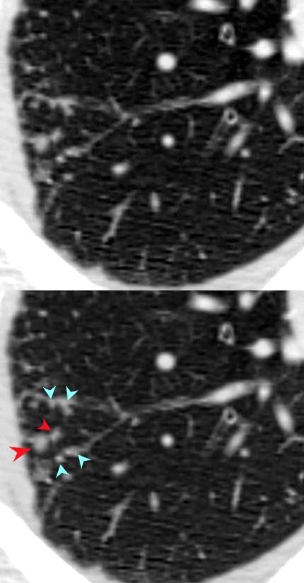
TB
Small Airway Disease and Nodular Thickening of the Interlobular Septum -Right Upper Lobe Active TB
CT in the axial plane shows extensive small airway disease dominant in the right upper lobe characterized by innumerable, ground glass micronodules, centrilobular nodules (teal blue arrowheads) and nodular thickening of the interlobular septa likely reflecting lymphatic involvement (red arrowheads)
Ashley Davidoff MD TheCommonVein.net 135825cL 192Lu

68 year old female presented with malaise night sweats weight loss QuantiFeron gold positive, with a past history of treated TB in her native country as a child. Axial CT images through the upper lobe shows a miliary pattern of disease affecting interlobular septa, centrilobular and tree in bud nodular patterns. Bronchoscopy isolated Mycobacterium complex. She was treated with good result
Ashley Davidoff MD TheCommonVein.net mycobacterium-complex-TB-68-008
Alveolar Septal Amyloidosis
CT scan in the axial projection at the base of the lungs show many features of amyloidosis including lung nodules (white arrowheads) and infiltrates (b), and diffuse deposition within the alveolar septa (red arrowheads, c) and centrilobular nodules(yellow arrow c)
Ashley Davidoff MD Boston Medical Center
TheCommonVein.net septal-amyloidosis-001b
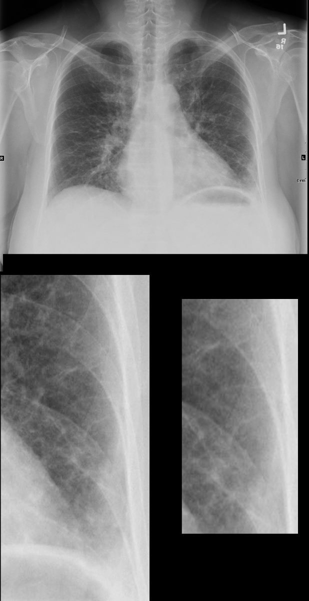
56 -year-old female with a history of amyloidosis (AL) presents for follow up following a pulmonary embolus. Frontal view of the chest shows diffuse reticular process best appreciated at the left base suggesting Kerley b lines (magnified in the lower panels). In the appropriate clinical setting these findings are compatible with the diagnosis of alveolar septal amyloidosis. Note the air fluid level in the stomach in this patient with known amyloidosis of the stomach with delayed gastric emptying.
Ashley Davidoff MD TheCommonVein.net 244 Lu 135741c05c
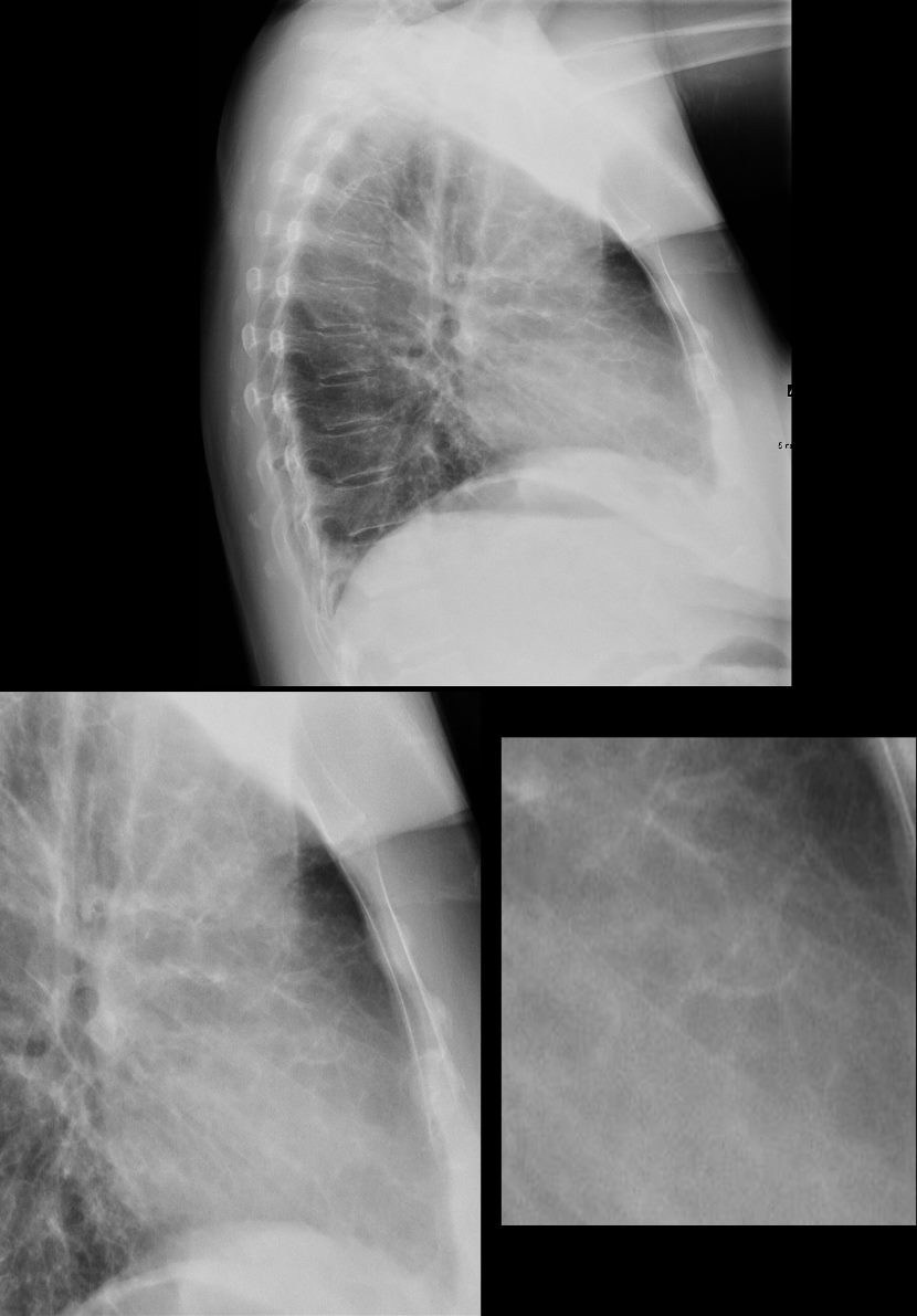
56 -year-old female with a history of amyloidosis (AL) presents for follow up following a pulmonary embolus. Lateral view of the chest shows diffuse reticular process best appreciated anteriorly suggesting thickening of the interlobular septa. (better appreciated in the magnified views in the lower panels). In the appropriate clinical setting these findings are compatible with the diagnosis of alveolar septal amyloidosis. Note the air fluid level in the stomach in this patient with known amyloidosis of the stomach with delayed gastric emptying.
Ashley Davidoff MD TheCommonVein.net 244 Lu 135741c07c
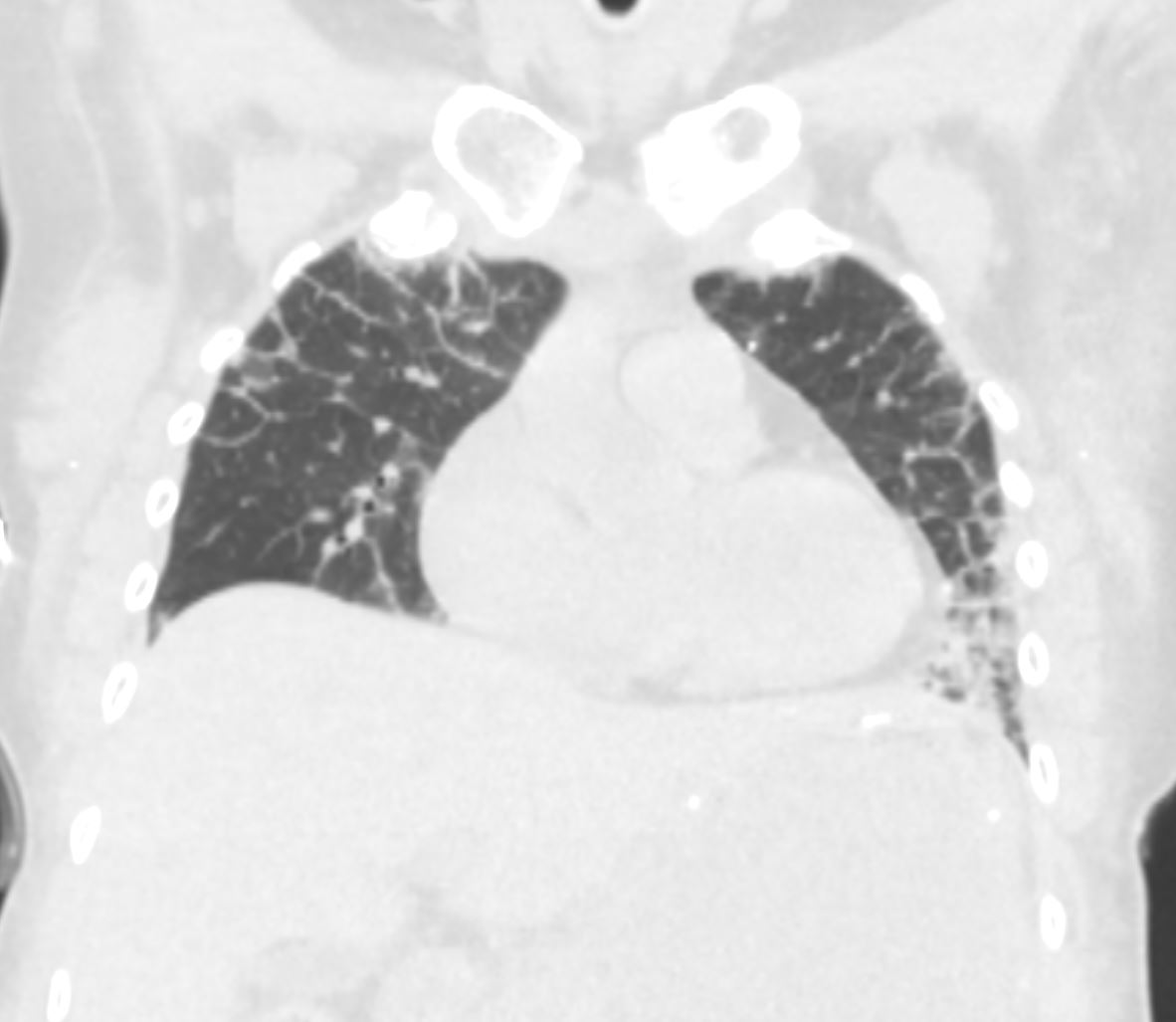
56 -year-old female with a history of amyloidosis (AL) presents for follow up. Coronal CT of the chest shows thickening of the interlobular septa most prominent in the periphery. In the appropriate clinical setting these findings are compatible with the diagnosis of alveolar septal amyloidosis.
Ashley Davidoff MD TheCommonVein.net 244 Lu 135741d15
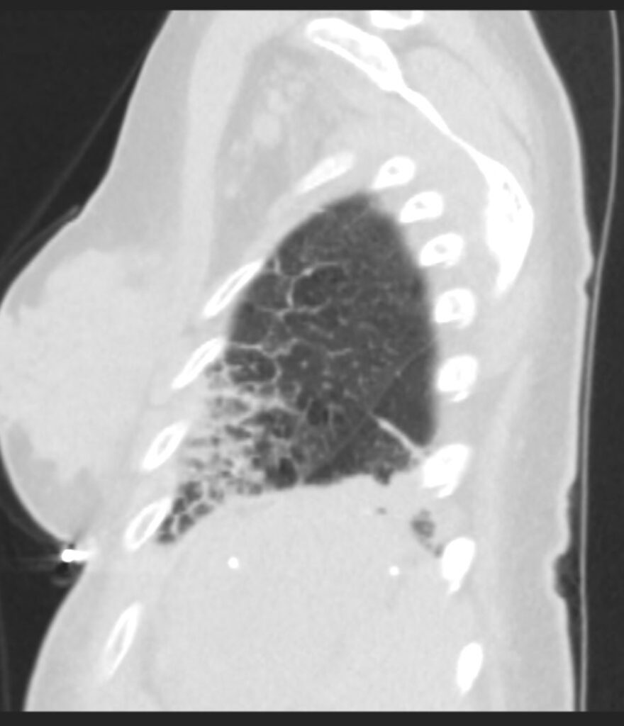
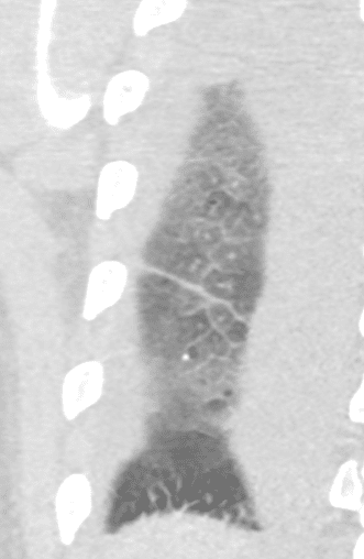
56 -year-old female with a history of amyloidosis (AL) presents for follow up. Sagittal CT of the chest shows thickening of the interlobular septa. Superiorly the septal thickening is smoother and finer and inferiorly the thickening is thicker and coarser. In the appropriate clinical setting these findings are compatible with the diagnosis of alveolar septal amyloidosis.
Ashley Davidoff MD TheCommonVein.net 244 Lu 135741d20
CT Alveolar Septal Amyloidosis Thin Smooth and Thick
CT Alveolar Septal Amyloidosis Interlobular Septal Thickening
56 -year-old female with a history of amyloidosis (AL) presents for follow up. Sagittal CT of the chest shows thickening of the interlobular septa. Superiorly the septal thickening is smoother and finer and inferiorly the thickening is thicker and coarser. In the appropriate clinical setting these findings are compatible with the diagnosis of alveolar septal amyloidosis.
Ashley Davidoff MD TheCommonVein.net 244 Lu 135741d20
Alveolar Septal Amyloidosis with Calcifications
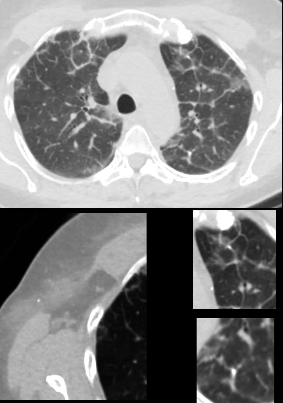
56 -year-old female with a history of amyloidosis (AL) presents for follow up. Axial CT of the chest shows diffuse reticular process best appreciated anteriorly with thickening of the interlobular septa. Punctate, dystrophic calcifications are seen in the interlobular septa (white arrowheads a, c and d), as well as in the pleura abutting the mediastinum (white arrowhead a). Image b shows similar calcifications, likely deposits of amyloid in the axilla with surrounding soft tissue changes
Ashley Davidoff MD TheCommonVein.net 244 Lu 135741d02cL
Septal Calcifications TB
INACTIVE SECONDARY TB WITH EXTENSIVE PARENCHYMAL AND LYMPHOVASCULAR INVOLVEMENT
48-year-old male with history of TB presents with back pain
AP view of the spine shows complex lesion in the right apex characterized by fibronodular opacities. There are scattered calcifications throughout the lungs but some are centered around the lymphatics, including the interlobular septa and centrilobular region
Ashley Davidoff MD TheCommonVein.net
CALCIFICATION ALONG LYMPHOVASCULAR BUNDLES (red arrows)
INACTIVE SECONDARY TB WITH EXTENSIVE PARENCHYMAL AND LYMPHOVASCULAR INVOLVEMENT
48-year-old male with history of TB presents with back pain
AP view of the spine shows complex lesion in the right apex characterized by fibronodular opacities. There are scattered calcifications throughout the lungs but some are centered around the lymphatics, including the interlobular septa and centrilobular region
Ashley Davidoff MD TheCommonVein.net
48-year-old male with history of TB
Axial CT through the mid chest shows calcified granulomas along an interlobular septum of a secondary lobule. There is also a centrilobular calcified granuloma These calcifications are likely centered around the lymphatics of the bronchovascular bundle and in the interlobular septum
Ashley Davidoff MD TheCommonvein.net 132604c
CT Alveolar Septal Amyloidosis with Calcifications
56 -year-old female with a history of amyloidosis (AL) presents for follow up. Axial CT of the chest shows diffuse reticular process best appreciated anteriorly with thickening of the interlobular septa. Punctate, dystrophic calcifications are seen in the interlobular septa (white arrowheads a, c and d), as well as in the pleura abutting the mediastinum (white arrowhead a). Image b shows similar calcifications, likely deposits of amyloid in the axilla with surrounding soft tissue changes
Ashley Davidoff MD TheCommonVein.net 244 Lu 135741d02cL
Links and References
- TCV