- 69 year old male with a history of
- HTN, HLD, pre-DM, COPD, s/p 1
- pw Left-sided pleuritic chest pain.
- left VATS with decortication and resection of
- large bullae after
- hydropneumothorax with empyema,
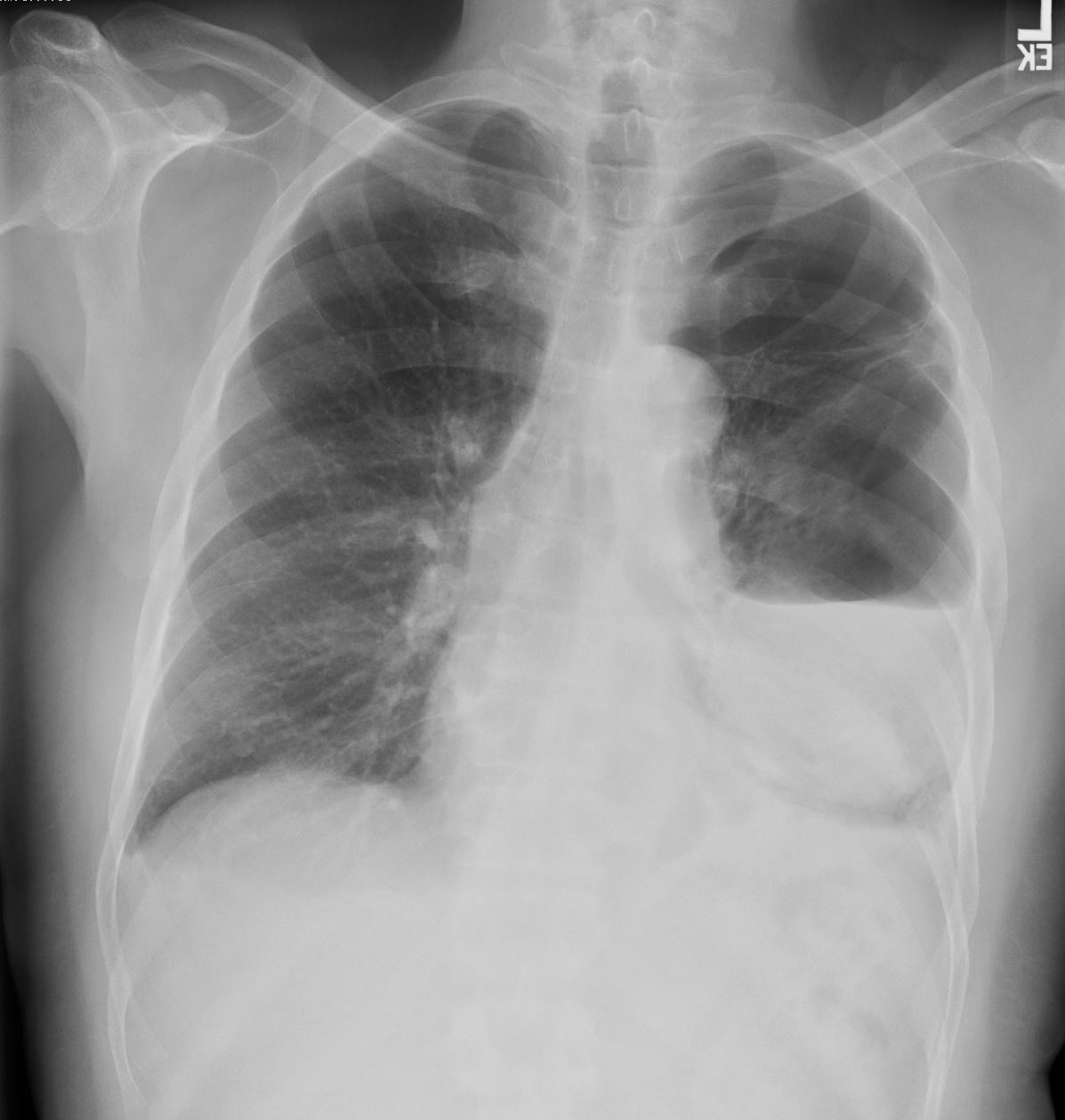
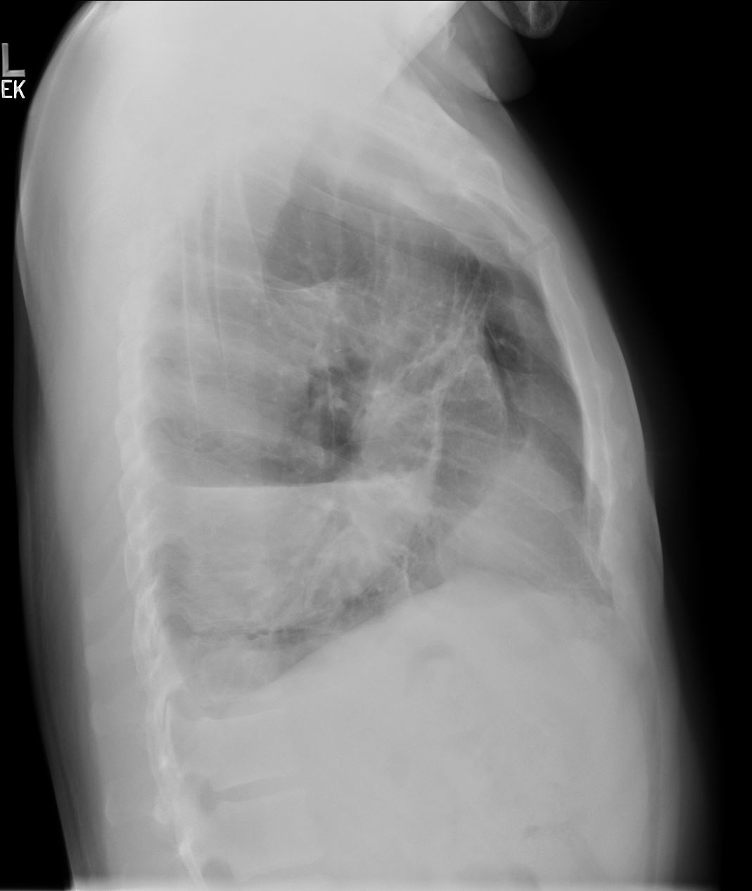
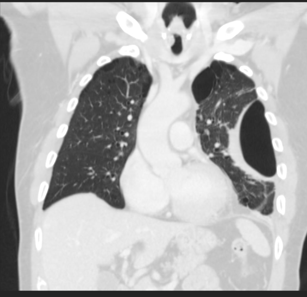
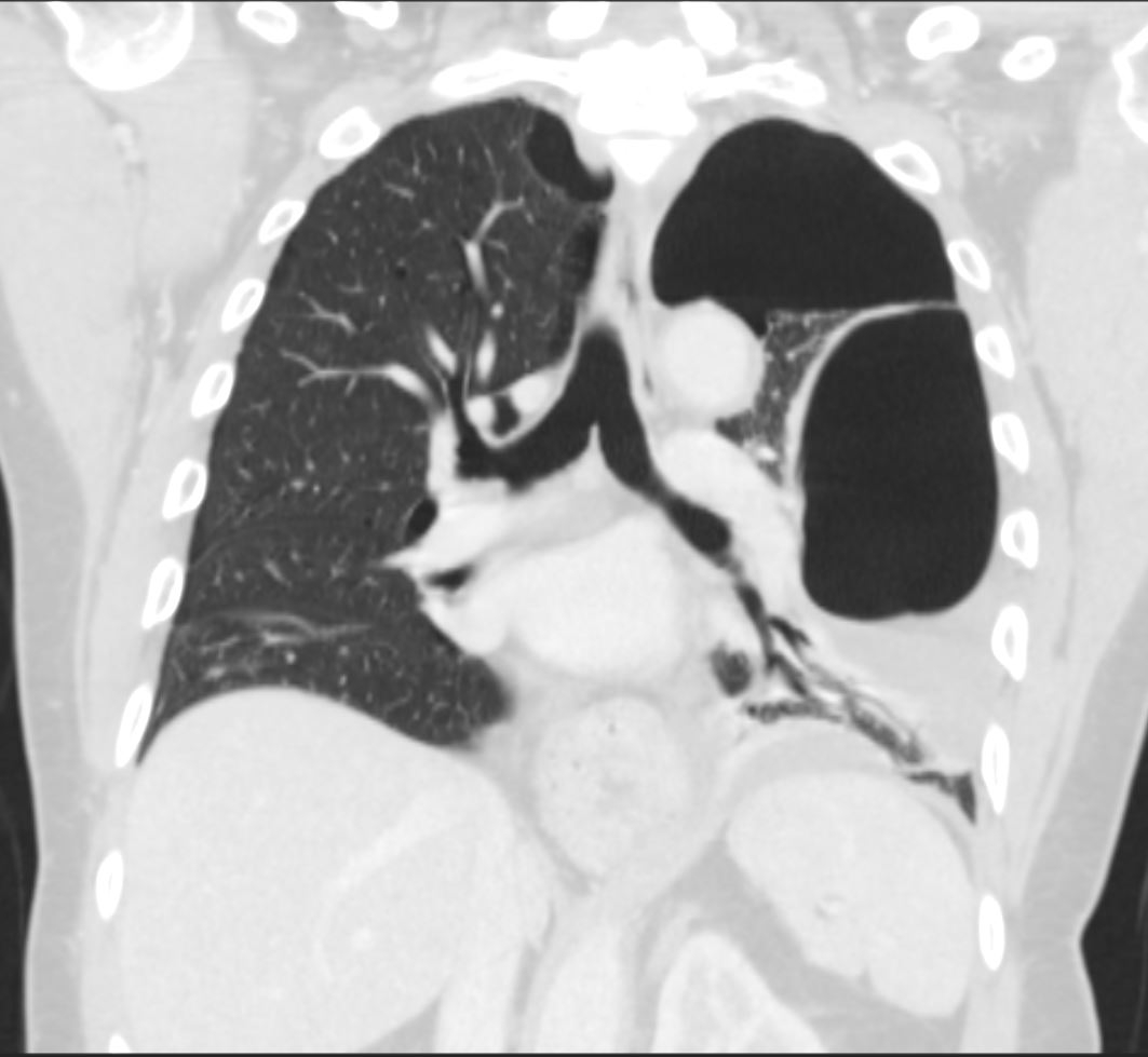
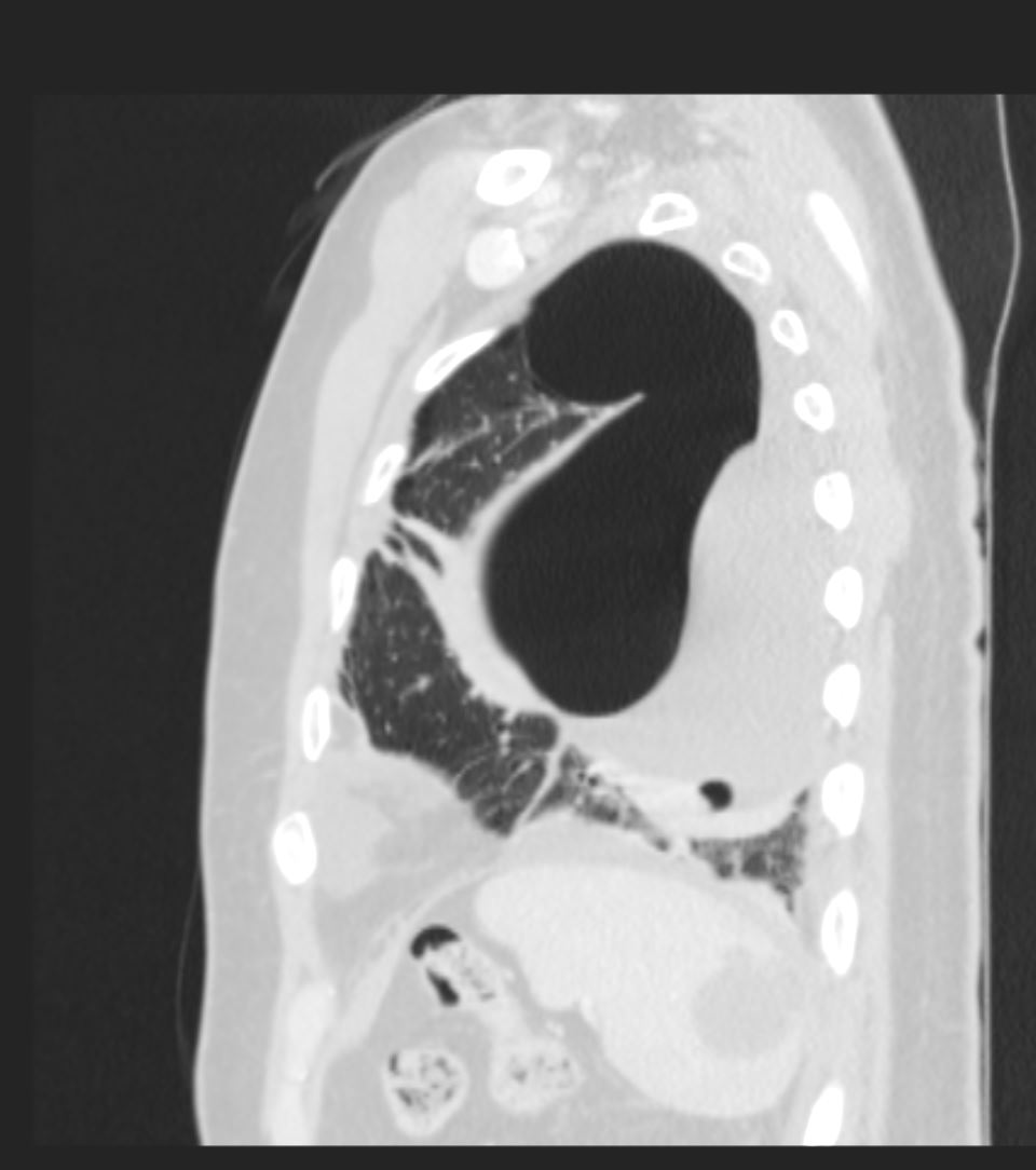
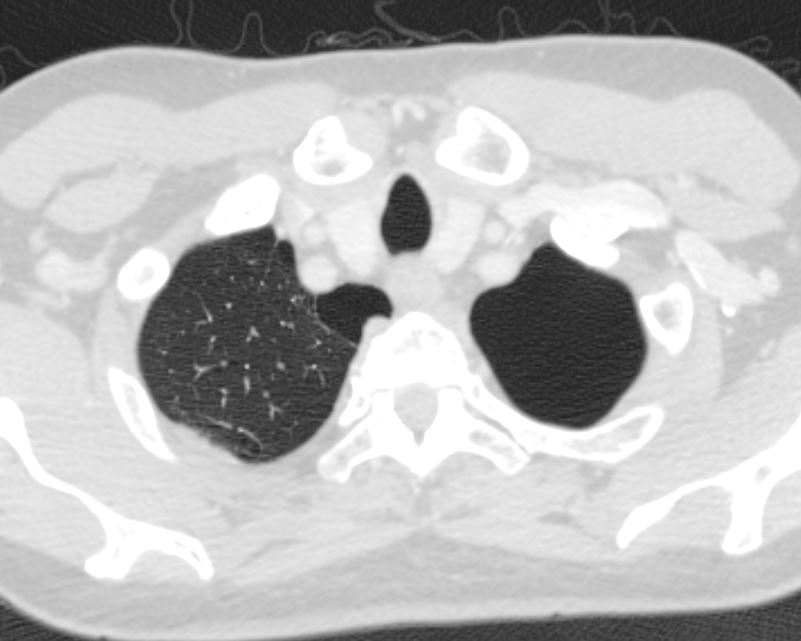
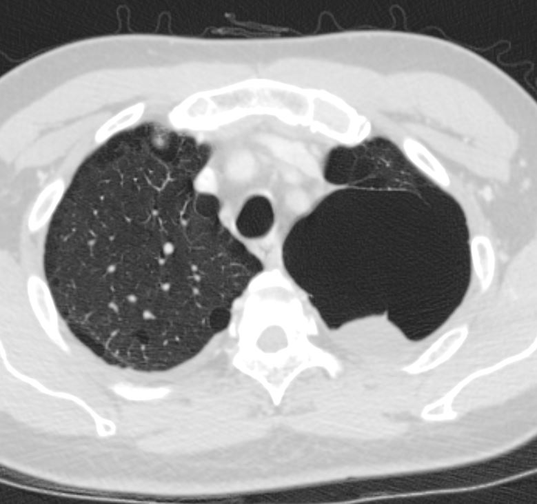
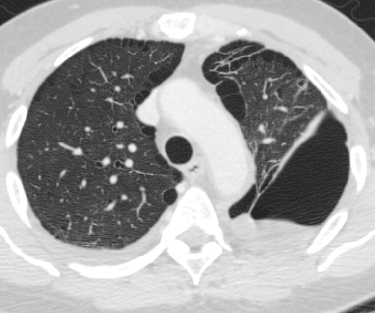
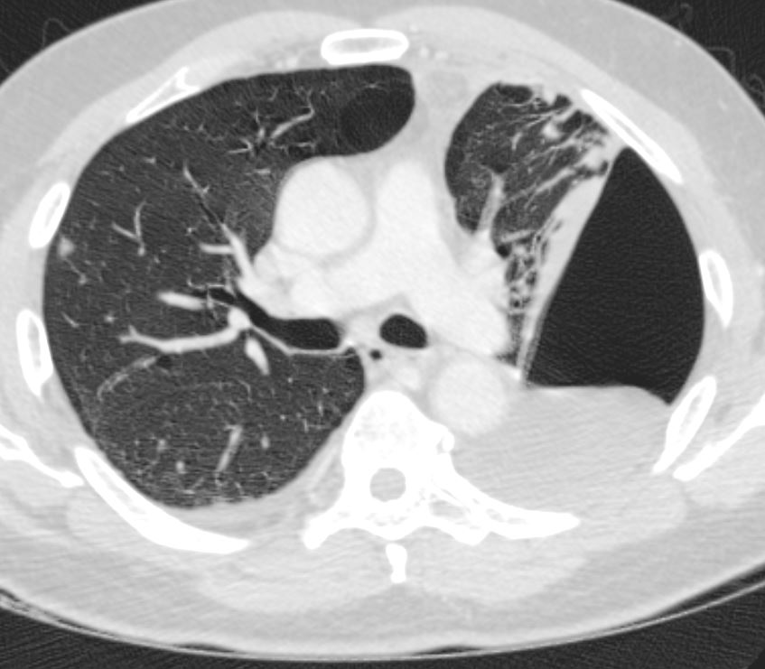
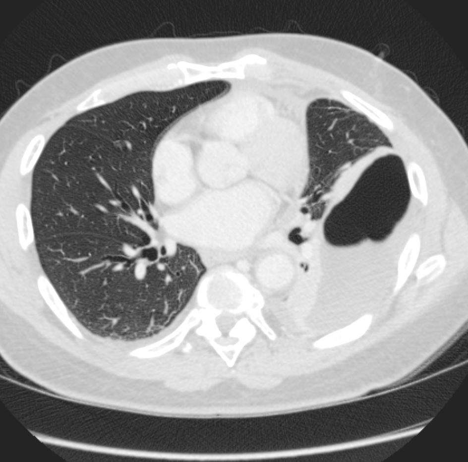
- There is extensive left-sided bulla disease with accompanying fluid collections in the left upper lobe as well as the left lower lobe. The large left upper lobe fluid collection represents fluid within a large bulla or, more likely, chronic hydropneumothorax secondary to a ruptured bulla. There is surrounding atelectasis of the lung parenchyma. There is a fluid collection in the left lower lobe, representing a loculated pleural effusion. On the right, to a much lesser extent than the left, there is bulla disease. No mediastinal lymphadenopathy is visualized.IMPRESSION:
1. Extensive left-sided bulla disease with a large left upper lobe fluid collection representing fluid within a large bulla or, more likely, chronic hydropneumothorax secondary to a ruptured bulla.
2. Loculated left lower lobe pleural effusion.
3 Multiple right-sided pulmonary nodules, the largest of which measures 6.3 mm. Short interval followup with a CT in 6 months is recommended.
4. 3.9 cm x 3.9 cm dominant cystic lesion in the spleen. Differential diagnosis includes posttraumatic pseudocyst, lymphangioma, and, less likely, abscess.
5. Asymmetric vocal cords. This may represent left-sided focal cord paralysis.
6. Small to moderate sized hiatal hernia.
Subsequent s/p left VATS decortication and bullae resection