
CASTLEMAN’S DISEASE
45 year old male presents for a chest X-ray after falling off a ladder A right lower lobe mass was identified as an incidental finding. Pathology showed Castleman’s disease
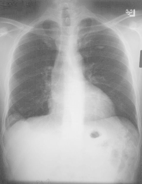
45 year old male presents for a chest X-ray after falling off a ladder A right lower lobe mass was identified as an incidental finding. Pathology showed Castleman’s disease
Courtesy Priscilla Slanetz MD MPH
TheCommonVein.net
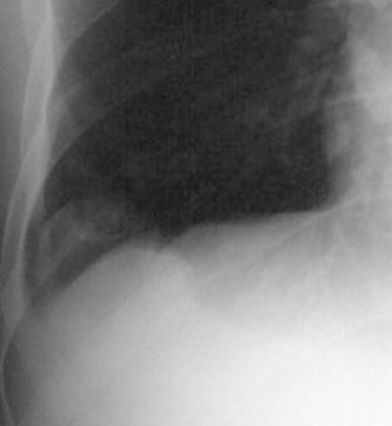
Magnification of the RLL Mass
45 year old male presents for a chest X-ray after falling off a ladder A right lower lobe mass was identified as an incidental finding. Pathology showed Castleman’s disease
Courtesy Priscilla Slanetz MD MPH
TheCommonVein.net
Non Contrast CT
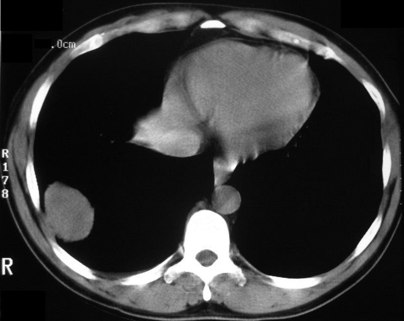
The non-contrast CT of the right lower lobe lung mass shows homogeneous soft tissue density . Pathology showed Castleman’s disease
Courtesy Priscilla Slanetz MD MPH
TheCommonVein.net
Contrast Enhanced CT
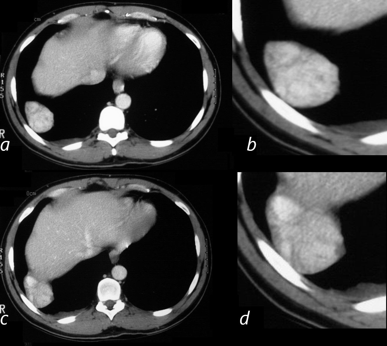
The contrast enhanced CT of the right lower lobe lung mass in the right lower lobe . Image a, magnified in b shows mostly homogeneous enhancement with a suggestion of nodular morphology . Image c shows a region anteriorly with slightly greater enhancement , and magnified in d.
Pathology showed Castleman’s disease
Courtesy Priscilla Slanetz MD MPH
TheCommonVein.net
Coronal CT Post Contrast
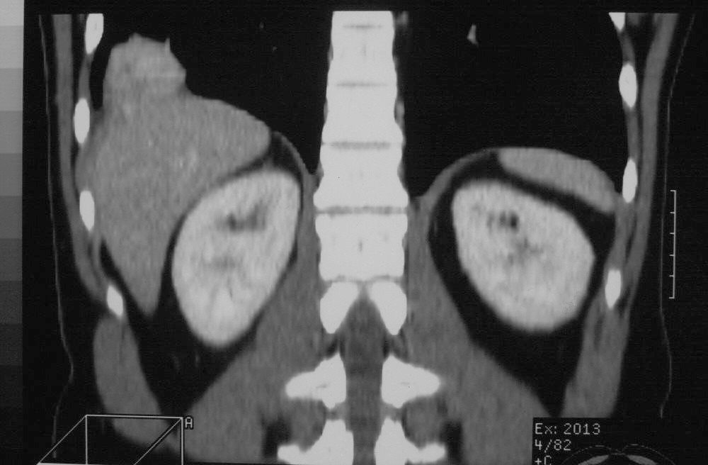
The contrast enhanced CT of the right lower lobe lung mass in the coronal plane shows mildly heterogeneous enhancement of the lymph node in close association with the pleura and diaphragm
Pathology showed Castleman’s disease
Courtesy Priscilla Slanetz MD MPH
TheCommonVein.net
More Posteriorly
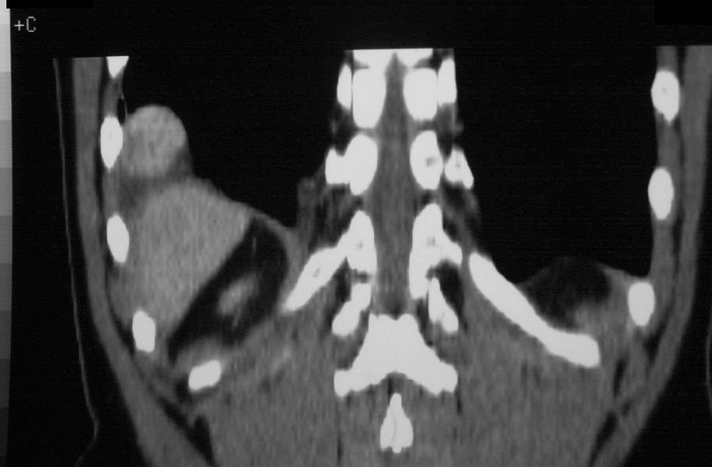
The contrast enhanced CT of the right lower lobe lung mass in the coronal plane shows mildly heterogeneous enhancement of the lymph node in close association with the pleura and diaphragm
Pathology showed Castleman’s disease
Courtesy Priscilla Slanetz MD MPH
TheCommonVein.net
Castleman’s disease, also known as Castleman disease or angiofollicular lymph node hyperplasia, is a rare and poorly understood disorder of the lymphatic system. It was first described by American pathologist Dr. Benjamin Castleman in the 1950s. Castleman’s disease is characterized by the abnormal growth of lymphocytes, a type of white blood cell, in lymph nodes. It can affect a single lymph node (unicentric Castleman’s disease) or multiple lymph nodes (multicentric Castleman’s disease).
Castleman’s disease can present with various features on a chest CT (computed tomography) scan, and the specific findings can vary depending on the subtype of the disease (unicentric or multicentric) and its stage. Here are some of the common features that might be observed on a chest CT scan in Castleman’s disease:
- Lymph Node Enlargement: Castleman’s disease typically involves enlarged lymph nodes. On a chest CT, you may see enlarged lymph nodes in the mediastinum (the area in the middle of the chest between the lungs). These nodes may vary in size and shape.
- Hilar Lymphadenopathy: Enlarged lymph nodes in the hilum, which is the central area of the lung where the bronchi, blood vessels, and lymph nodes are located, can be a characteristic feature.
- Enhancement with Contrast: In some cases, the enlarged lymph nodes may show contrast enhancement, indicating increased blood flow or vascularity within the nodes. This enhancement can vary in degree.
- Calcifications: Castleman’s disease lymph nodes may occasionally contain calcifications, which can appear as dense, white areas within the nodes on a CT scan.
- Compression of Adjacent Structures: Enlarged lymph nodes in the mediastinum can compress nearby structures, such as the trachea (windpipe) or major blood vessels, potentially leading to symptoms like shortness of breath or difficulty swallowing.
- Perinodal Inflammation: Inflammatory changes around the affected lymph nodes, including fat stranding and increased density of the surrounding tissue, may be present.
- Focal Lesions: In some cases, Castleman’s disease can present as solitary or multiple nodules within the lung parenchyma (lung tissue). These nodules may have features that mimic other lung conditions.
- Pleural Effusion: In advanced cases or in certain subtypes of Castleman’s disease, there may be pleural effusion (fluid accumulation around the lungs), which can also be visible on a chest CT.