
61 year old male with a history of treated mycobacterial infections and chronic cough
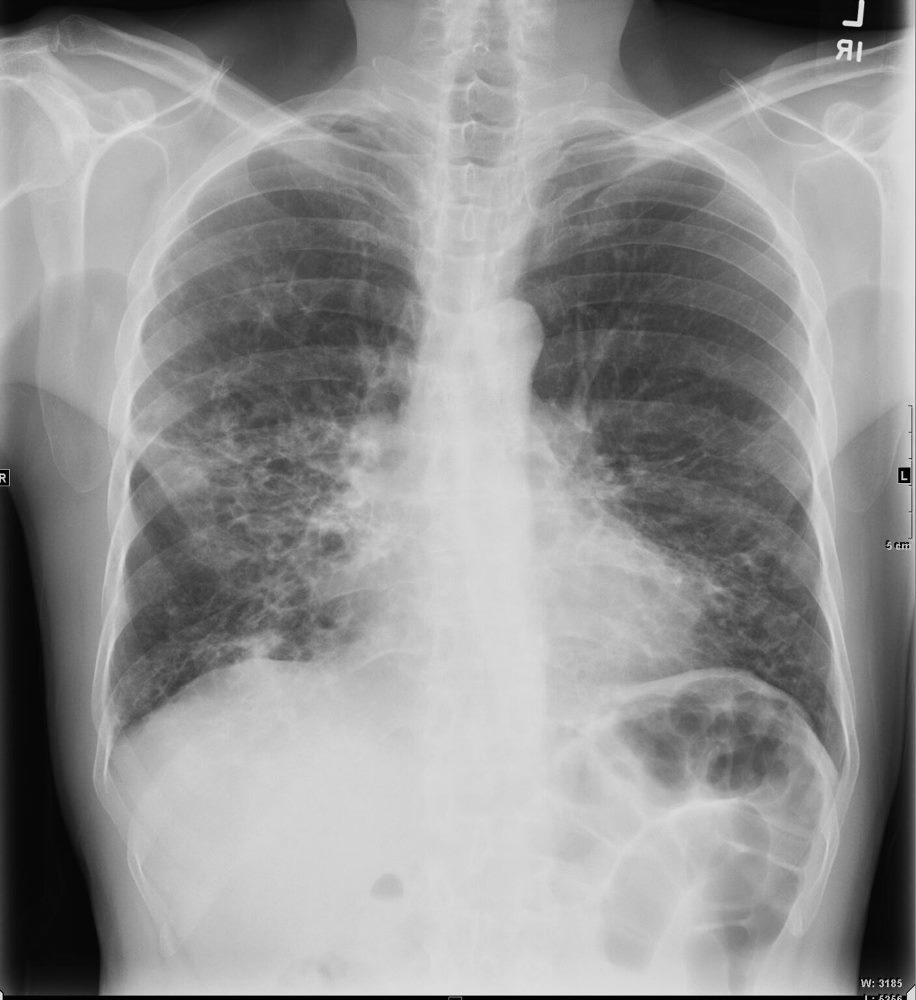
Frontal view shows shaggy heart borders with bibasilar cystic changes consistent with bronchiectasis in the middle lobe and lingula
Ashley Davidoff MD TheCommonVein.net 250Lu 135871
61-year-old male with a history of treated mycobacterial infections including MAC and chronic cough.
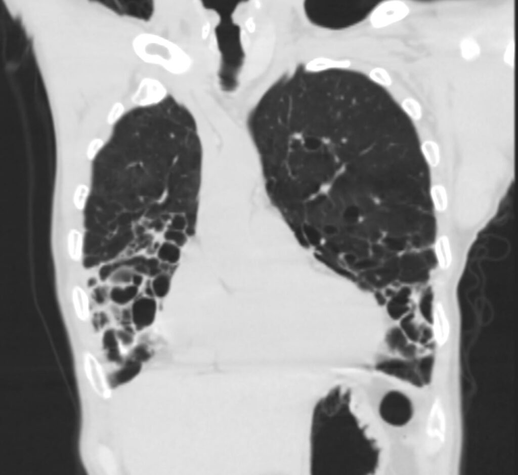
Coronal CT at the level of the heart shows significant bronchiectasis to the middle lobe and lingula and as a result abut the right and left heart border accounting for the CXR findings of a “shaggy heart border”. There is a relative paucity of mucus in the ectatic airways. The history of MAC and the distribution of the bronchiectasis in the middle lobe and lingula are reminiscent of the diagnosis of Lady Windermere syndrome
Ashley Davidoff MD TheCommonVein.net 250Lu 135879
61-year-old male with a history of treated mycobacterial infections including MAC and chronic cough.
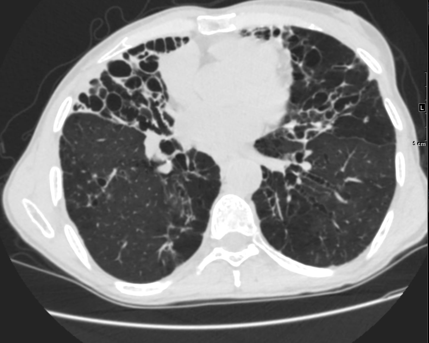
Axial CT at the level of the mid to lower chest shows mildly ectatic segmental airways to the lower, and middle lobe bronchi but significant bronchiectasis to the middle lobe and lingula involving the subsegmental airways. There is a relative paucity of mucus in the ectatic airways. The history of MAC and the distribution of the bronchiectasis in the middle lobe and lingula are reminiscent of the diagnosis of Lady Windermere syndrome
Ashley Davidoff MD TheCommonVein.net 250Lu 135876
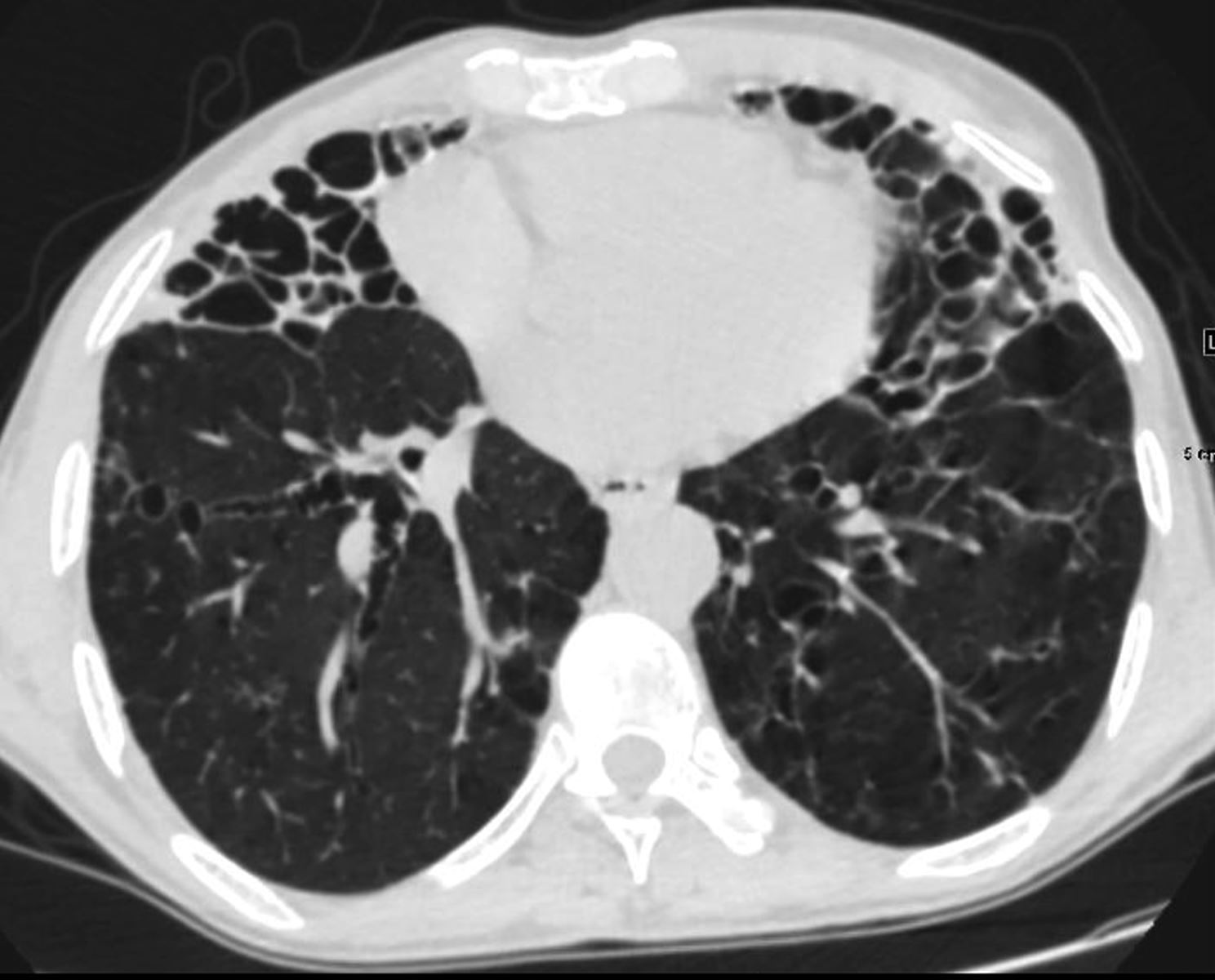
61-year-old male with a history of treated mycobacterial infections including MAC and chronic cough.
Axial CT at the level of the mid to lower chest shows mildly ectatic segmental airways to the lower, and middle lobe bronchi but significant bronchiectasis to the middle lobe and lingula involving the subsegmental airways. There is a relative paucity of mucus in the ectatic airways. The history of MAC and the distribution of the bronchiectasis in the middle lobe and lingula are reminiscent of the diagnosis of Lady Windermere syndrome
Ashley Davidoff MD TheCommonVein.net 250Lu 135876
61-year-old male with a history of treated mycobacterial infections including MAC and chronic cough.
Axial CT at the level of the mid to lower chest shows mildly ectatic segmental airways to the lower, and middle lobe bronchi but significant bronchiectasis to the middle lobe and lingula involving the subsegmental airways. There is a relative paucity of mucus in the ectatic airways. The history of MAC and the distribution of the bronchiectasis in the middle lobe and lingula are reminiscent of the diagnosis of Lady Windermere syndrome
Ashley Davidoff MD TheCommonVein.net 250Lu 135877
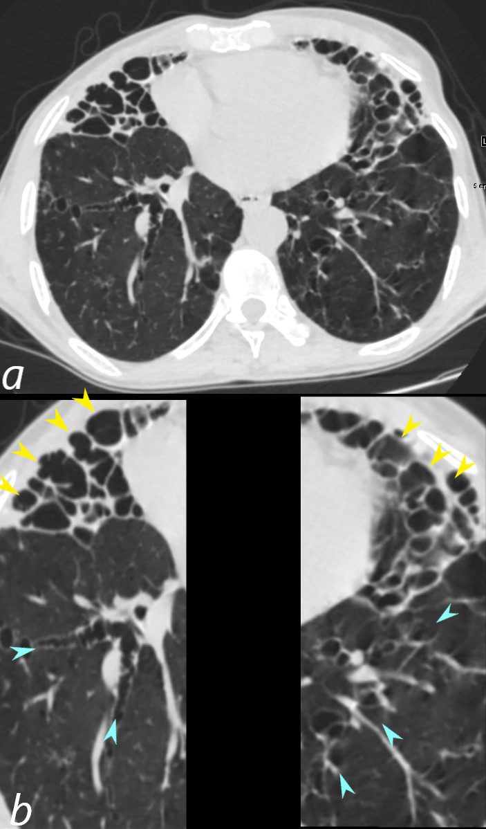
61-year-old male with a history of treated mycobacterial infections including MAC and chronic cough.
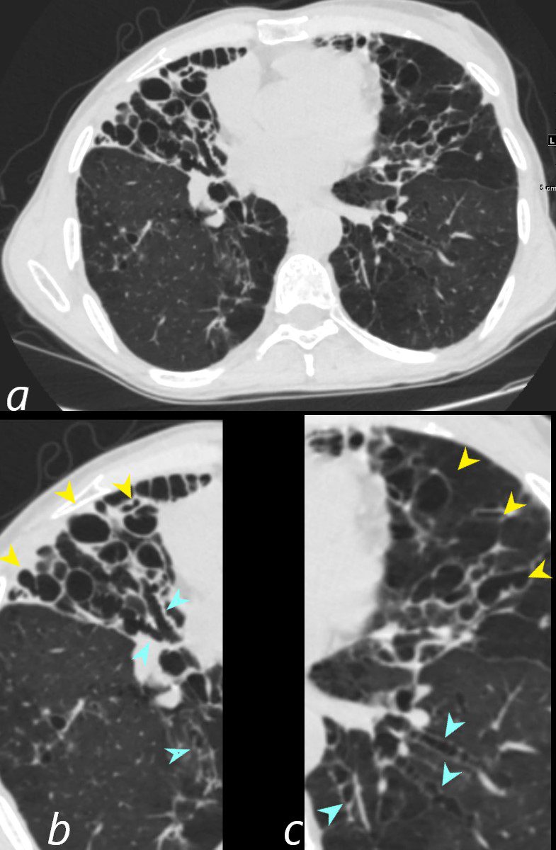
Axial CT at the level of the mid to lower chest shows mildly ectatic segmental airways to the lower, and middle lobe bronchi (teal arrowheads (b and c) but significant bronchiectasis to the middle lobe and lingula involving the subsegmental airways (yellow arrowheads b and c). There is a relative paucity of mucus in the ectatic airways. The history of MAC and the distribution of the bronchiectasis in the middle lobe and lingula are reminiscent of the diagnosis of Lady Windermere syndrome
Ashley Davidoff MD TheCommonVein.net 250Lu 135877cL