
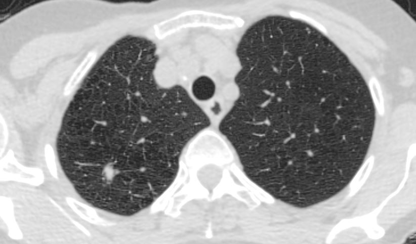
The PET scan showed faint FDG uptake which was below blood pool
Pathology revealed inflammatory pseudotumor
Ashley Davidoff MD TheCommonVein.net
Inflammatory pseudotumor of the lung, also known as inflammatory myofibroblastic tumor (IMT) of the lung, is a rare non-cancerous (benign) mass or lesion that can develop within the lung tissue. It is characterized by an overgrowth of inflammatory cells and myofibroblasts, which are cells involved in wound healing and tissue repair.
The exact cause of inflammatory pseudotumor of the lung is not fully understood. It may arise as a response to inflammation, infection, or an autoimmune reaction. Some cases have been associated with previous lung infections, trauma, or surgery. However, in many cases, the cause remains unknown.
Inflammatory pseudotumor of the lung can affect individuals of any age, but it tends to be more common in children and young adults. The condition can present with a variety of symptoms, including cough, chest pain, shortness of breath, fever, and weight loss. However, some individuals may be asymptomatic, and the tumor is incidentally discovered on imaging studies performed for other reasons.
Diagnosis of inflammatory pseudotumor of the lung involves a combination of imaging studies, such as chest X-rays, computed tomography (CT) scans, and PET scans These imaging techniques help visualize the mass and its characteristics. In some cases, a biopsy may be necessary to confirm the diagnosis.
Treatment of inflammatory pseudotumor of the lung typically involves surgical removal of the tumor. In most cases, this leads to a complete cure. However, in some instances, the tumor may recur, and additional treatment may be necessary. Rarely, if the tumor is inoperable or has spread extensively, other treatment options such as radiation therapy or corticosteroids may be considered.
The prognosis for inflammatory pseudotumor of the lung is generally favorable. With appropriate treatment, the majority of individuals have a good outcome. Regular follow-up with imaging studies is recommended to monitor for any recurrence or new lesions.