
Linear Atelectasis

Ashley Davidoff TheCommonVein.net

60 year old male with linear (discoid) atelectasis in the middle lobe and the left upper lobe on CT. Note moderate sized bilateral pleural effusion. Minor compressive atelectasis caused by the left effusion.
Ashley Davidoff MD TheCommonVein.net
56-year-old male with linear atelectasis in the medial segment of the middle lobe of the lung
Ashley Davidoff MD The CommonVein.net 56 M lungs atelectasis 001
Crescentic
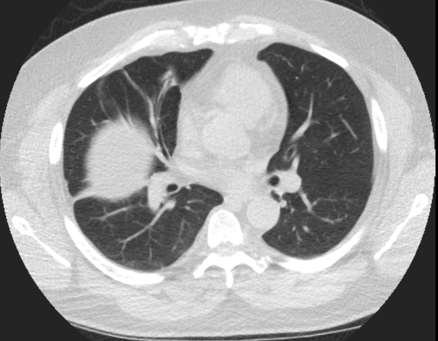
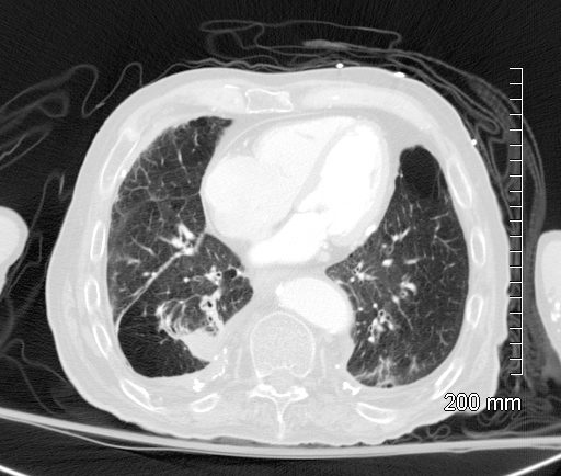
Mild Bilateral Crescentic Pneumonic Consolidation
58year old male presents with dyspnea. CT scan shows bilateral pleural effusions with crescentic region of compressive atelectasis in the left lower lobe
Ashley Davidoff MD TheCommonVein.net
Crescents
Luftsichel Sign
Left Upper Lobe Collapse and Luftsichel Sign

Left Upper Lobe Atelectasis
Female patient with central squamous cell carcinoma of the lung with left upper lobe collapse and hyperinflation of the left lower lobe resulting in a Luftsichel sign
Ashley Davidoff MD TheCommonVein.net 152Lu
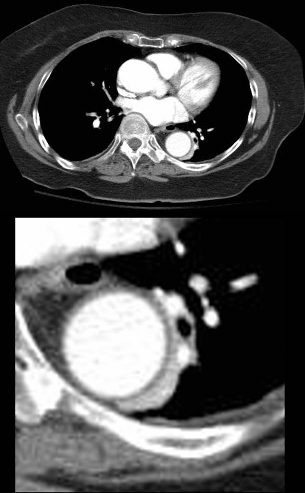
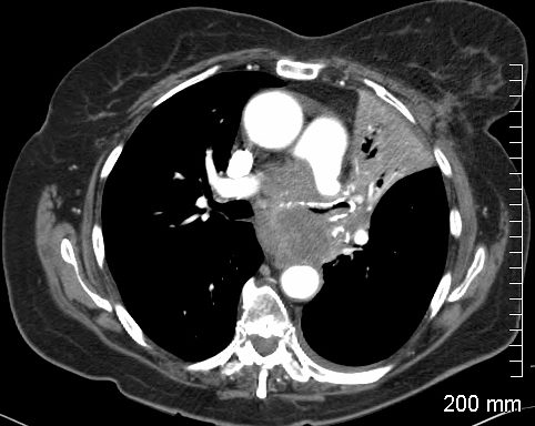
68 year old male with a cough.
CT shows Compressive Atelectasis alongside the pulsating aorta
Ashley Davidoff MD TheCommonVein.net
37493
Right Upper Lobe
PA Chest X-Ray Triangular Shape on the PA View

CXR shows right upper lobe (RUL) atelectasis. Final diagnosis was a central RUL proximal squamous cell carcinoma with extensive filling of the distal bronchi-ectatic segmental and subsegmental airways
Ashley Davidoff TheCommonVein.net
Triangular Shape

Ashley Davidoff TheCommonVein.net
Triangular Shaped Left Upper Lobe Collapse
82-year-old female with dyspnea presents with an obstructing and infiltrating central squamous cell carcinoma of the left main stem bronchus with secondary post obstructive atelectasis of the left upper lobe of the lung. In addition, there is encasement of the left pulmonary artery and a small left effusion. A spiculated lesion at the base of the left breast in close association with the left pectoralis muscle The lesion also extends beyond the muscle to abut the rib. There is a small amount of fluid in the pericardial recess, and an small left pleural effusion.
Ashley Davidoff MD TheCommonVein.net RnD case
Triangular Shaped Right Lower Lobe
Right Lower Lobe Atelectasis and ABPA
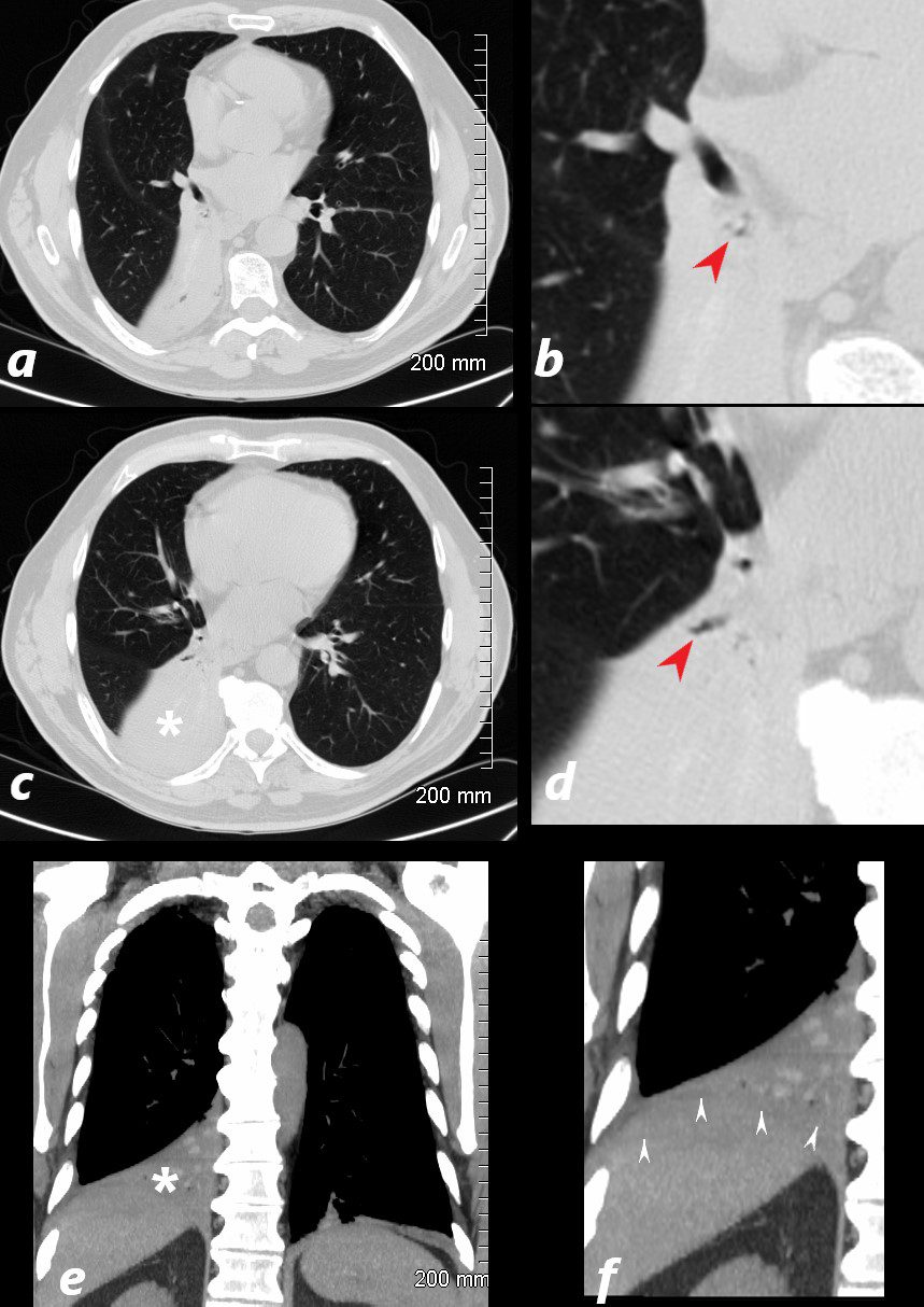
77 year old male presents chest discomfort
CT scan without contrast shows atelectasis of the right lower lobe )asterisk c and r) and also seen axial projection (a) magnified in (b) and in (c) magnified in {d) Red arrowheads in b and d show airways filled with material. Aspergillus was isolated at bronchoscopy. Coronal imaging (e magnified in f) show silhouetting of the right hemidiaphragm by the atelectatic lung (white arrowheads
Ashley Davidoff TheCommonVein.net 117786cL
Pie Shaped Lateral

CXR shows right upper lobe (RUL) atelectasis. Final diagnosis was a central RUL proximal squamous cell carcinoma with extensive filling of the distal bronchi-ectatic segmental and subsegmental airways
Ashley Davidoff TheCommonVein.net
Rectangular Shape Lingula
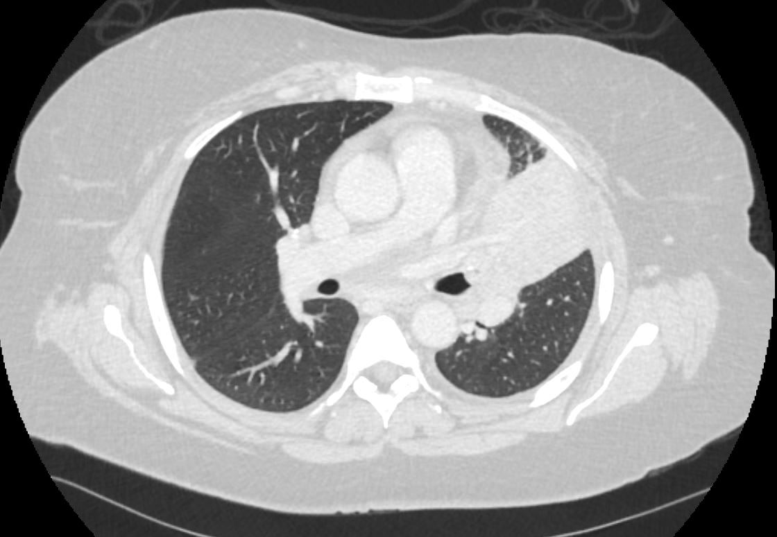
58-year-old female presents with a cough. CT in the axial plane shows an obstructing lesion in the left mainstem bronchus of the lung with post obstructive atelectasis of the lingula and a small portion of aerated left upper lobe anteriorly. T he major fissure is displaced anteriorly.
Pathology revealed findings consistent with a carcinoid tumor of the left bronchus.
Ashley Davidoff MD TheCommonVein.net 257Lu 136110
Middle Lobe
Right Lower Lobe
Left Upper Lobe
Lingula
Silhouetting Left Heart Border
Veiling Effect
Horizontal Main Stem and
Vertical LLL bronchovascular Bundle
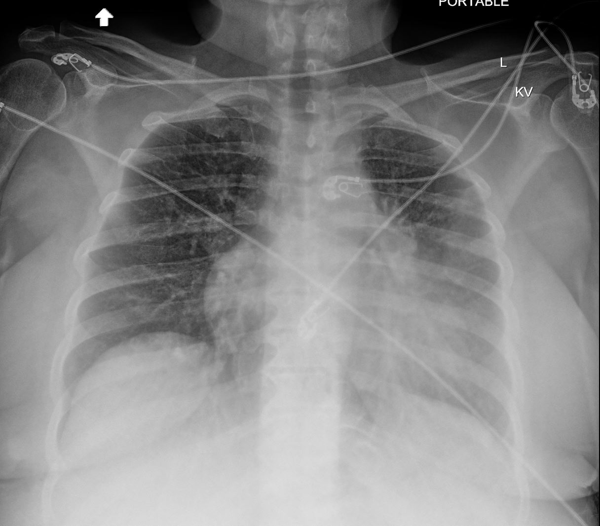
58-year-old female presents with a cough Frontal CXR shows silhouetting of the left heart border with hazy or veiling opacity extending out from the left hilum and fading out inferiorly . The left hilum is pulled superiorly, resulting in an almost horizontal course of the left main bronchus and vertical orientation of the left lower lobe bronchus
Ashley Davidoff MD TheCommonVein.net 257Lu 136109
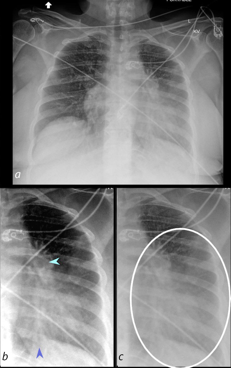
58-year-old female presents with a cough Frontal CXR shows silhouetting of the left heart border with hazy or veiling opacity extending out from the left hilum and fading out inferiorly (white circle c). The left hilum is pulled superiorly (teal arrowhead b) , resulting in an almost horizontal course of the left main bronchus and vertical orientation of the left lower lobe bronchovascular bundle (dark blue arrowhead b)
Ashley Davidoff MD TheCommonVein.net 257Lu 136109cL01
Silhouetting of the Left Heart Border
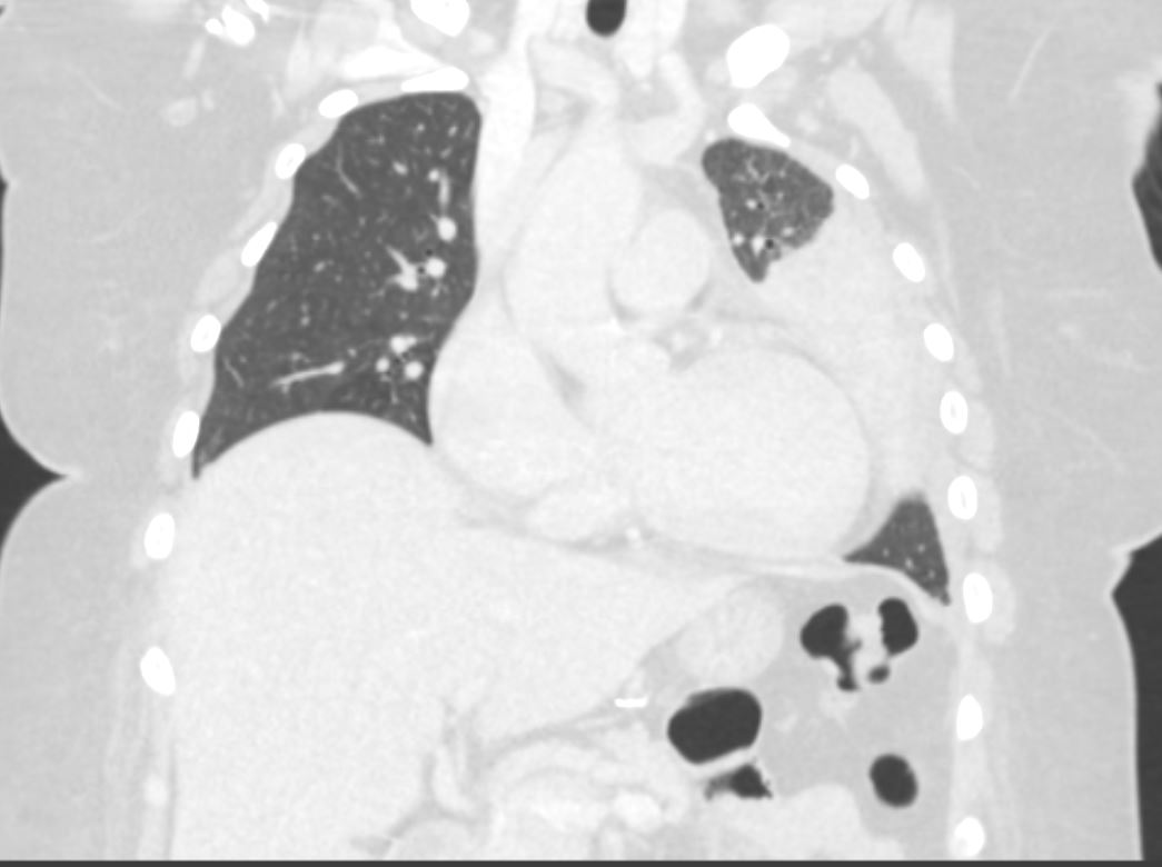
58-year-old female presents with a cough. CT in the coronal plane shows post obstructive atelectasis of the lingula which silhouettes the left heart border. A small portion of aerated left upper lobe is noted in the left apex.
Pathology revealed findings consistent with a carcinoid tumor of the left bronchus.
Ashley Davidoff MD TheCommonVein.net 257Lu 136115
Left Lower Lobe
Segmental
Wedge Shaped Right Middle Lobe Lateral Segment
Sub Segmental Atelectasis
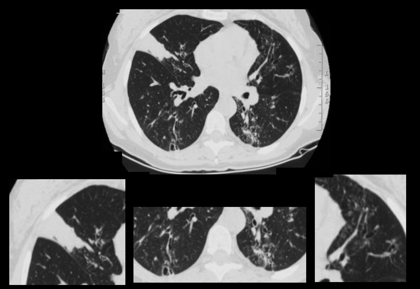
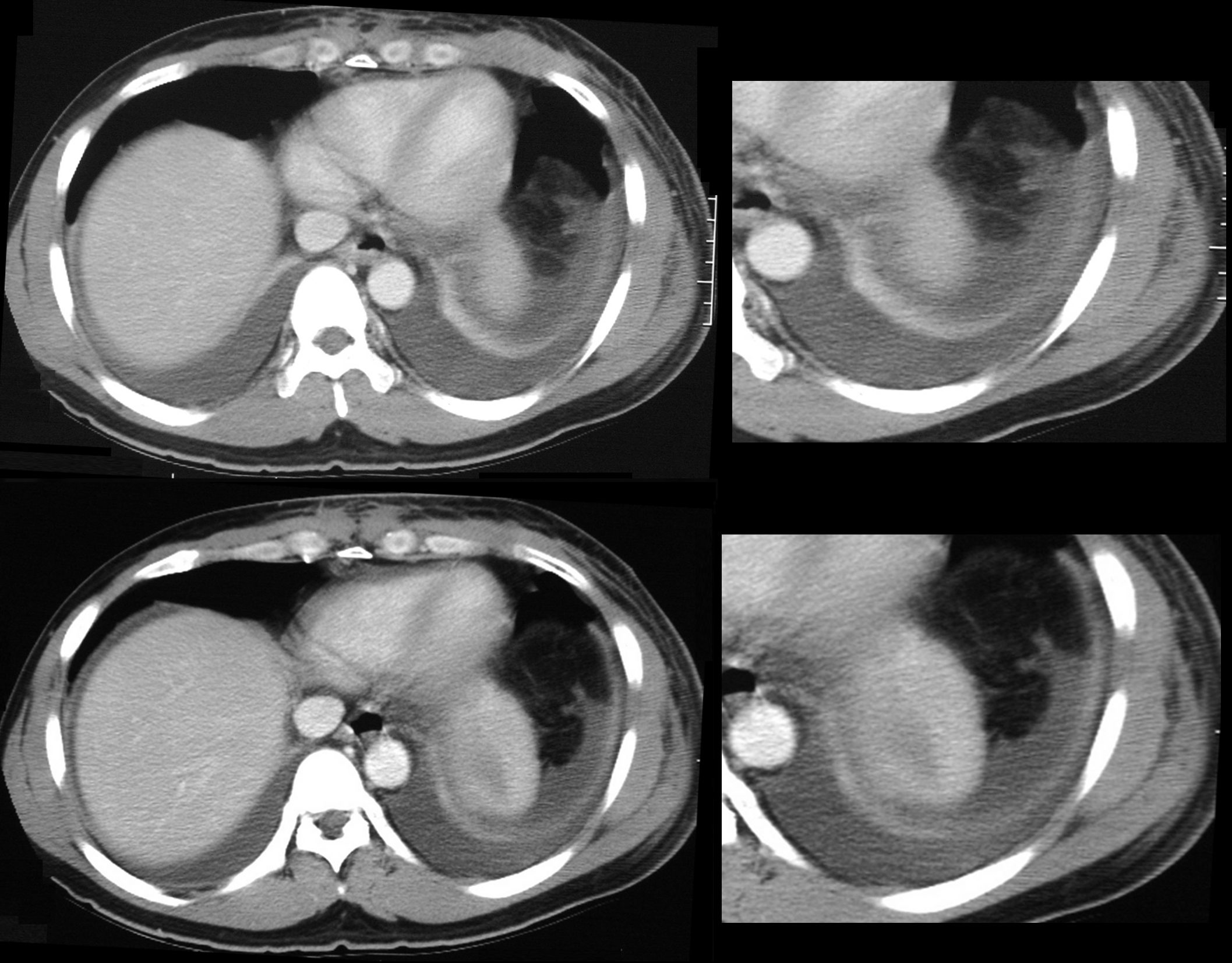
Atelectasis, Mild Bronchial Wall Thickening, and Bronchiolectasis in the RML, Lingula and Bilateral Lower Lobes
CT Allergic Bronchopulmonary Aspergillosis (ABPA)
48 year old female with a history of asthma presents with productive cough. CT scan 18 months prior confirms atelectasis in the middle lobe (upper panel and right lower panel) . There is diffuse mild multicentric foci of bronchial wall thickening in the segmental and subsegmental airways of the middle lobe, lingula and the lower lobes bilaterally (upper panel magnified in lower 3 panels).
Ashley Davidoff MD TheCommonVein.net
Rounded Atelectasis
72-year-old male with a history of asbestos exposure presents with a cough. Axial CTscan shows a pleural based nodule with a comet tail and a series of lung markings folded into the nodule. There is subsegmental compensatory hyperinflation of the lateral segment of the right lower lobe Noted bilateral pleural thickening and pleural based calcification which is reminiscent of asbestos related disease. Early evolution of rounded atelectasis is also noted in the left lower lobe
Ashley Davidoff MD TheCommonVein.net RnD 240Lu
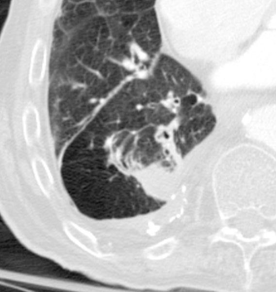
72-year-old male with a history of asbestos exposure presents with a cough. Axial CTscan shows a magnified view of a pleural based nodule with a comet tail and a series of lung markings folded into the nodule. There is subsegmental compensatory hyperinflation of the lateral segment of the right lower lobe Noted pleural thickening and pleural based calcification which is reminiscent of asbestos related disease.
Ashley Davidoff MD TheCommonVein.net RnD 240Lu