
- 60 y.o. female with a history of GERD, and HTN and
- admission 3 years prior
- for chronic cough,
- fevers,
- dyspnea, and
- fatigue
- biopsy proven
- subacute and
- chronic
- organizing eosinophilic PNA
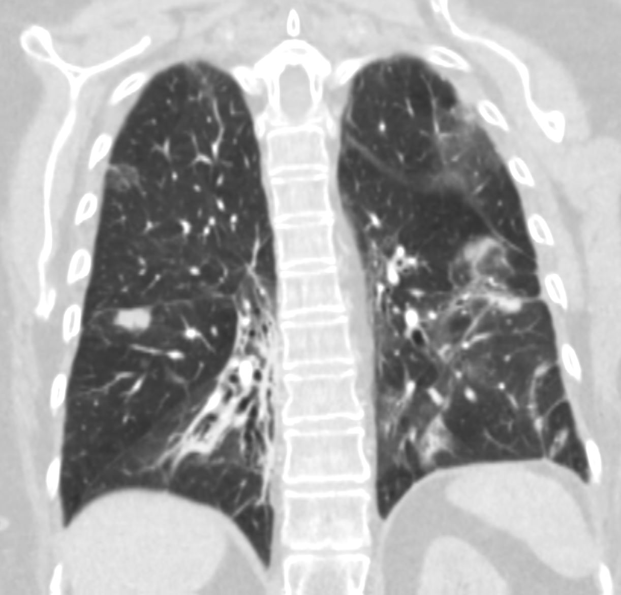
- progressive phase of subacute to chronic eosinophilic pneumonia, including
- interstitial fibrosis,
- intraalveolar fibrin deposits with
- eosinophils and
- lymphohistiocytoses and
- fibroblastic proliferation foci.
- ORGANIZING PNEUMONIA SHOWING SUBACUTE AND CHRONIC PHASES, FIBRINOUS AIRSPACE EXODUATES WITH ADMIXED EOSINOPHILS AND HISTIOCYTES.
THE DISTRIBUTION IS MULTIFOCAL AND PATCHY.
NO EVIDENCE OF VASCULITIS OR GRANULOMA SEEN.
- started on prednisone
- started on mepolizumab 1 year later and
- weaned off prednisone 2 years ago
- with improvement of symptoms and
- CXR now presenting for a follow up.
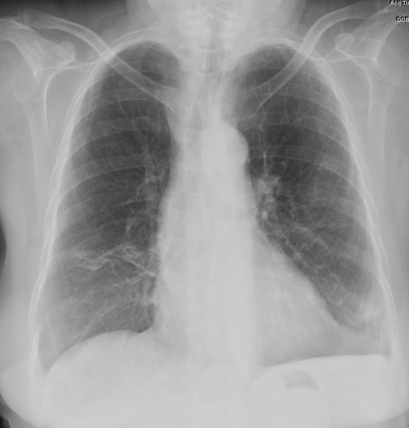
1 year Prior to Illness
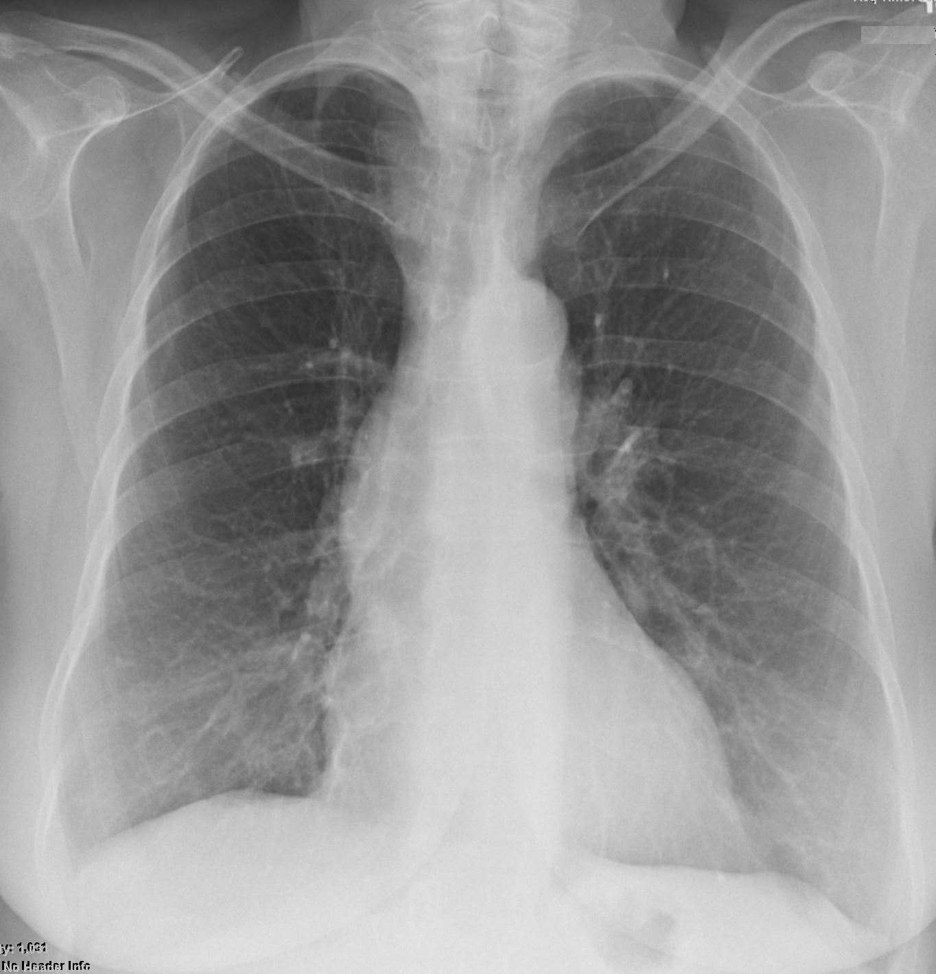
CXR from 1 and half years prior
Ashley Davidoff MD TheCommonVein.net
eosinophillic-pneumonia-001
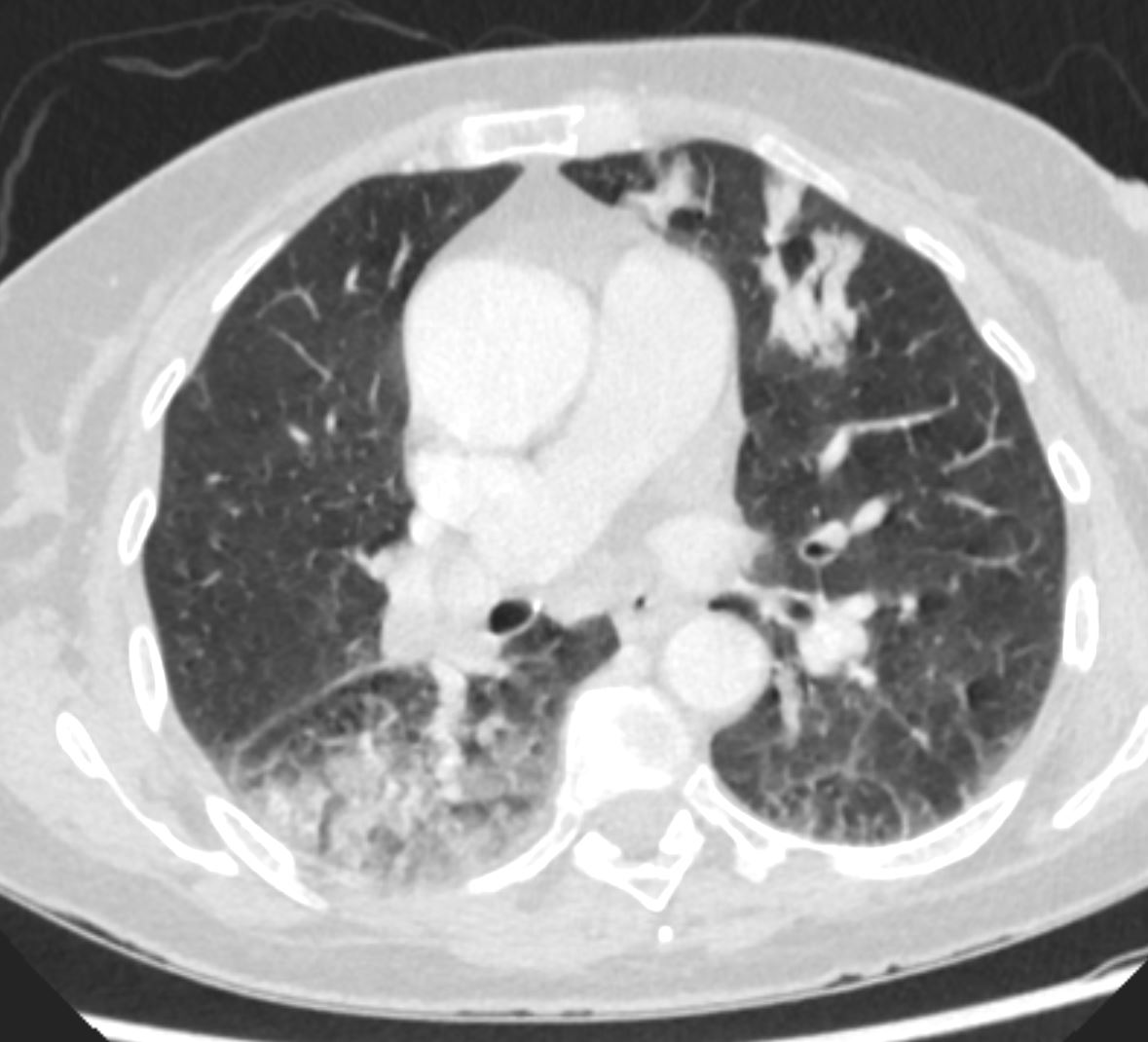
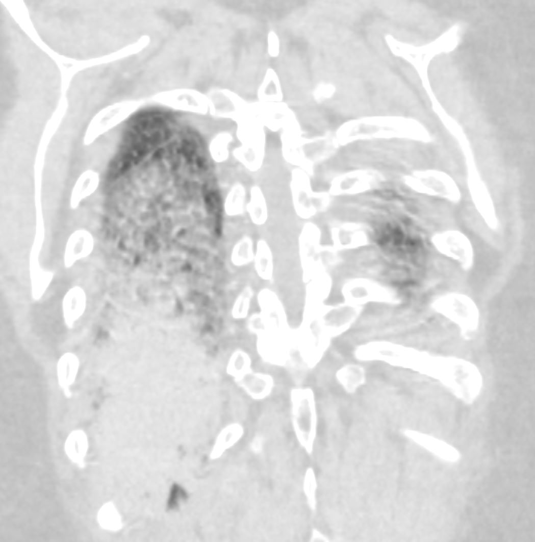
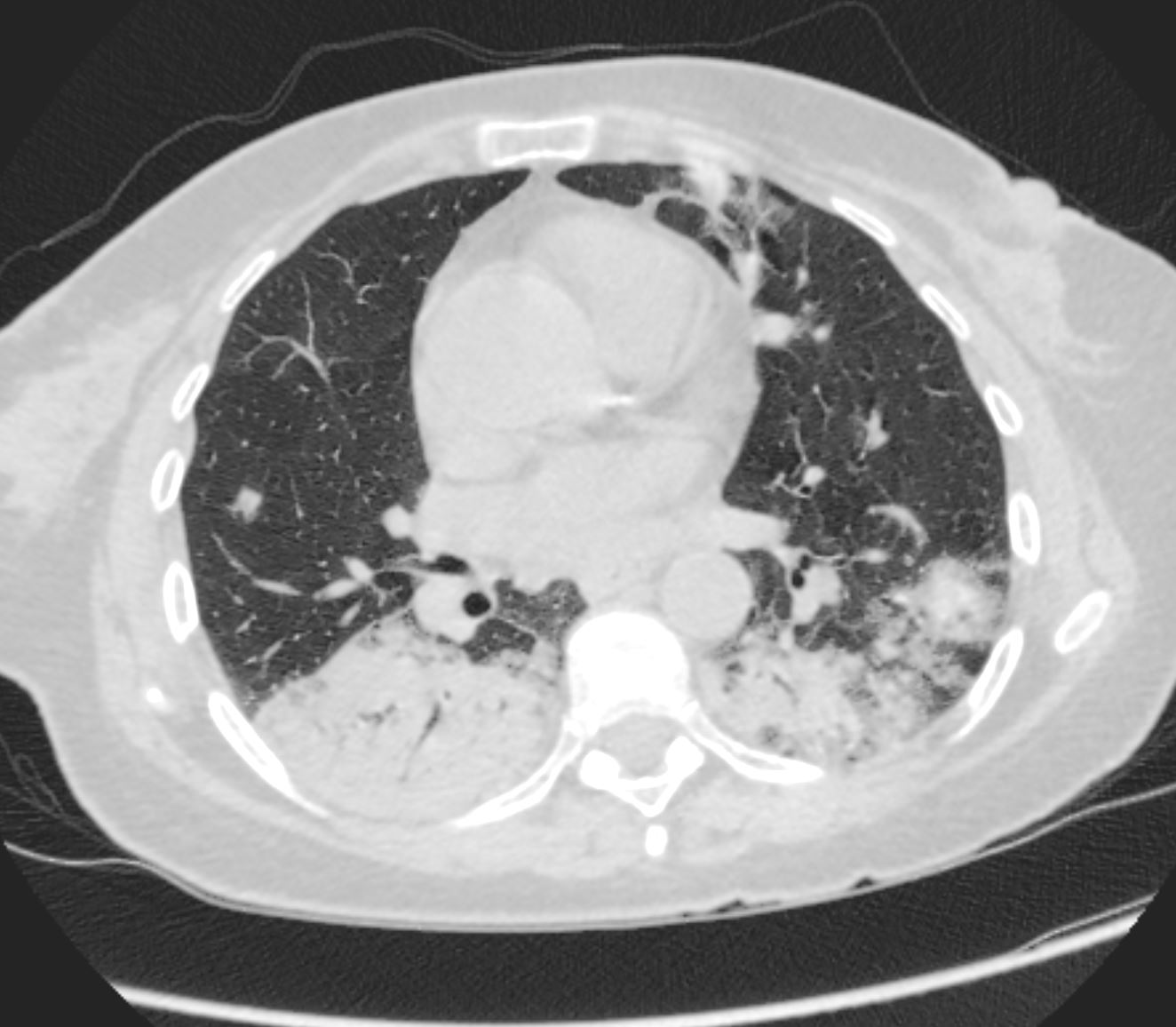
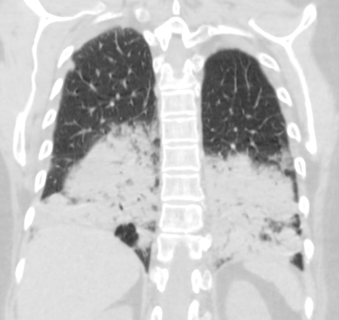
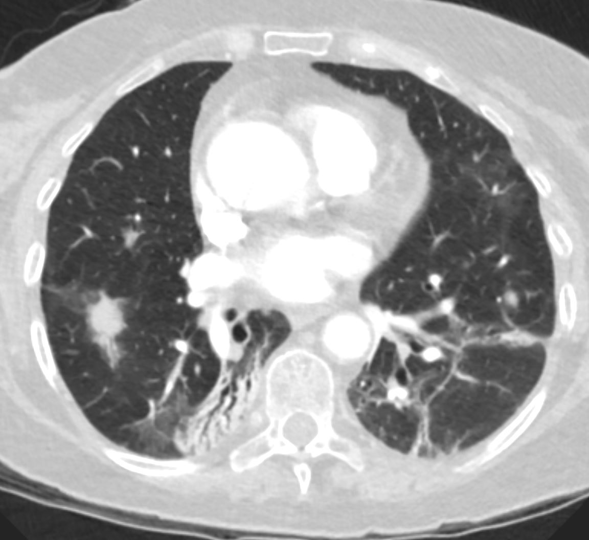
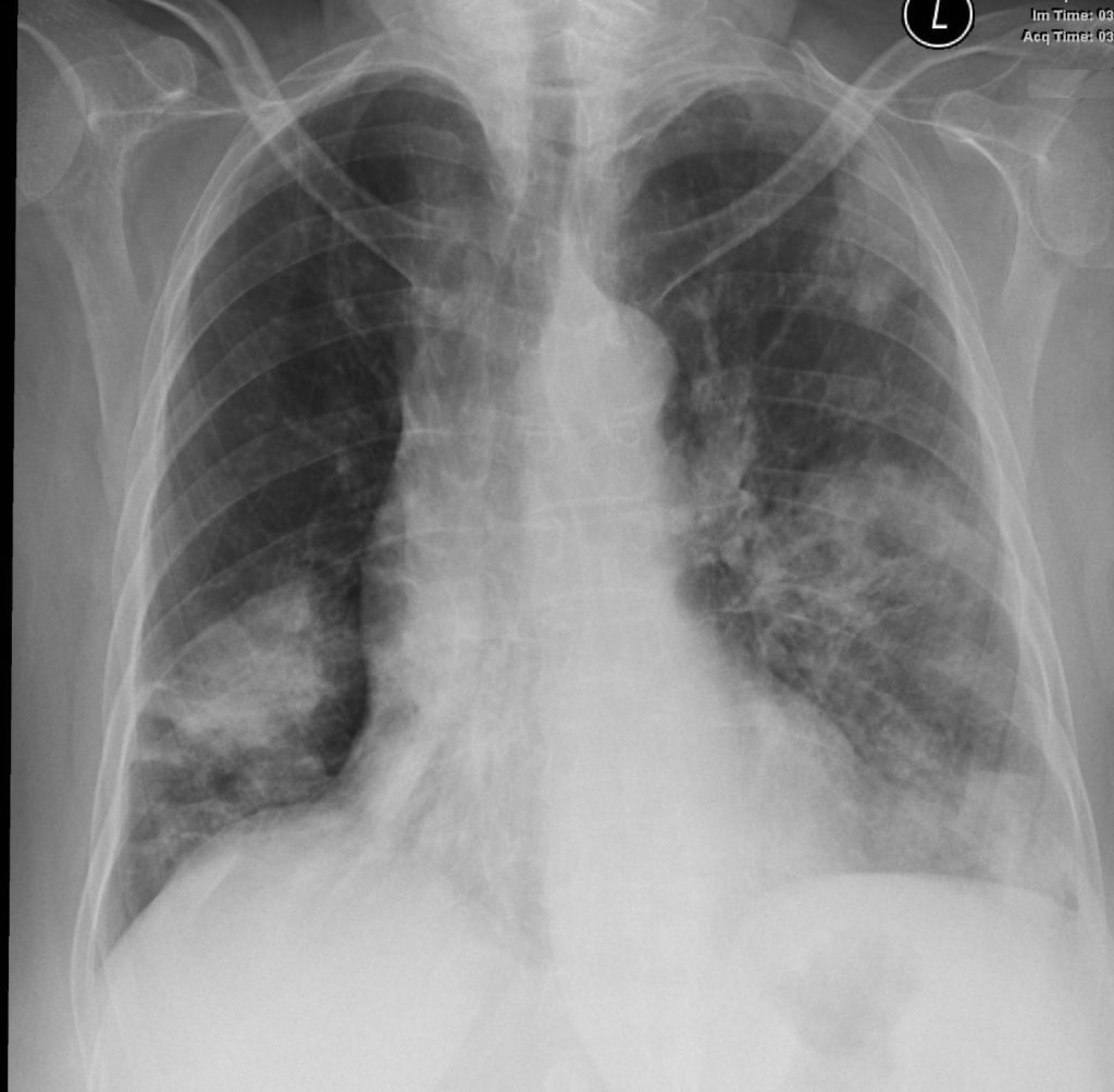
Presents with Cough and Dyspnea
Bibasilar Infiltrates and Multifocal Nodular Infiltrates
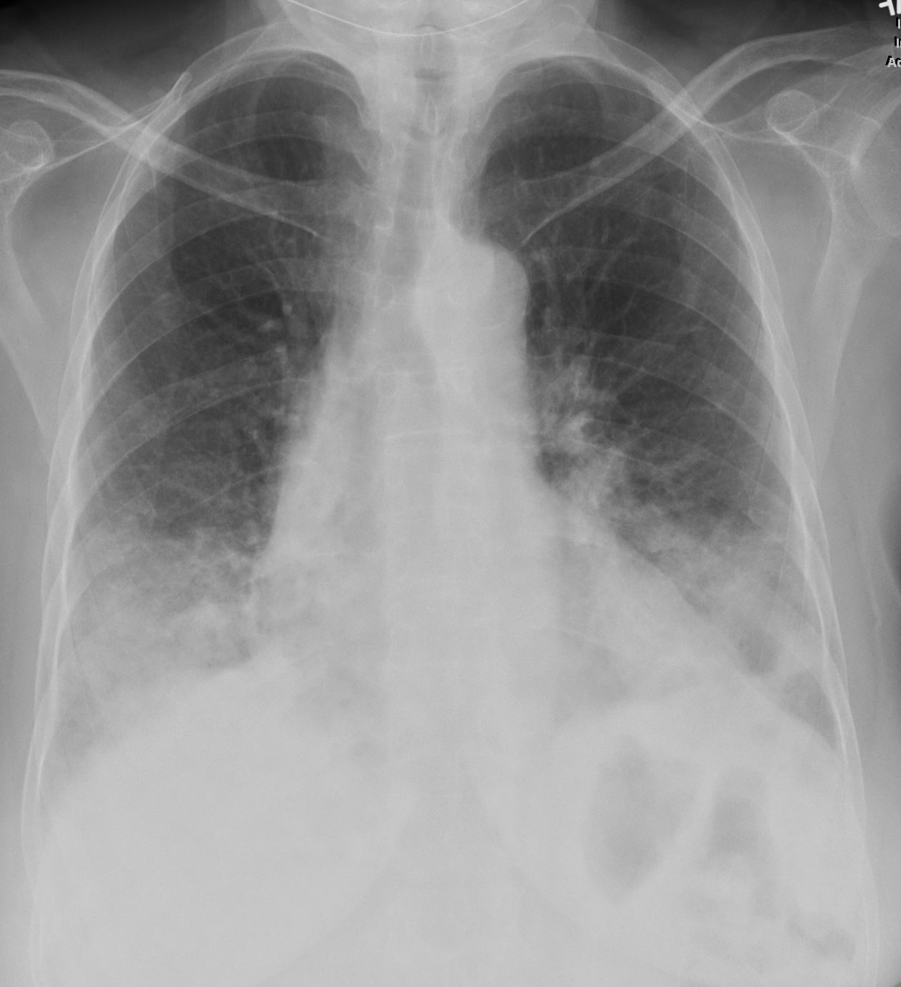
CXR at presentation shows dense bibasilar consolidations
Ashley Davidoff MD TheCommonVein.net
eosinophillic-pneumonia-002
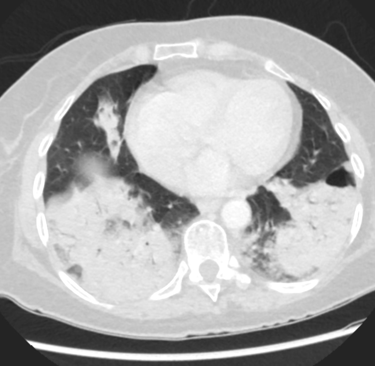
Ashley Davidoff MD TheCommonVein.net
eosinophillic-pneumonia-003
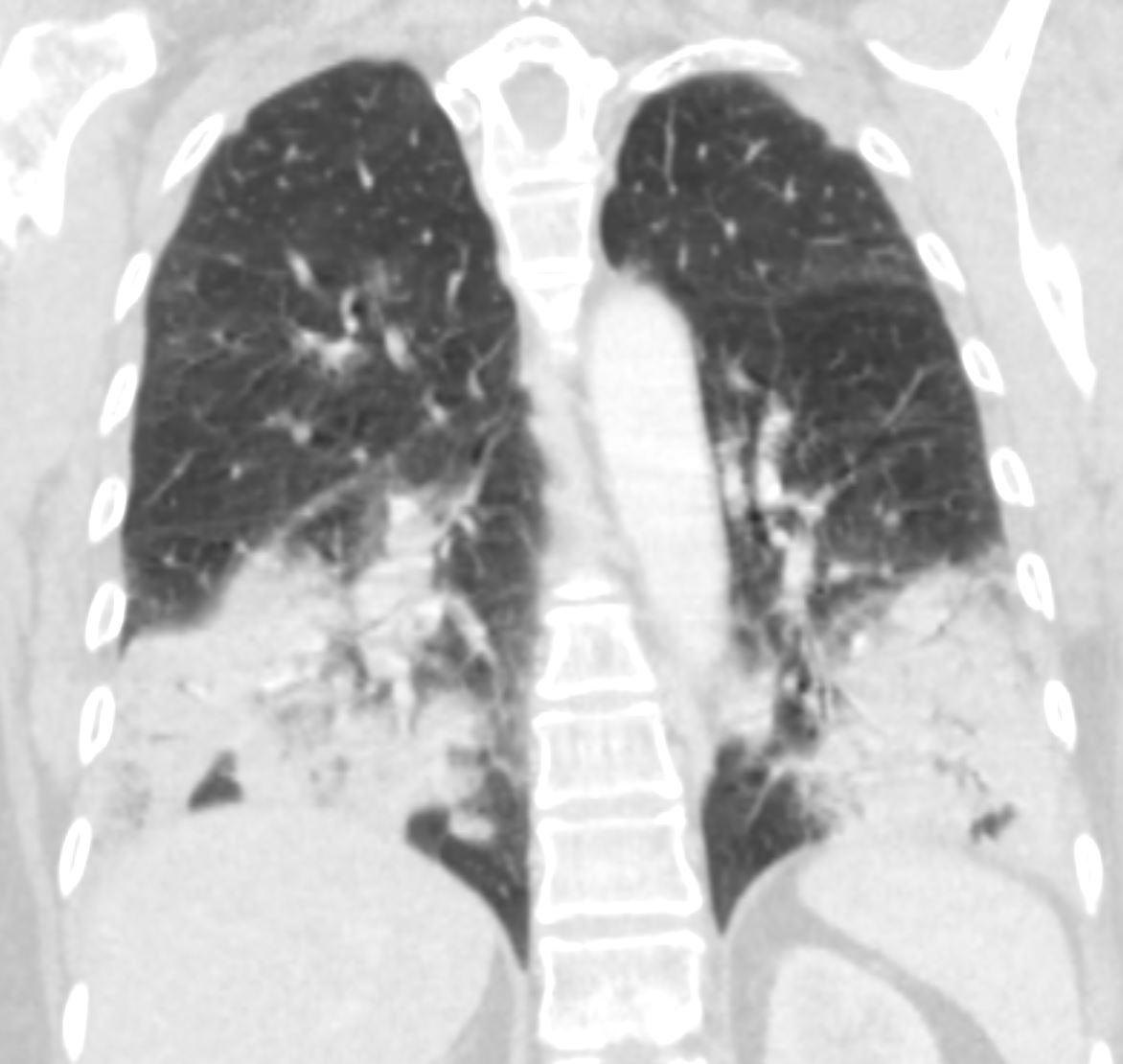
Ashley Davidoff MD TheCommonVein.net
eosinophillic-pneumonia-006
Ashley Davidoff MD TheCommonVein.net
eosinophillic-pneumonia-007
Ashley Davidoff MD TheCommonVein.net
eosinophillic-pneumonia-007b
2 Weeks Later
Ashley Davidoff MD TheCommonVein.net
eosinophillic-pneumonia-009
Ashley Davidoff MD TheCommonVein.net
eosinophillic-pneumonia-011
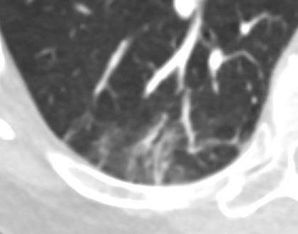
Bronchiolectasis
Ashley Davidoff MD TheCommonVein.net
eosinophillic-pneumonia-0112b
3 Months After Presentation
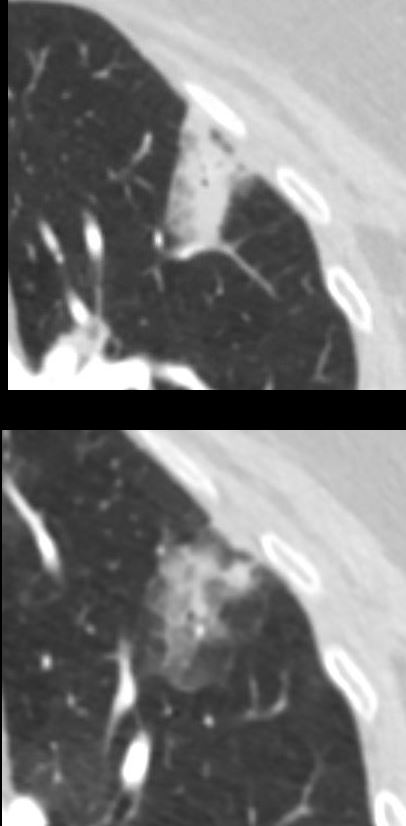
3 months later (lower image)
Ashley Davidoff MD TheCommonVein.net
eosinophillic-pneumonia-007b
Ashley Davidoff MD TheCommonVein.net
eosinophillic-pneumonia-012c
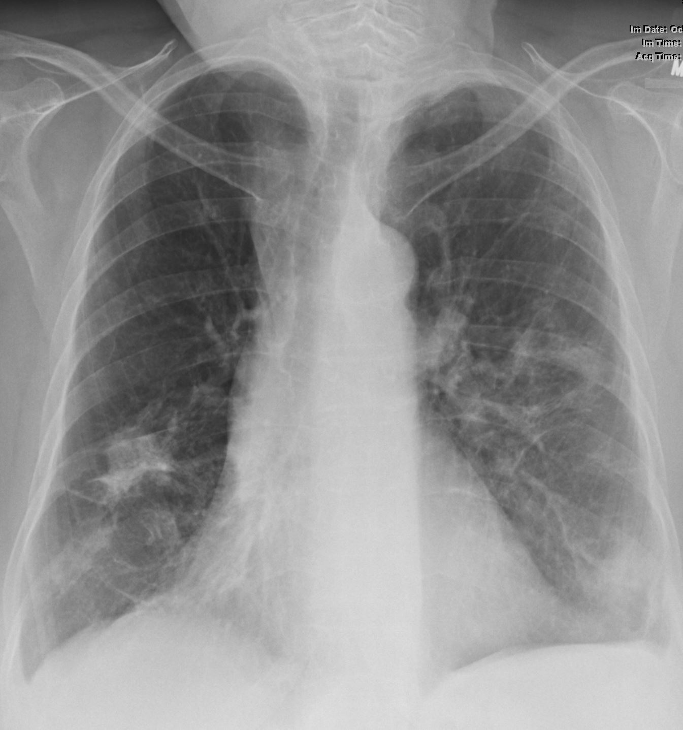
7 Months After Presentation Improving Bibasilar Infiltrates
Ashley Davidoff MD TheCommonVein.net
eosinophillic-pneumonia-014
Ashley Davidoff MD TheCommonVein.net
eosinophillic-pneumonia-016
Ashley Davidoff MD TheCommonVein.net
eosinophillic-pneumonia-017
8 Months After Presentation
Ashley Davidoff MD TheCommonVein.net
eosinophillic-pneumonia-018
9 Months After Presentation
Ashley Davidoff MD TheCommonVein.net
eosinophillic-pneumonia-020
Links and References
TCV Case 703