Etymology
- Derived from the Latin word linea, meaning “line,” and the Greek word atelectasis, meaning “incomplete expansion.” The term refers to a thin, plate-like area of lung collapse visible on imaging.
AKA
- Discoid atelectasis
- Plate-like atelectasis
Definition
What is it?
- Linear atelectasis refers to a form of partial lung collapse characterized by thin, linear, or plate-like opacities on imaging, typically involving the subsegmental lung areas. It is often transient and related to shallow breathing or reduced lung expansion.
Caused by
- Hypoventilation due to:
- Postoperative state
- Prolonged immobility
- Sedation or anesthesia
- Compression from pleural effusion or adjacent mass
- Resorption of trapped air distal to an obstructed airway
- Chest wall splinting due to pain (e.g., rib fractures)
Resulting in
- Alveolar collapse without significant volume loss in adjacent lung regions
- Reduced ventilation in the affected area
Structural Changes
- Collapse of alveoli in subsegmental regions, often along lung fissures
- Minimal impact on overall lung volume
Pathophysiology
- Linear atelectasis occurs when alveoli fail to expand fully due to hypoventilation or external compression. The lack of alveolar inflation leads to localized collapse of lung parenchyma, which appears as thin, linear opacities on imaging. The process is often reversible with interventions that promote lung expansion.
Pathology
- Collapsed alveoli with minimal inflammation
- Potential mucus plugging in associated airways
- Reversible changes unless associated with underlying scarring or chronic conditions
Radiology in Detail
CXR
Findings
- Thin, linear opacities extending across lung fields, often parallel to the pleura
- Commonly located in dependent lung regions or along interlobar fissures
Associated Findings
- May coexist with small pleural effusions
- No significant mediastinal shift or volume loss
CT
Parts
- Subsegmental lung regions, often in dependent areas or adjacent to pleura
Size
- Typically involves small portions of lung tissue
Shape
- Thin, linear, or plate-like opacities
- These opacities are often parallel to the diaphragm or pleura because the collapse occurs along the natural anatomical planes of least resistance, such as interlobar fissures or the pleural surface. The linear configuration reflects the uniform, localized alveolar collapse in these regions.
Position
- Commonly in the lower lobes or along interlobar fissures
Character
- Non-specific linear opacities with minimal surrounding tissue reaction
Time
- Acute onset; often resolves within hours to days with appropriate interventions
Associated Findings
- May be accompanied by subtle ground-glass changes or small effusions
Other Imaging Modalities
MRI/PET CT/NM/US/Angio
- MRI: Rarely used for linear atelectasis but may identify associated pleural or parenchymal abnormalities
- Ultrasound: Useful for detecting small pleural effusions contributing to atelectasis
Key Points and Pearls
- Linear atelectasis is often transient and related to shallow breathing or reduced lung expansion.
- It is commonly seen postoperatively or in bedridden patients.
- Radiologically, it appears as thin, plate-like opacities, typically in dependent lung regions or along fissures.
- Interventions such as incentive spirometry, mobilization, and addressing pain can resolve linear atelectasis effectively.
- Differentiating linear atelectasis from chronic scarring or fibrotic changes is essential to avoid misdiagnosis.

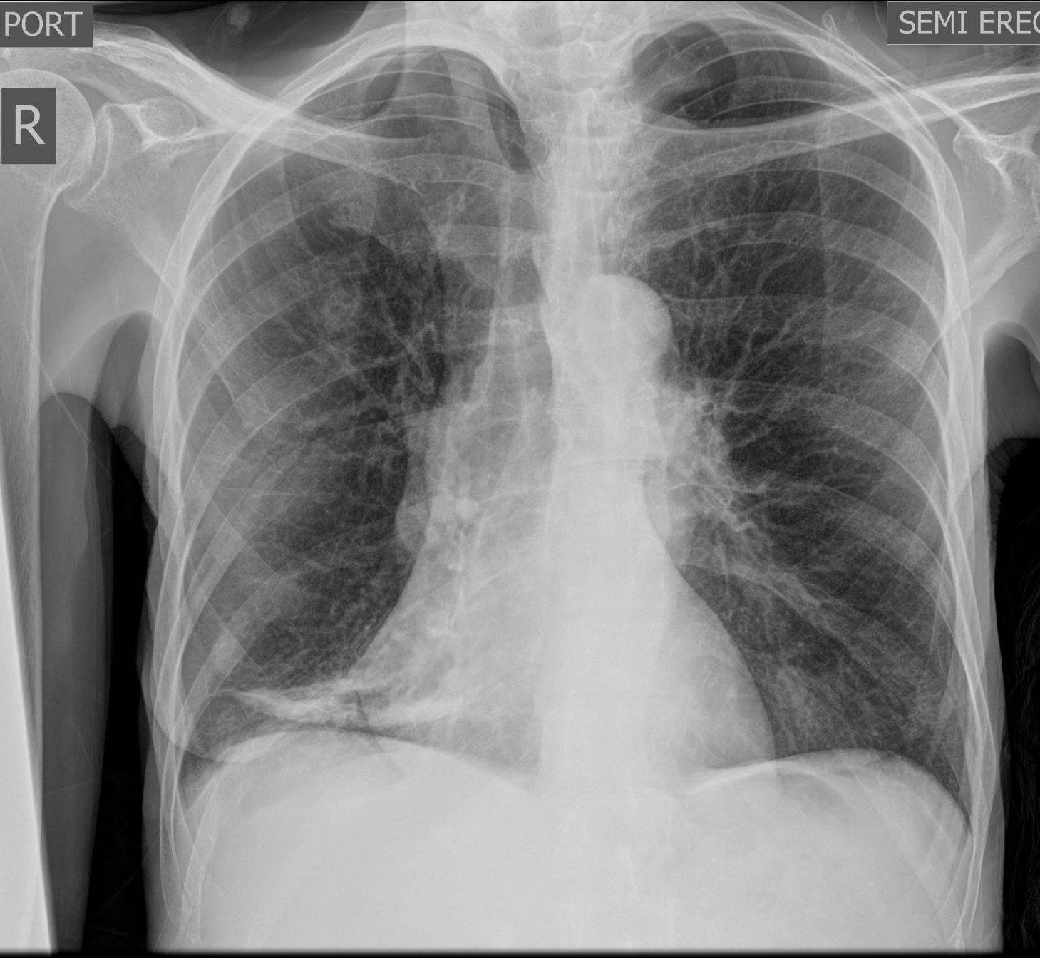
Frontal Chest Xray of a 55-year-old female shows a region of discoid atelectasis (aka linear atelectasis plate atelectasis ) in the right lower lung zone
Courtesy Ashley Davidoff MD TheCommonVein.net 136548

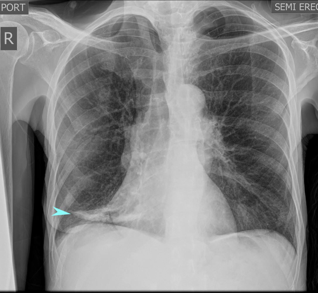
Frontal Chest Xray of a 55-year-old female shows a region of discoid atelectasis (aka linear atelectasis plate atelectasis ) in the right lower lung zone (teal arrow)
Courtesy Ashley Davidoff MD TheCommonVein.net 136548

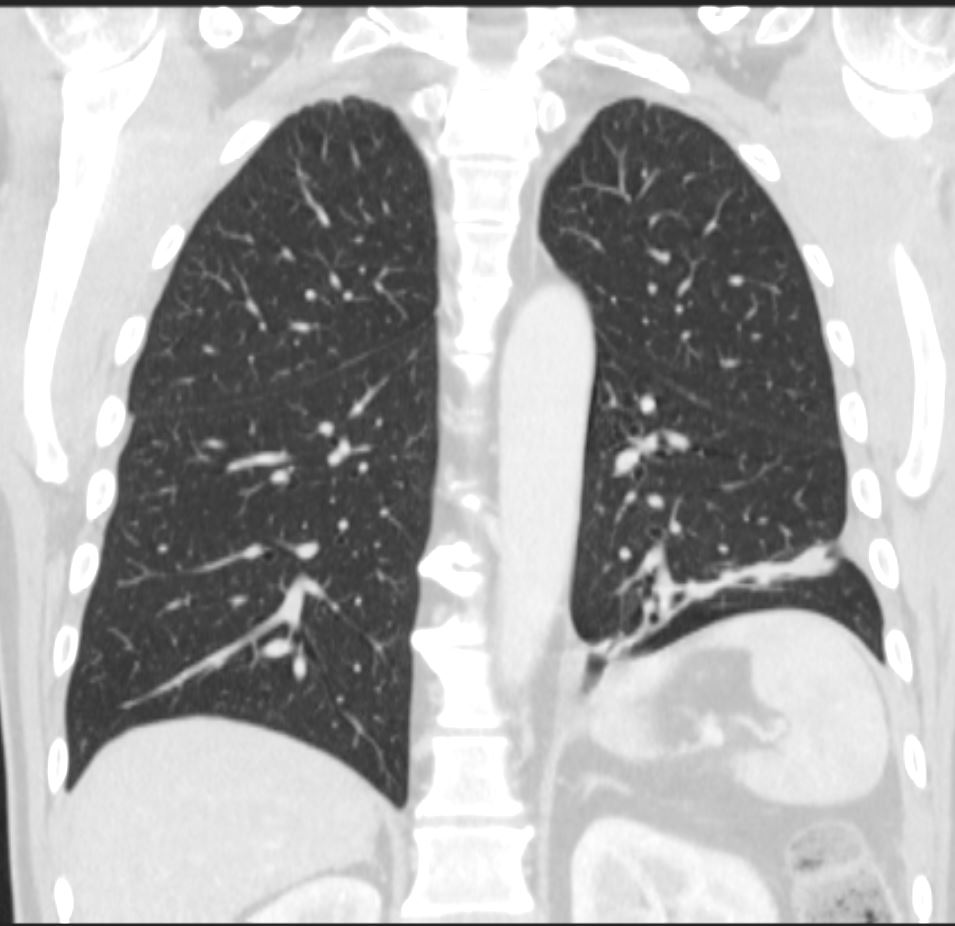
CT scan in the coronal plane 3 months later shows significant improvement of the atelectasis involving a basal segment of the left lower lobe associated with persistent elevation of the left hemidiaphragm indicating volume loss. The atelectasis now has a discoid, linear, or plate-like appearance
Ashley Davidoff MD TheCommonVein.net 276Lu 136238
aka discoid atelectasis aka plate-like atelectasis

66 year old male with linear (discoid) atelectasis in the left lower lobe on CT
Ashley Davidoff MD TheCommonVein.net


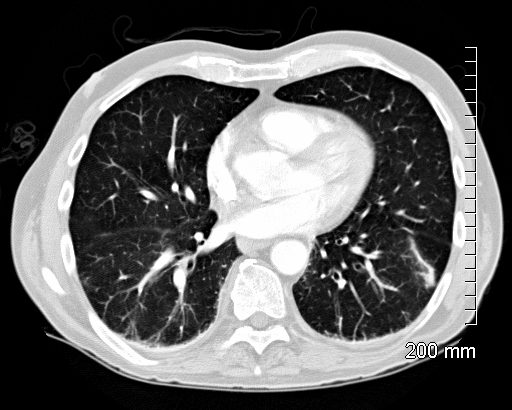
60 year old male with linear (discoid) atelectasis in the middle lobe and the left upper lobe on CT. Note moderate sized bilateral pleural effusion. Minor compressive atelectasis caused by the left effusion.
Ashley Davidoff MD TheCommonVein.net


66 year old male with linear (discoid) atelectasis in the left lower lobe on CT
Ashley Davidoff MD TheCommonVein.net
Radiographic Appearance
- Linear Opacity: Appears as a thin, straight or slightly curvilinear band of increased density, usually 1–3 cm wide.
- Location: Often seen in the lower lobes of the lungs, particularly along the costophrenic angles.
- Orientation: Usually parallel to the pleural surface, often horizontal or oblique in alignment.
Common Causes
Linear or discoid atelectasis can result from various factors that cause localized alveolar collapse, including:
- Hypoventilation: Commonly occurs post-operatively or in patients with prolonged bed rest.
- Shallow Breathing: Seen in patients with pleuritic pain, rib fractures, or abdominal discomfort, as these conditions limit deep inspiration.
- Obstruction: Mucus plugging in smaller airways can cause localized atelectasis.
- Compression: External pressure from pleural effusion, a mass, or pneumothorax can collapse nearby alveoli.
Clinical Significance
Linear or discoid atelectasis is generally a benign finding and often transient. It does not usually indicate significant pathology, although it can sometimes be a marker of poor ventilation or underlying lung disease. It is important to differentiate it from more concerning findings, such as infiltrates or fibrotic bands, which may indicate chronic or progressive disease.
Summary
In radiographic terms, linear or discoid atelectasis is a thin, linear opacity representing areas of localized alveolar collapse, often due to hypoventilation or compression, and is typically a temporary and benign finding.
Links and References
Chat GPT – What is radiographic definition of linear or discoid atelectasis?
