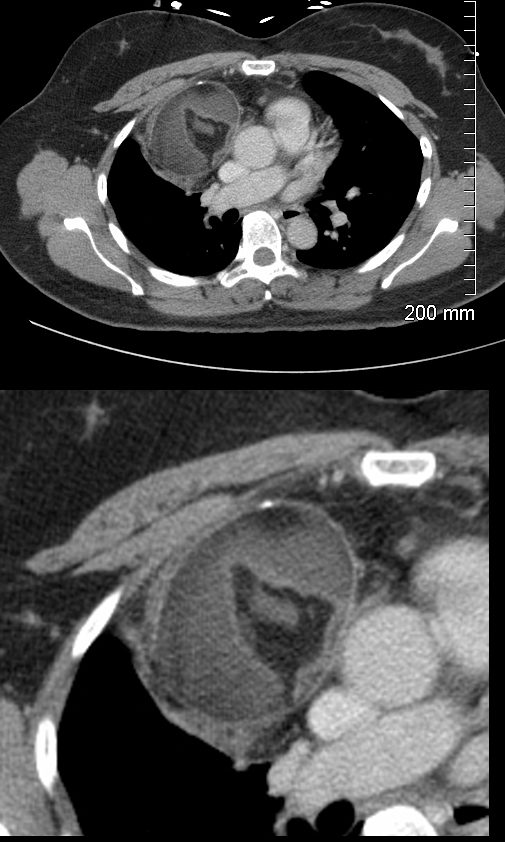
47-year-old female presents with an abnormal CXR
CT scan in the axial projection shows a 6.3X 5.9cms mass in the anterior mediastinum that shows central fatty elements, surrounded by soft tissue elements and a anterior peripheral calcification. These findings are consistent with a teratoma
Ashley Davidoff MD TheCommonVein.net 275Lu 136333c
- Mediastinal Teratoma:
- are germ cell tumors that can
- contain tissues derived from the three germ cell layers
-
- ectoderm,
- mesoderm, and
- endoderm.
- composed therefore of various cell types,
- hair,
- skin,
- teeth, or
- more complex structures.
-
- Causes:
- The exact cause of teratomas is unknown.
- thought to arise from primordial germ cells,
- Genetic factors during embryonic development may contribute to the formation of teratomas.
- Functional and Structural Changes:
-
- Compression of nearby structures
- may result in symptoms such as
- chest pain,
- cough,
- dyspnea, or
- superior vena cava syndrome if large enough.
-
- Clinical Diagnosis:
-
- No pathognomonic clinical symptoms or signs
-
- Lab Diagnosis:
-
- tumor markers, which can be elevated in some germ cell tumors.
- alpha-fetoprotein (AFP) and
- beta-human chorionic gonadotropin (β-hCG),
- tumor markers, which can be elevated in some germ cell tumors.
-
- Imaging Characteristics:
-
- Chest X-ray (CXR):
- May show a mass in the mediastinum,
- Chest X-ray (CXR):
-
- CT (Computed Tomography):
-
- CT scans are commonly used for better visualization of the tumor’s
- size,
- location, and
- characteristics.
- heterogeneous masses with areas of
-
- fat,
- calcification, and
- cystic changes.
-
- heterogeneous masses with areas of
- tumor’s relationship with surrounding structures.
- CT scans are commonly used for better visualization of the tumor’s
-
- MRI (Magnetic Resonance Imaging):
-
- Tissue characterization
- heterogeneous masses with areas of
-
- fat,
- calcification, and
- cystic changes.
-
- heterogeneous masses with areas of
- Tissue characterization
-
- PET (Positron Emission Tomography) Scan:
-
- Benign teratomas
- not highly PET positive
- Can be employed to determine the metabolic activity of the tumor and its extent. PET scans are helpful in staging and planning treatment.
- Benign teratomas
-
- Diagnosis,
- Distinguishing between benign and malignant usually requires tissue examination
- PET has limitations in definitively differentiating between
- benign and malignant teratomas.
- Benign Teratomas:
- generally composed of well-differentiated tissues
- metabolic activity tends to be lower, and
- usually do not show intense uptake on PET scans.
- Malignant Teratomas:
- some malignant teratomas
- may exhibit increased metabolic activity,
- it’s not a universal rule.
- Benign Teratomas:
- benign and malignant teratomas.
- Hence PET scan
-
- not definitive in
- distinguishing between
- benign and malignant teratomas.
- Pathological examination through biopsy or surgical excision is usually required .
- Histological evaluation for malignancy , shows
-
- immature or undifferentiated components,
-
-
- Staging: Staging involves determining the extent of the tumor’s spread. It may include imaging studies such as CT, MRI, and PET scans.
- Treatment:
-
- involves surgical removal of the tumor.
- extent of surgery will depend on the
- tumor size, location, and involvement of adjacent structures.
- In some cases, chemotherapy or radiation therapy may be recommended, especially for malignant teratomas.
-
CT Anterior Mediastinal Benign Teratoma

47-year-old female presents with an abnormal CXR
CT scan in the axial projection shows a 6.3X 5.9cms mass in the anterior mediastinum that shows central fatty elements, surrounded by soft tissue elements and a anterior peripheral calcification. These findings are consistent with a teratoma
Ashley Davidoff MD TheCommonVein.net 275Lu 136333c

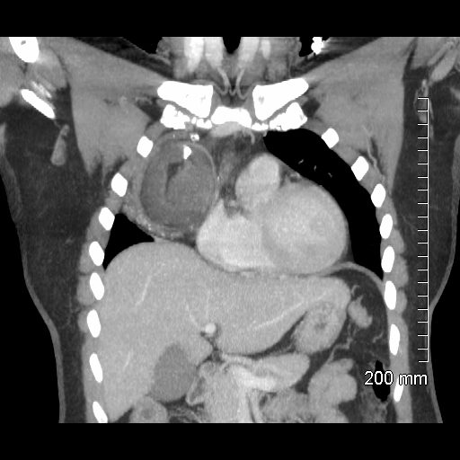
47-year-old female presents with an abnormal CXR
CT scan in the coronal projection shows a 6.3X 5.9 x 6.8cms mass in the anterior mediastinum that shows central fatty elements, surrounded by intermediate density of homogeneous soft tissue elements and a prominent calcification with a shape reminiscent of a tooth, and a more heterogeneous infero-lateral rim of soft tissue density with punctate calcifications.. These findings are consistent with a teratoma
Ashley Davidoff MD TheCommonVein.net 275Lu 136340

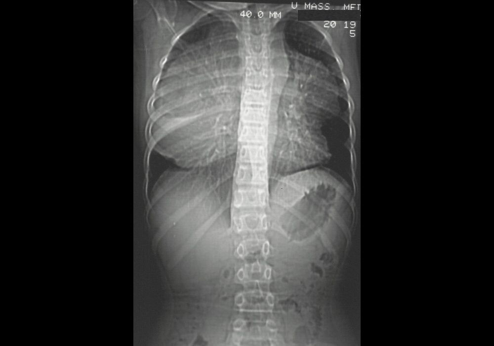
Scout CT a large mass occupying almost the entire thoracic cavity.
Ashley Davidoff MD TheCommonVein.net 24649

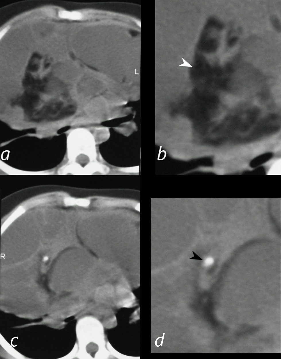
CT scan in the axial projection shows a large complex mass occupying the entire anterior mediastinum that shows a large fatty component (magnified in b, white arrowhead), and a punctate calcification (magnified in d, black arrowhead) in the context of multiple large cystic masses separated by bands of soft tissue components.
Ashley Davidoff MD TheCommonVein.net 24650cL
