017Lu 27F LIP HIV AIDS Lymphoma
27 year old male with a history of perinatal HIV with intermittent highly active antiretroviral therapy (HAART) compliance with a CD4 count of < 50 with biopsy confirmed B cell lymphoma of the liver, s/p CHOP therapy , chronic esophageal strictures s/p dilatations, esophageal candidiasis, LIP, bronchiectasis pancreatitis, and portal vein and splenic vein thrombosis.
Initial Chest X-ray shows a diffuse reticular pattern with cystic changes dominant at the bases.

Initial Chest X-ray shows a diffuse reticular pattern with architectural distortion and cystic changes dominant at the bases.
Ashley Davidoff MD TheCommonVein.net 139266 017Lu
CT at this time confirmed the presence of diffuse cystic changes with the largest cysts at the lung base but also involving the mid lung regions. Ascites and splenomegaly were also present

27 year old male with a history of perinatal HIV and lymphocytic interstitial pneumonitis
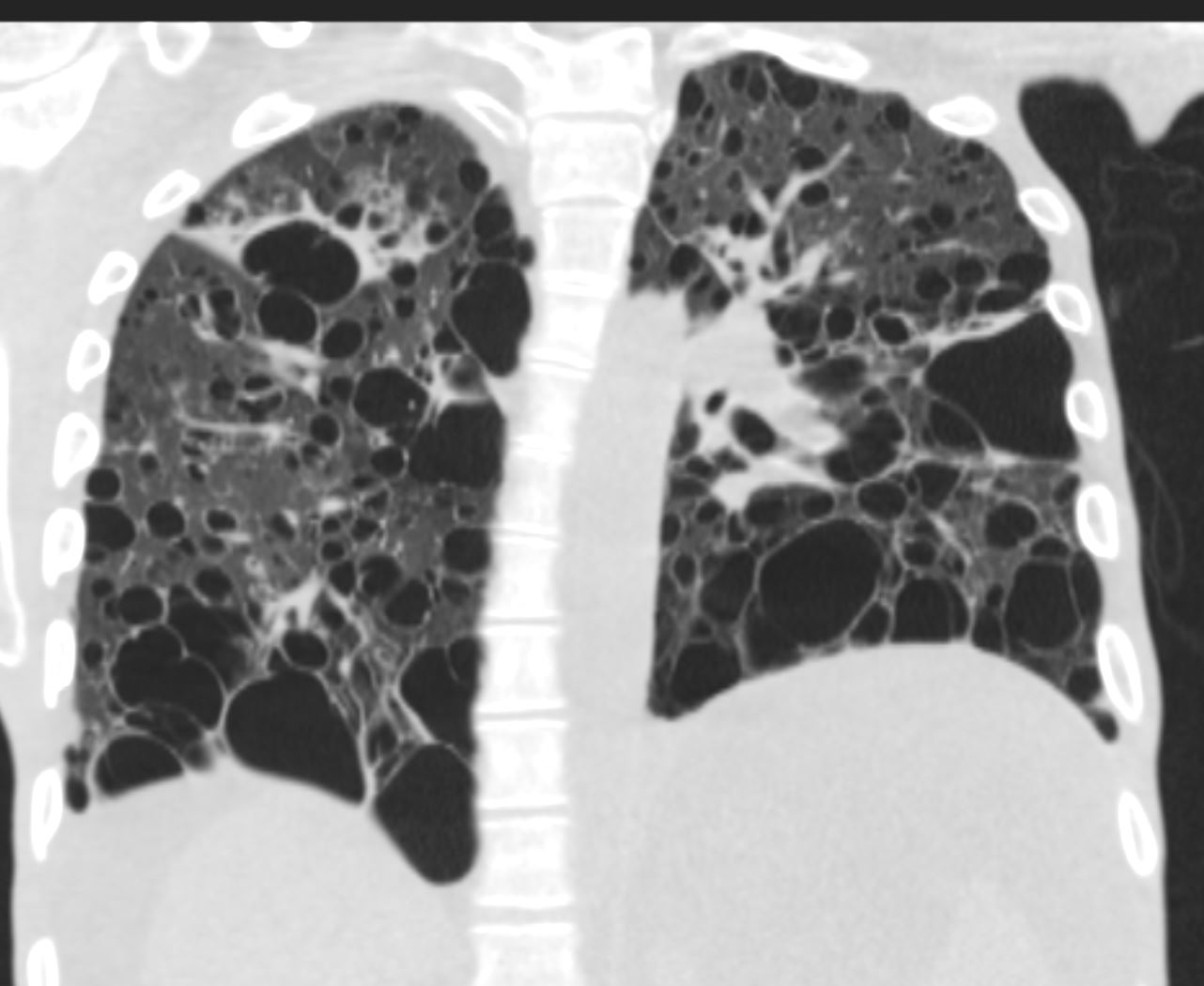
CT in the coronal plane confirms the presence of diffuse cystic changes with the largest cysts at the lung bases. Ascites and splenomegaly are also present
Ashley Davidoff MD TheCommonVein.net 139268 017Lu

27 year old male with a history of perinatal HIV and lymphocytic interstitial pneumonitis
CT in the coronal plane confirms the presence of diffuse cystic changes with the largest cysts in the lower and mid lung regions.
Ashley Davidoff MD TheCommonVein.net 139267 017Lu

27 year old male with a history of perinatal HIV and B cell lymphoma
Coronal reconstruction of the CT shows splenomegaly and moderate ascites. Ascites and splenomegaly are also present
Ashley Davidoff MD TheCommonVein.net 139269 017Lu
One Month Later
presents with fever and neutropenia.

Scout CT shows a diffuse reticular pattern with architectural distortion and cystic changes dominant at the bases.
Ashley Davidoff MD TheCommonVein.net 139271 017Lu

27 year old male with a history of perinatal HIV and lymphocytic interstitial pneumonitis
CT in the coronal plane confirms the presence of diffuse cystic changes with the largest cysts at the lung bases but also involving the mid and upper lung fields .
Ashley Davidoff MD TheCommonVein.net 139272 017Lu

27 year old male with a history of perinatal HIV and lymphocytic interstitial pneumonitis
CT in the coronal plane confirms the presence of diffuse cystic changes with the largest cysts at the lung bases but also involving the mid and upper lung fields .
Ashley Davidoff MD TheCommonVein.net 139273 017Lu

Focal band of ground glass opacity noted in the right upper lobe
Ashley Davidoff MD TheCommonVein.net 139274 017Lu

.Focal band of ground glass opacity noted in the right upper lobe
Ashley Davidoff MD TheCommonVein.net 139275 017Lu

27 year old male with a history of perinatal HIV and lymphocytic interstitial pneumonitis
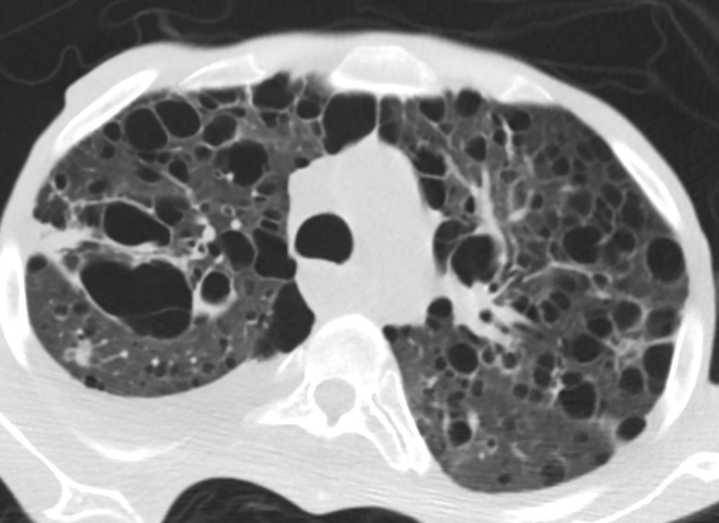
CT in the axial plane confirms the presence of diffuse thin walled cystic changes . Focal band of ground glass opacity noted in the right upper lobe
A mixed density 8mm nodule is noted in the posterior segment of the right upper lobe. There is a new small to moderate right sided pleural effusion.
Ashley Davidoff MD TheCommonVein.net 139276 017Lu

27 year old male with a history of perinatal HIV and lymphocytic interstitial pneumonitis
CT in the axial plane confirms the presence of diffuse thin walled cystic changes . Focal band of ground glass opacity noted in the right upper lobe
There is a new small to moderate right sided pleural effusion.
Ashley Davidoff MD TheCommonVein.net 139276 017Lu

27 year old male with a history of perinatal HIV B cell lymphoma of the liver presents with a fever and neutropenia
CT in the axial plane shows a thick walled cyst or abscess cavity in the right upper lobe that was felt to be the source of his fever.
Ashley Davidoff MD TheCommonVein.net 139287 017Lu

27 year old male with a history of perinatal HIV B cell lymphoma of the liver presents with a fever and neutropenia
CT in the axial plane shows a thick walled cyst or abscess cavity with an air-fluid level in the right upper lobe that was felt to be the source of his fever. Ipsilateral right hilar adenopathy is present There is a new small to moderate effusion
Ashley Davidoff MD TheCommonVein.net 139286 017Lu

27 year old male with a history of perinatal HIV and lymphocytic interstitial pneumonitis
CT in the axial plane confirms the presence of diffuse thin walled cystic changes some of which are associated with bronchovascular bundles . Small right loculated effusion is noted
Ashley Davidoff MD TheCommonVein.net 139278 017Lu

27 year old male with a history of perinatal HIV with biopsy confirmed B cell lymphoma of the liver, chronic esophageal strictures s/p dilatations, esophageal candidiasis,
Initial Chest X-ray shows a diffuse reticular pattern with cystic changes dominant at the bases.
CT through the heart reveals low density blood indicating anemia and a distended distal esophagus
Ashley Davidoff MD TheCommonVein.net 139281 017Lu

27 year old male with a history of perinatal HIV with biopsy confirmed B cell lymphoma of the liver, chronic esophageal strictures s/p dilatations, esophageal candidiasis,
CT through the heart reveals a fluid filled distended distal esophagus, likely due to stricture with hyperemic wall suggesting esophagitis
Ashley Davidoff MD TheCommonVein.net 139284 017Lu

27 year old male with a history of perinatal HIV biopsy confirmed B cell lymphoma of the liver, chronic esophageal strictures s/p dilatations, esophageal candidiasis.
Initial Chest X-ray shows a diffuse reticular pattern with cystic changes dominant at the bases.
CT in the axial plane through the upper abdomen shows with hyperemic mucosa open GE junction, loss of gastric folds suggesting atrophic gastritis. There is a large volume of ascites.. Attenuation of the hepatic veins may be related to an infiltrative process
Ashley Davidoff MD TheCommonVein.net 139285 017Lu
