49 year old male with COPD HIV
11 Years Prior
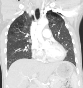
CT shows mild upper lobe centrilobular emphysema
Ashley Davidoff MD The CommonVein.net 139243 28Lu
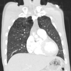
CT shows mild upper lobe centrilobular emphysema
Ashley Davidoff MD
The CommonVein.net 139241 28Lu
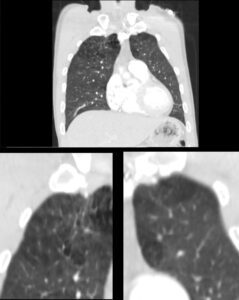
CT shows mild upper lobe centrilobular emphysema
The lower images are magnifications and show the classical Swiss cheese appearance of emphysema with a few of the areas revealing centrilobular structures.
Ashley Davidoff MD The CommonVein.net 139241c 28Lu
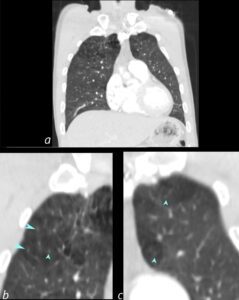
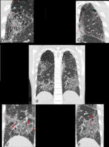
CT shows mild upper lobe centrilobular emphysema
The lower images (b and c) are magnifications and show the classical Swiss cheese appearance of emphysema with a few of the areas revealing centrilobular structures (teal arrowheads).
Ashley Davidoff MD The CommonVein.net 139241cL 28Lu
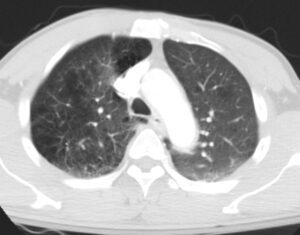
CT shows mild upper lobe centrilobular emphysema
Ashley Davidoff MD The CommonVein.net 139238 28Lu
11 Years Later
60year old Male with Emphysema and HIV presents with increasing dyspnea
CXR Shows Increasing
Diffuse Interstitial Prominence with
Reticular Pattern
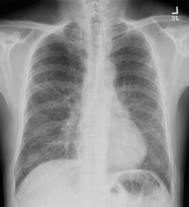
Frontal CXR shows diffuse interstitial prominence with mild upper lobe lucency likely related to upper lobe centrilobular emphysema
Ashley Davidoff MD The CommonVein.net 139244 28Lu
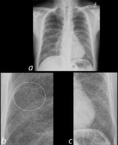
Frontal CXR shows diffuse interstitial prominence with a reticular pattern (ringed in b)
Ashley Davidoff MD
The CommonVein.net 139244cL 28Lu
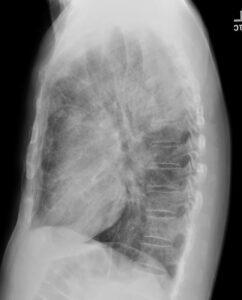
lateral CXR shows mild hyperinflation with flattening of the hemidiaphragms and diffuse interstitial prominence
Ashley Davidoff MD The CommonVein.net 139245 28Lu
Multifocal Ground Glass Changes with
Thickening of the Interlobular Septa Reticular Pattern and Centrilobular Emphysema
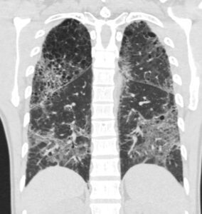
CT scan in the coronal plane shows mild progressive centrilobular emphysema with new multifocal regions of ground glass opacity in both the lower and upper lobes, thickening of the interlobular septa and the presence of new small basilar cysts
LIP (lymphocytic interstitial pneumonia) is included in the radiological differential diagnosis
Ashley Davidoff MD The CommonVein.net 139260 28Lu
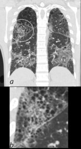
CT scan in coronal projection shows multifocal regions of ground glass opacity in both the lower and upper lobes, mild centrilobular emphysema. The right upper lobe changes are magnified in b and show a reticular pattern as a result of interlobular septal thickening
Ashley Davidoff MD
The CommonVein.net 139260c01 28Lu
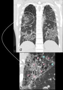
CT scan in coronal projection (a) shows multifocal regions of ground glass opacity in both the lower and upper lobes, associated with mild centrilobular emphysema. The right upper lobe changes are magnified in b and show a reticular pattern as a result of interlobular septal thickening (pink arrowheads in b) The cystic changes (teal blue arrowheads) are part of an underlying emphysematous process.
Ashley Davidoff MD
The CommonVein.net 139260c01L 28Lu
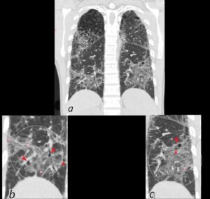
CT scan in coronal projection shows multifocal regions of ground glass opacity in both the lower and upper lobes, and mild centrilobular emphysema. The lower lobe changes are magnified in b and c and show multifocal regions of ground glass opacity , with thickening of the interlobular septa and the presence of new small bibasilar cysts (red arrowheads b and c)
LIP (lymphocytic interstitial pneumonia) is included in the radiological differential diagnosis
Ashley Davidoff MD
The CommonVein.net 139260c02L 28Lu
CT scan in shows multifocal regions of ground glass opacity in both the lower and upper lobes, mild progressive centrilobular emphysema but the presence of some small basilar cysts
LIP (lymphocytic interstitial pneumonia) is included in the radiological differential diagnosis
Ashley Davidoff MD The CommonVein.net 139260cL 28Lu
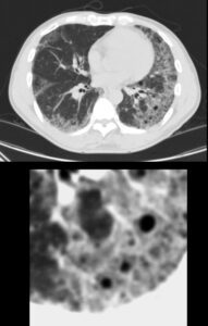
CT scan in the axial plane shows new multifocal regions of ground glass opacity thickening of the interlobular septa and the presence of new small basilar cysts
LIP (lymphocytic interstitial pneumonia) is included in the radiological differential diagnosis
Ashley Davidoff MD The CommonVein.net 139252-01 28Lu
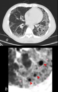
CT scan in the axial plane shows new multifocal regions of ground glass opacity (white circles, a ) thickening of the interlobular septa (pink arrowheads b), and the presence of new small basilar cysts (red arrowheads, b)
LIP (lymphocytic interstitial pneumonia) is included in the radiological differential diagnosis
Ashley Davidoff MD The CommonVein.net 139252-01L 28Lu
