Progressive chronic parenchymal lung disease of unclear cause”, originally thought to have invasive adenocarcinoma, however, at lung tumor conference, the report was amended, plan was to proceed with VATS biopsy.
3 years ago Suspected PE
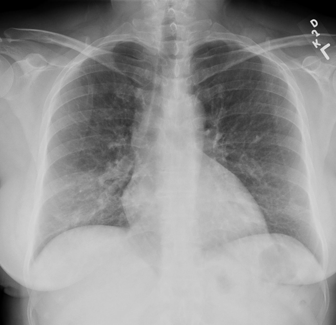
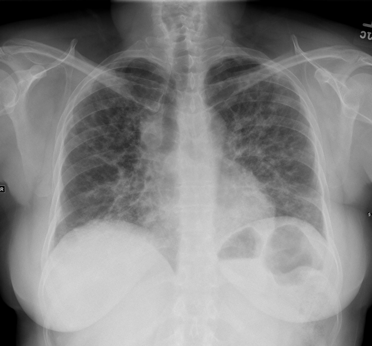
CXR
Lungs and pleura: Again seen are bilateral patchy and streaky
opacities in both lungs, unchanged since suggestive of
pneumonia. No pneumothorax. No pleural effusions.
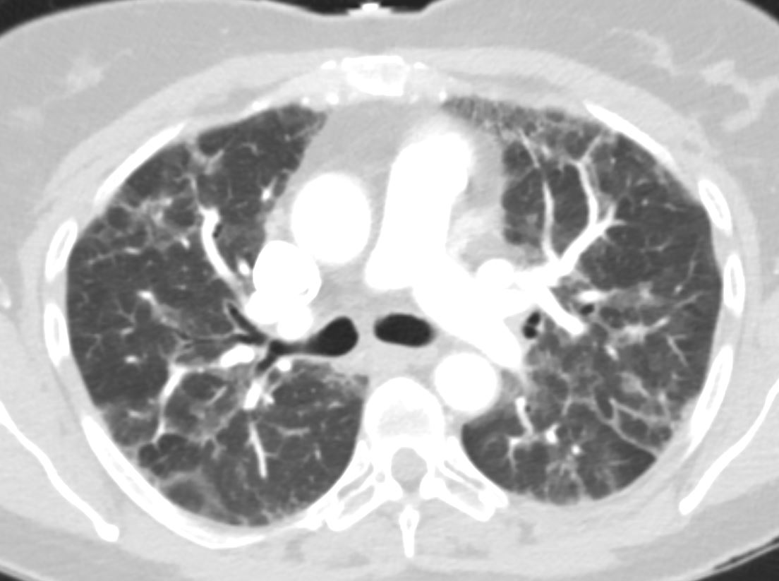
CT
Diffuse groundglass reticular and patchy groundglass opacities
suggestive of infectious/inflammatory process. Findings are
compatible with nonspecific infection, and the pattern would be
concordant with COVID-19 infection, as per features described by the Society of Thoracic Radiology.
2 Years AGo
3 Months Ago
 Path
Path
RIGHT LOWER LOBE TRANSBRONCHIAL BIOPSY:
Pulmonary alveolar parenchyma with atypical reactive pneumocyte proliferation and alveolar histiocytes consistent with clinico-radiologic impression of an infectious/ inflammatory process.
No tumor identified.
Reactive sloughed pneumocytes stain with CK7 and TTF-1(strong) while histiocytes stain with CD163. Both stain with Napsin-A. p40 is negative.
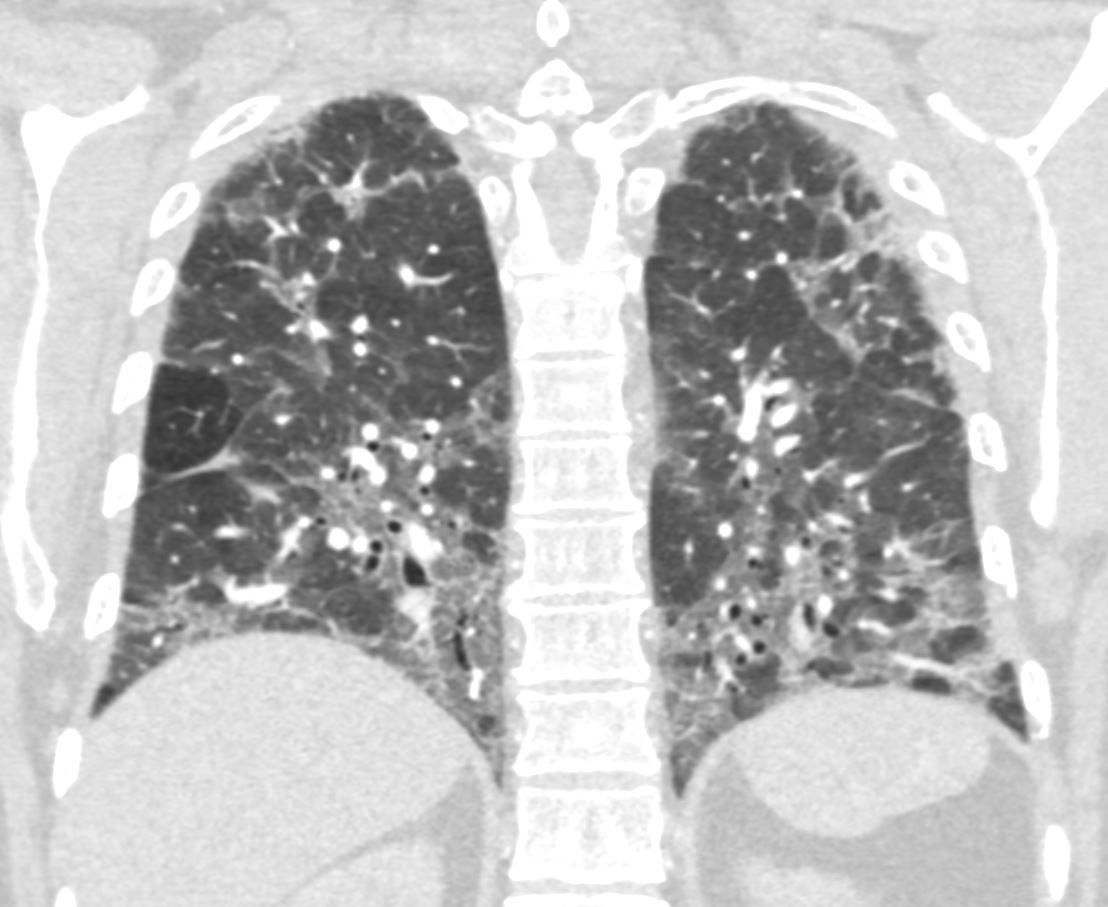
Current CT
Reticular and patchy groundglass opacities throughout both lungs is againseen with interval mild improvement of groundglass opacities in both lower
lobes s


