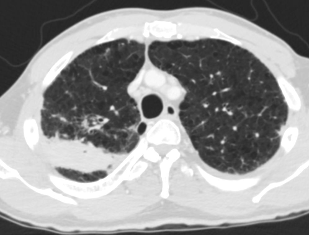
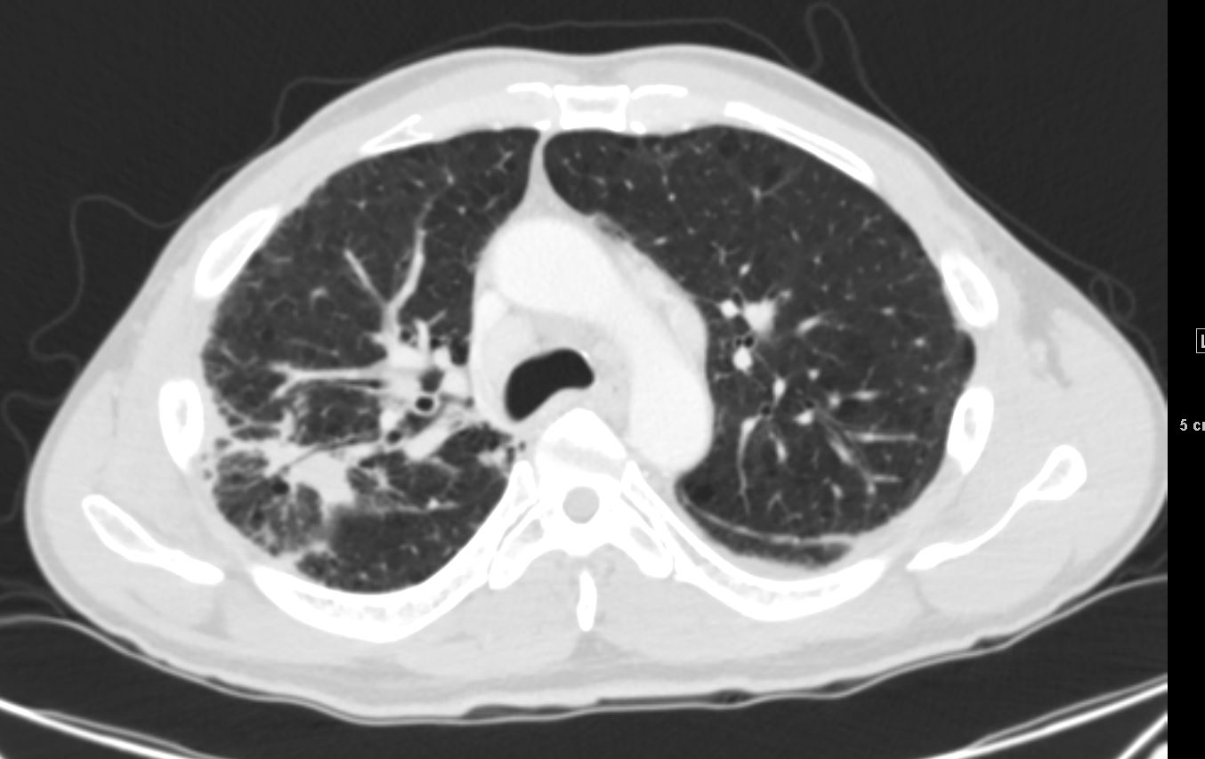
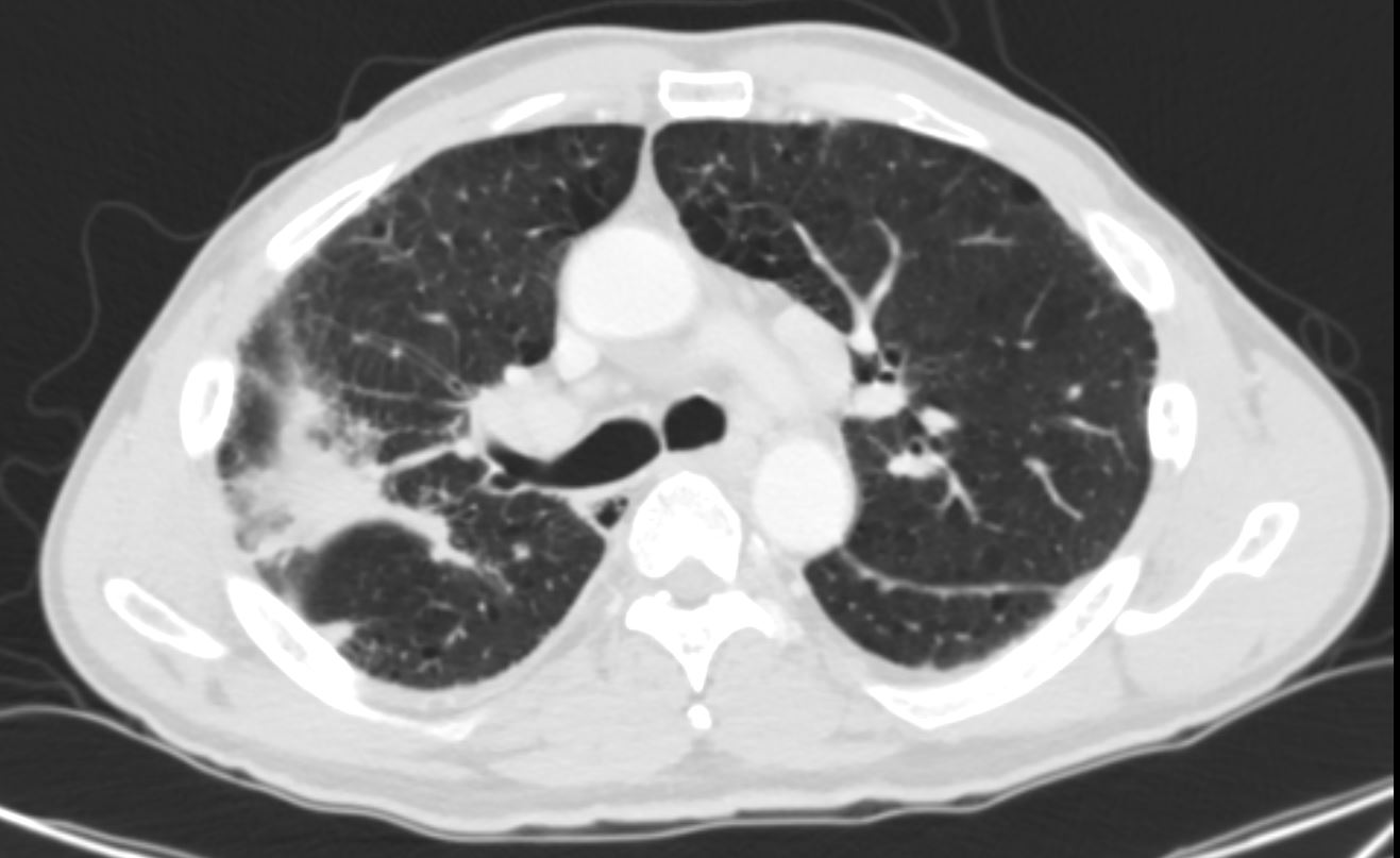
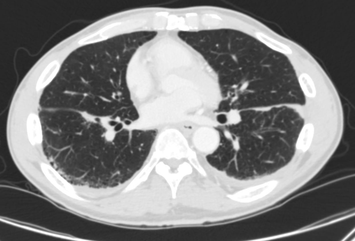
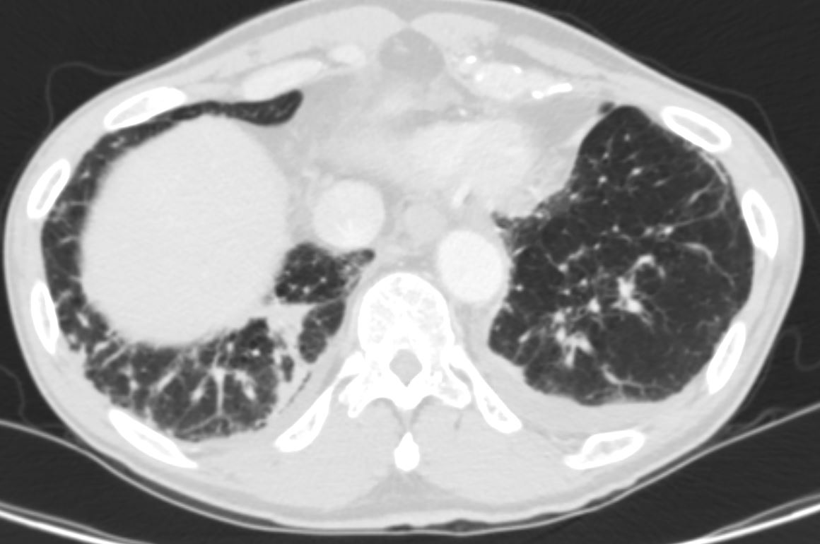
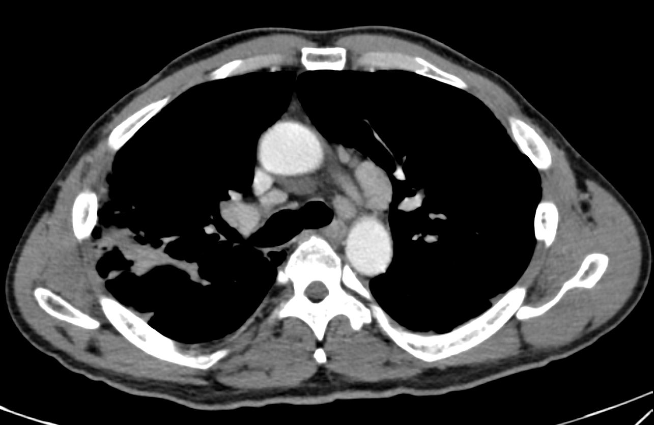
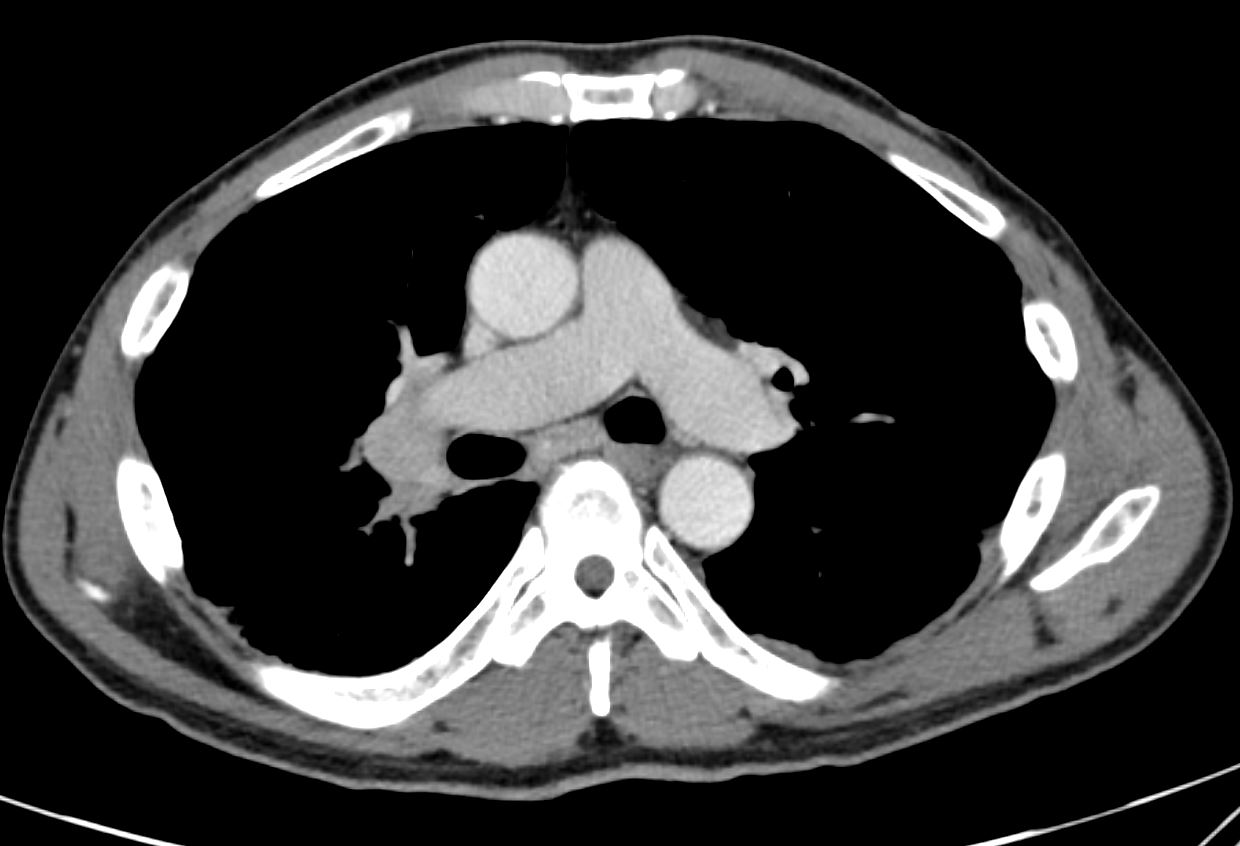
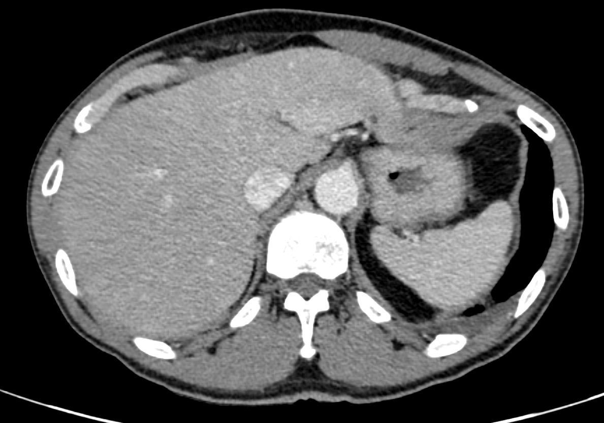
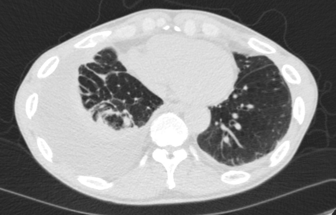
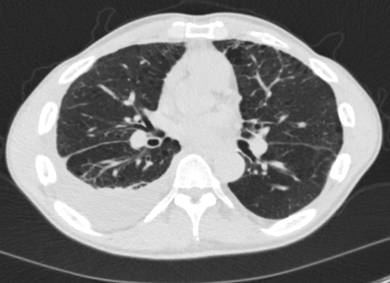
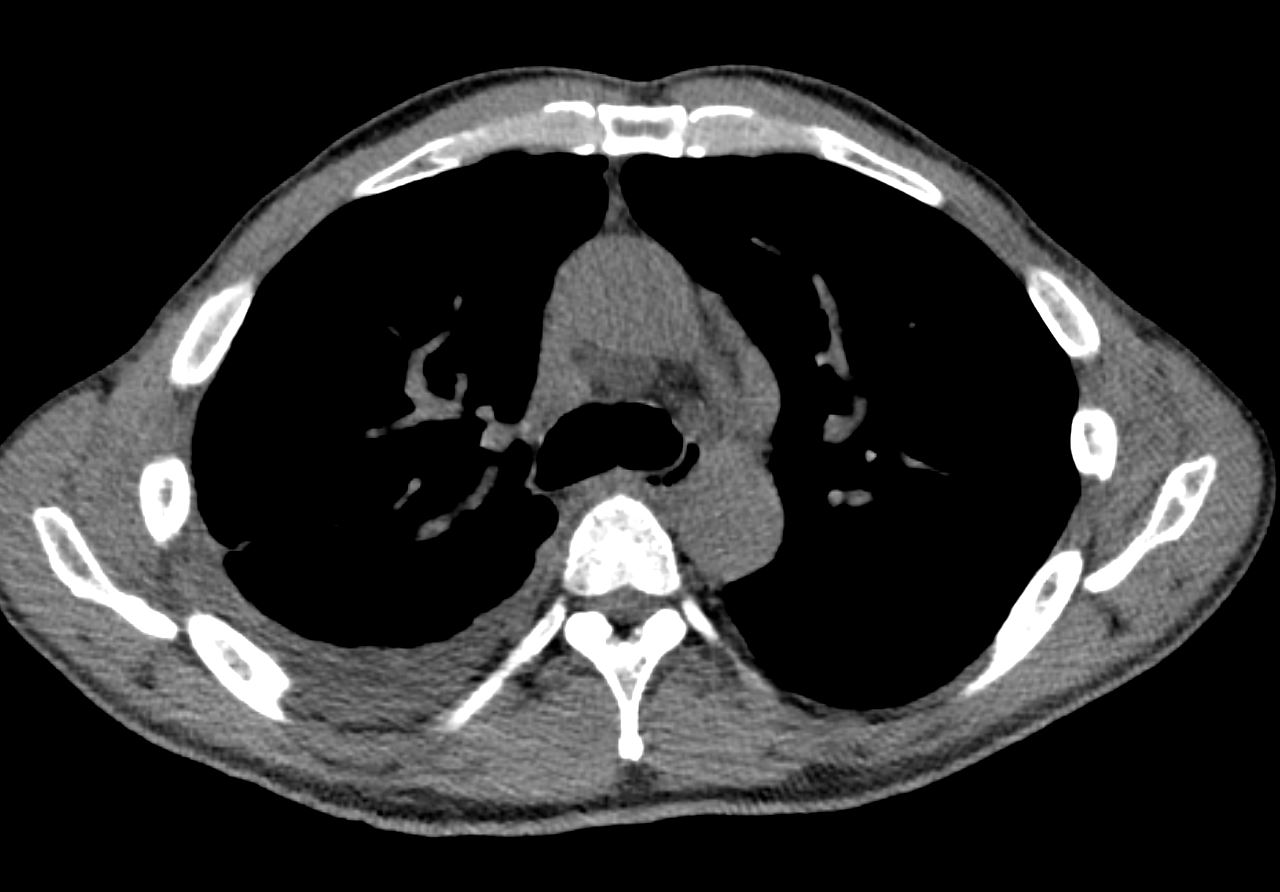
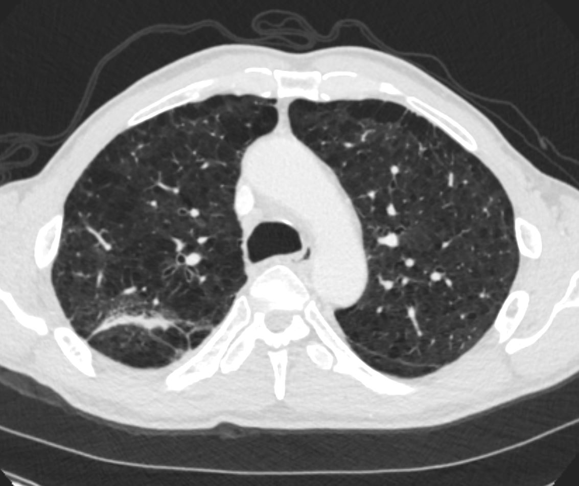
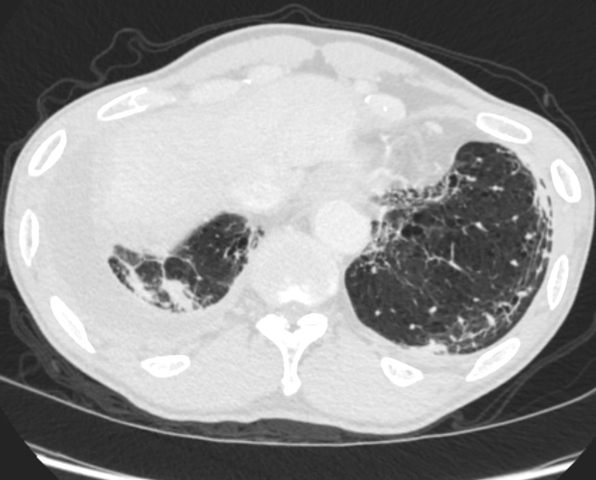
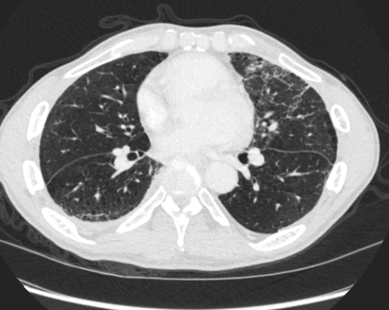
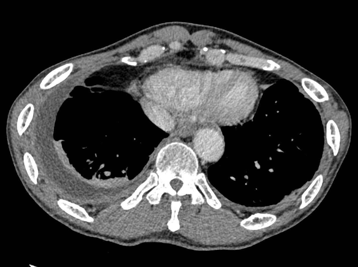
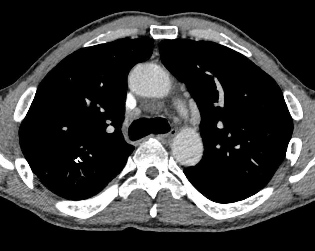
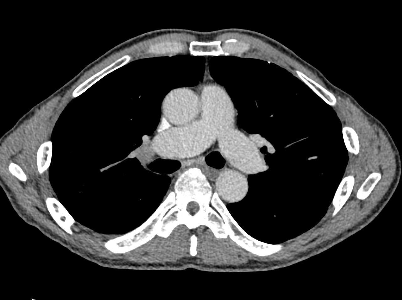
52y.o. male, with PMH tobacco use (60py), COPD, spinal surgery, sarcoidosis
7 years ago asymptomatic
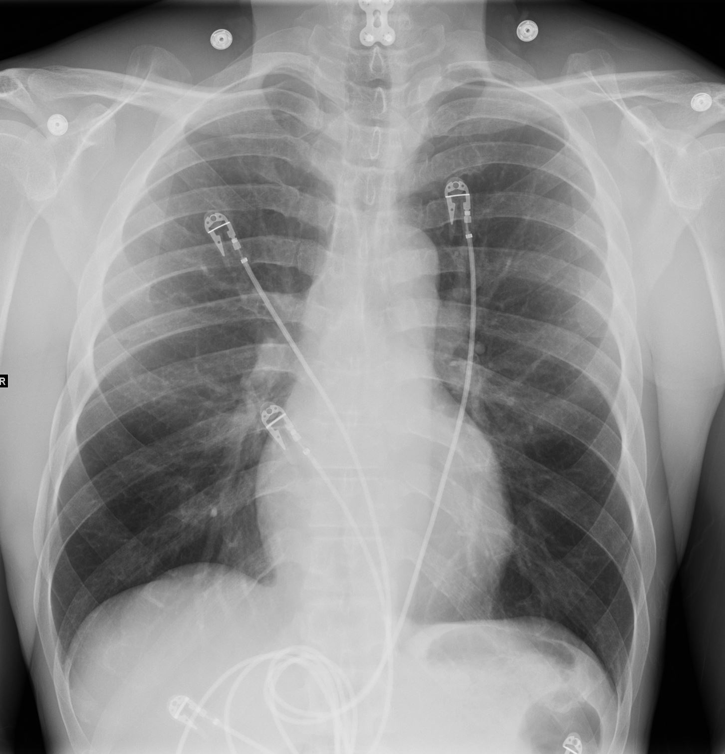
Normal CXR 7 years ago
Courtesy Paul Kohanteb MD
TheCommonVein.net
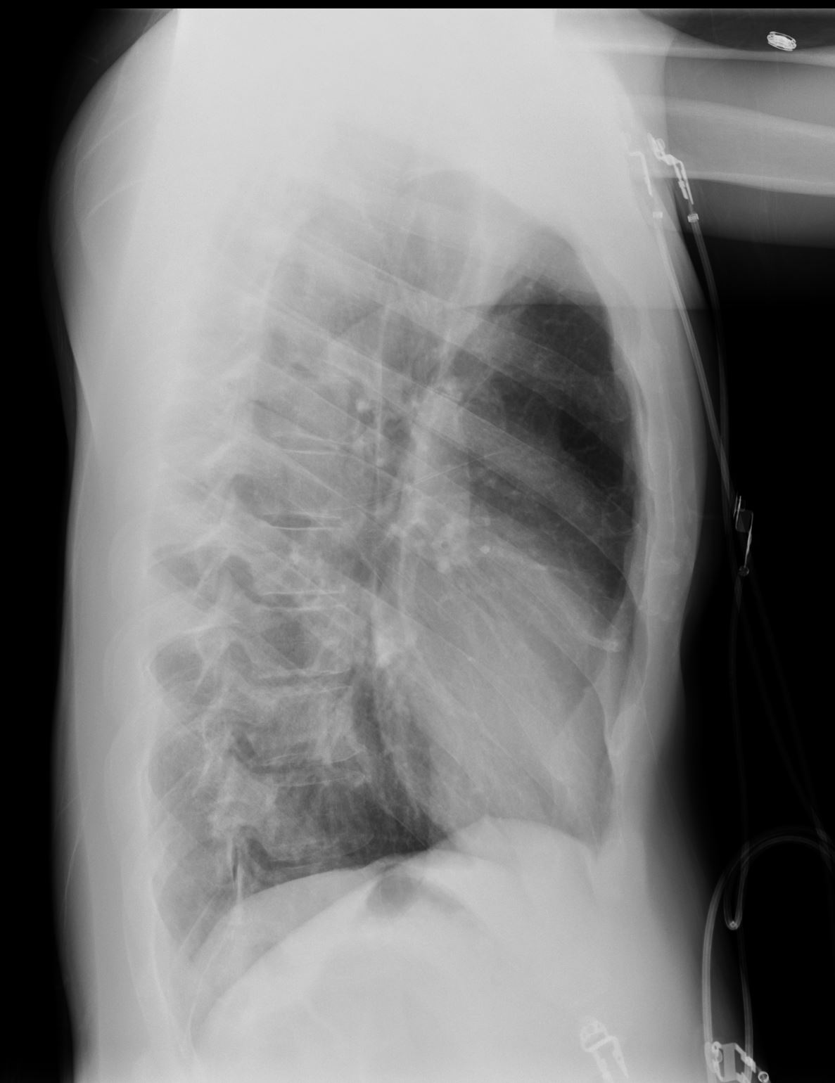
Normal CXR 7 years ago
Courtesy Paul Kohanteb MD
TheCommonVein.net
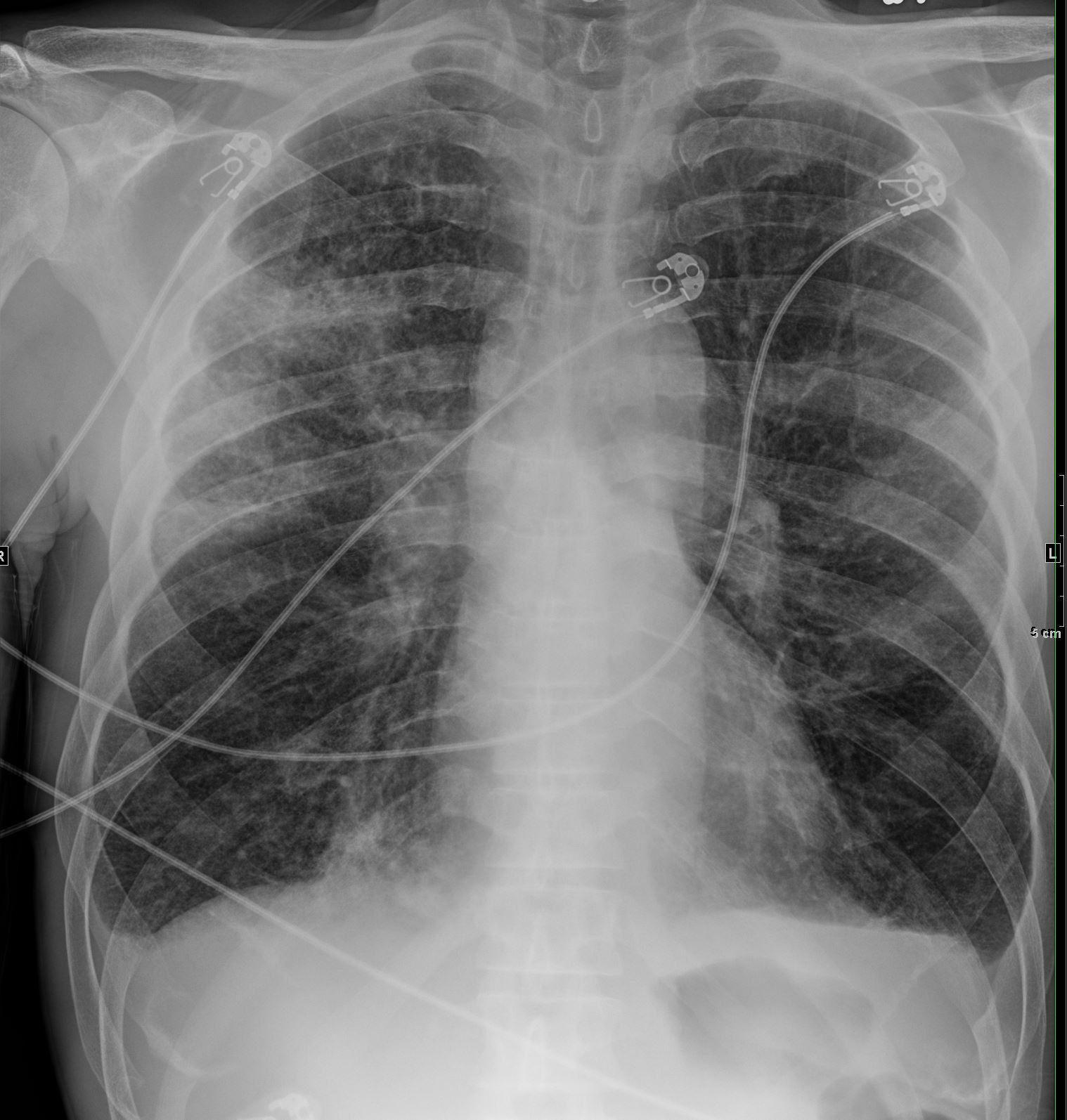
2 years ago presented with dyspnea for 2 weeks
CXR
Courtesy Paul Kohanteb MD
TheCommonVein.net
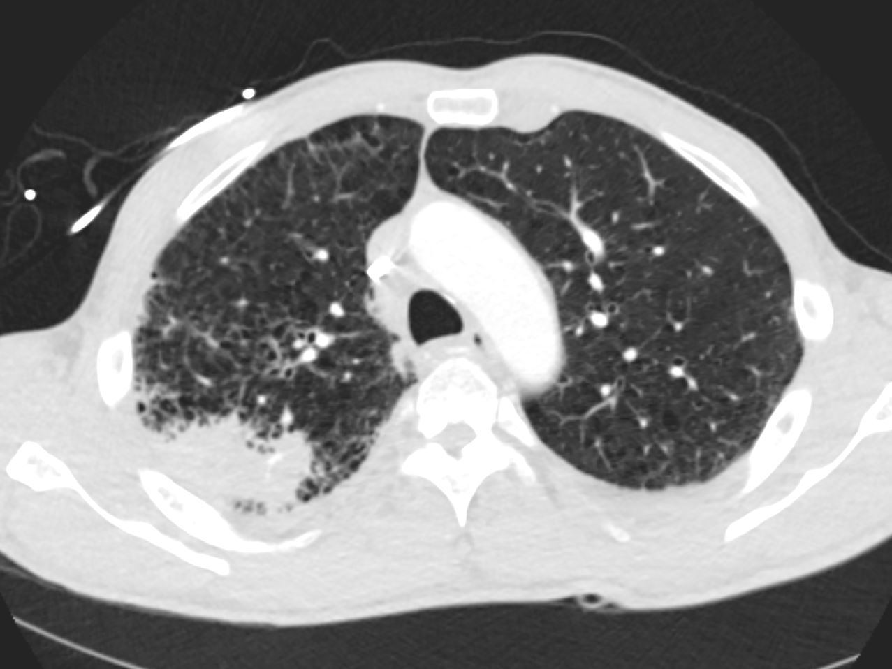
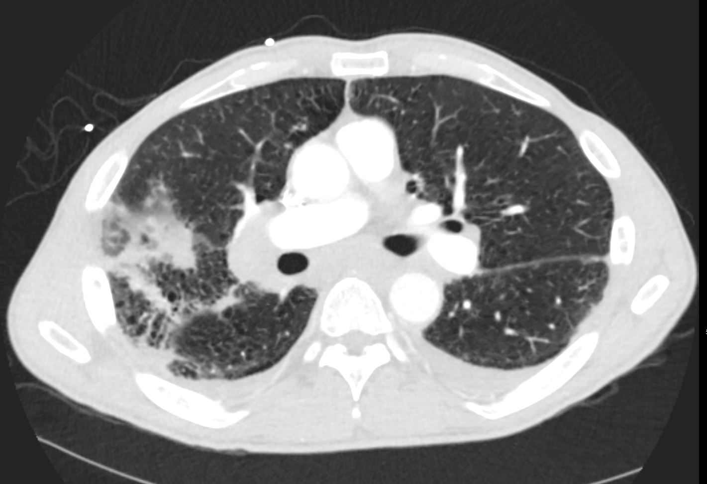
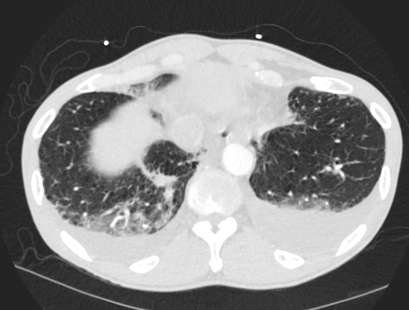
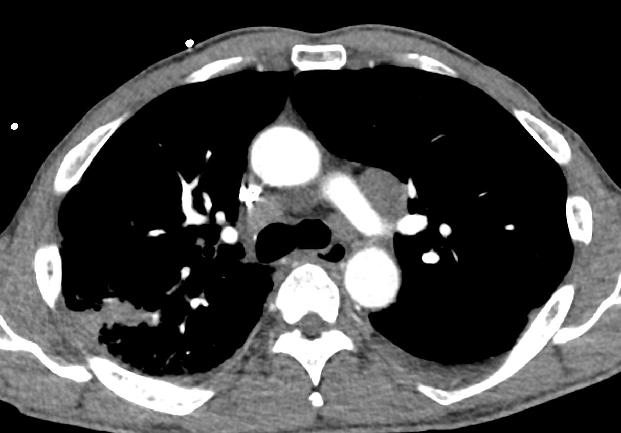
Emphysematous changes. Multifocal patchy opacities
with consolidation in the right upper lung concerning for multifocal
pneumonia.
CT showed
methotrexate
Courtesy Paul Kohanteb MD
TheCommonVein.net
Courtesy Paul Kohanteb MD
TheCommonVein.net
Courtesy Paul Kohanteb MD
TheCommonVein.net
Courtesy Paul Kohanteb MD
TheCommonVein.net
Courtesy Paul Kohanteb MD
TheCommonVein.net
Courtesy Paul Kohanteb MD
TheCommonVein.net
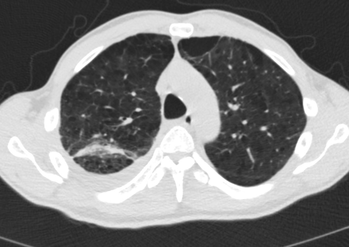
- 2 Months later
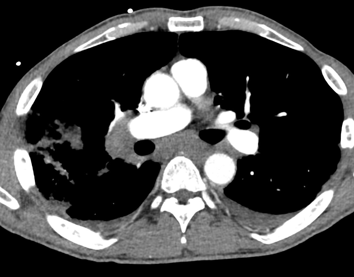
- right sided lung mass pleural involvement, and extensive adenopathy with
- RUL Mass
-

RUL infiltrate increased CT 2 months later
Courtesy Paul Kohanteb MD TheCommonVein.net- RUL Mass Mass is Bronchocentric with Obstruction

RLL Bronchocentric mass increased in size
CT 2 months later
Courtesy Paul Kohanteb MD TheCommonVein.netRLL Mass
-

RLL Bronchocentric mass increased in size
CT 2 months later
Courtesy Paul Kohanteb MD TheCommonVein.netBilateral fissural thickening CT 2 months later
Courtesy Paul Kohanteb MD TheCommonVein.netThickened Interlobular Septa
Interlobular septal thickened in the RLL CT 2 months later
Courtesy Paul Kohanteb MD TheCommonVein.net - Lymphadenopathy

Mediastinal and hilar adenopathy
Courtesy Paul Kohanteb MD TheCommonVein.netLymphadenopathy

Mediastinal and hilar adenopathy
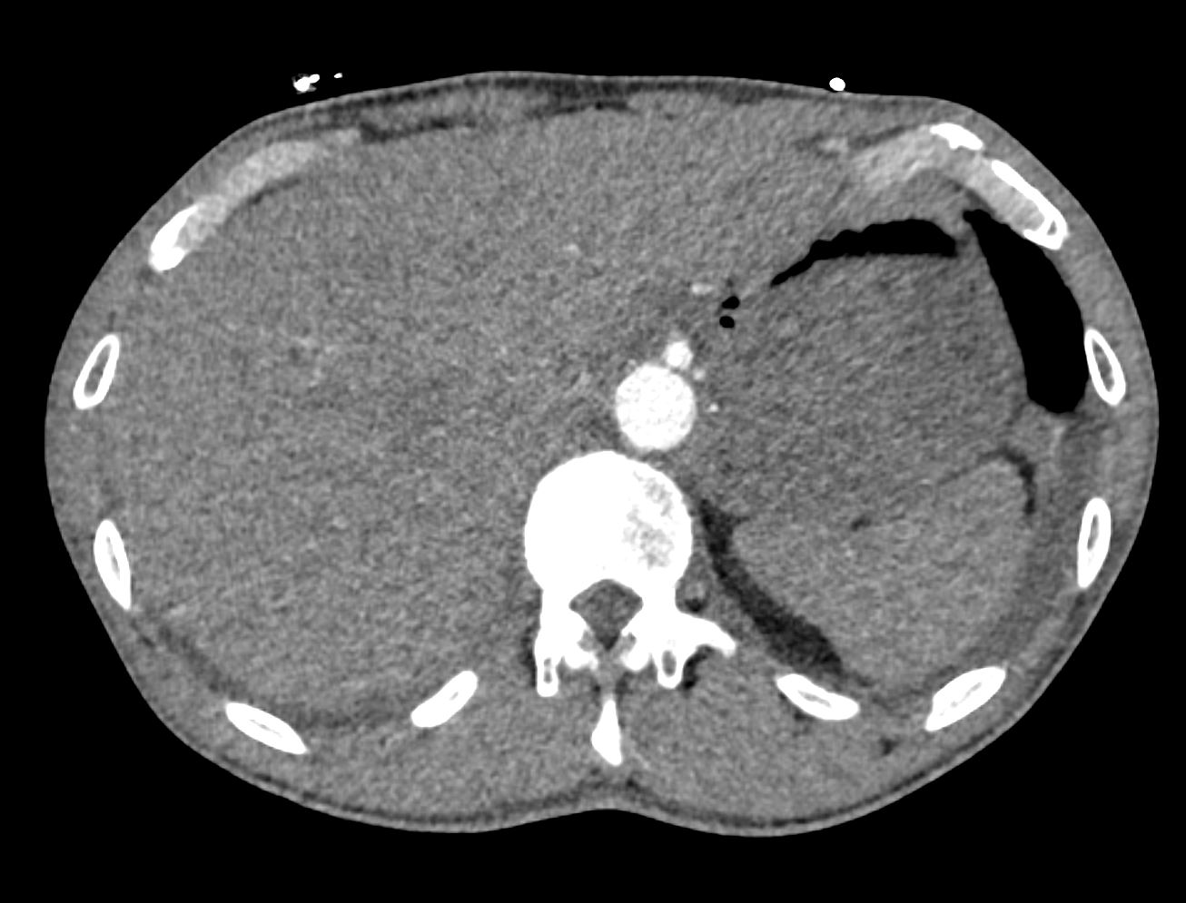
Courtesy Paul Kohanteb MD TheCommonVein.netSpleen and Liver Negative

Normal appearing spleen
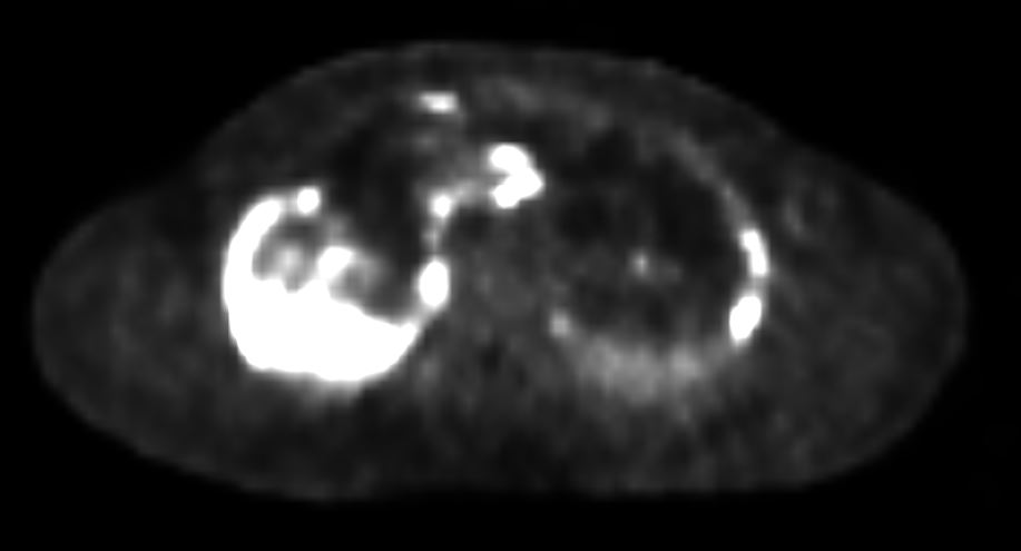
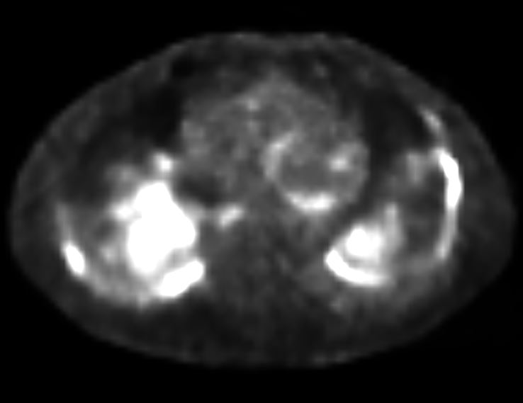
Courtesy Paul Kohanteb MD TheCommonVein.net - PET positivity suggesting lung cancer with pleural involvement
- RUL Mass Hyperintense and Lymphadenopathy

PET positive RUL mass pleura and lynph nodes bilaterally
Courtesy Paul Kohanteb MD TheCommonVein.net- RLL Mass Hyperintense

PET positive RLL mass pleura and lynph nodes bilaterally
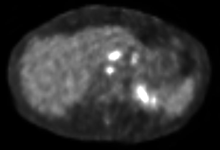
Courtesy Paul Kohanteb MD TheCommonVein.netLymphadenopathy

PET positive foregut lymph nodes
Courtesy Paul Kohanteb MD TheCommonVein.net1 year prior PET scan showed
- extensive hypermetabolic activity associated with
extensive predominantly - pleural-based malignancy in
- all lobes of thebilateral lungs with
- some parenchymal involvement and
- interlobularseptal thickening concerning for
- lymphangitic carcinomatosis,
- lymphadenopathy and bilateral pleural effusions which have overall progressed.
- extensive hypermetabolic activity associated with
- RLL Mass Hyperintense
- Pathology form an EBUS 2years ago
- revealed non-caseating granulomata without malignancy,
- consistent with sarcoidosis.
- started on Methotrexate
- dyspnea worsened .
- Prednisone 15mg, now down to 10mg.
- started on Methotrexate
-
1 year ago
- RUL Mass Scar Like
-

RUL mass hass shrunk
Courtesy Paul Kohanteb MD
TheCommonVein.netRLL Mass Poorly Visualized Because of New Effusion

RLL mass hass shrunk
Courtesy Paul Kohanteb MD
TheCommonVein.netFissures Resolved and New Effusion

New Effusion
Courtesy Paul Kohanteb MD
TheCommonVein.netLymphadenopathy Improved

Decreasedm Adenopathy
Courtesy Paul Kohanteb MD
TheCommonVein.net -
Current on Methotrexate and Prednisone
- RUL Mass Scar Like
-

RUL mass remains scar like
Courtesy Paul Kohanteb MD
TheCommonVein.netRLL Mass Poorly Visualized Because Complex Effusion

RLL mass persists but smaller
Courtesy Paul Kohanteb MD
TheCommonVein.net -
Fissures Resolved

RLL mass hass shrunk
Changes in the Fissures have Resolved
Courtesy Paul Kohanteb MD
TheCommonVein.netImproving Complex Effusion

Effusion smaller but persists and is complex
Courtesy Paul Kohanteb MD
TheCommonVein.net - Lymphadenopathy Improved

Decreased Adenopathy
Courtesy Paul Kohanteb MD
TheCommonVein.netLymphadenopathy Improved

Decreased Adenopathy
Courtesy Paul Kohanteb MD
TheCommonVein.net

