Sarcoidosis
59f
Path 14 years ago
Wedge biopsy lung designated “left upper lobe”: Noncaseating granulomas consistent with sarcoidosis (comment)
Comment: Clinical correlation is required to arise at a diagnosis of sarcoidosis. AFB and fungal PAS stains are negative. The differential diagnosis also includes berylliosis and other occupational lung diseases, talc granulomatosis and hypersensitivity pneumonia.
MICROSCOPIC DESCRIPTION
Sections of the gross lung nodules reveal innumerable confluent and small isolated noncaseating granulomas containing numerous multinucleated giant cells. Individual granulomas are scattered throughout the remainder of the lung tissue. An occasional granuloma demonstrates central necrosis. An occasional Schaumann body is identified. The granulomas are rimmed by lymphocytes. The granulomas are noted both peribronchial and within the interstitium. A few of the granulomas are collagenized. Fungal PAS and AFB stains are negative.Wedge biopsy lung designated “left upper lobe”: Noncaseating granulomas consistent with sarcoidosis (comment)
Comment: Clinical correlation is required to arise at a diagnosis of sarcoidosis. AFB and fungal PAS stains are negative. The differential diagnosis also includes berylliosis and other occupational lung diseases, talc granulomatosis and hypersensitivity pneumonia.
are negative.
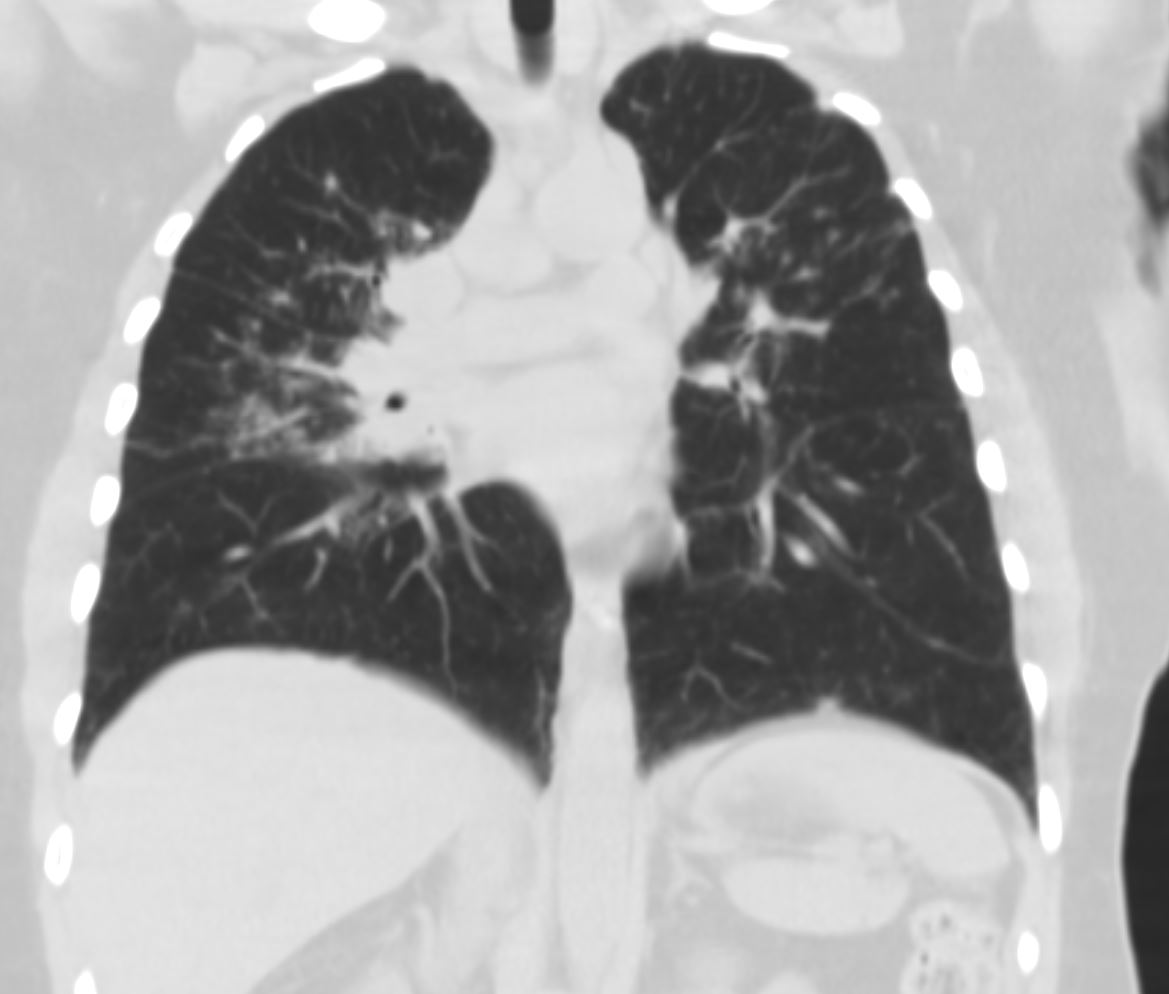
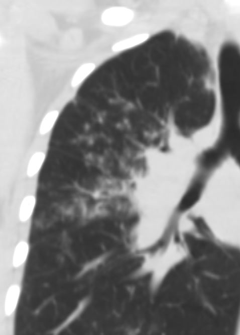
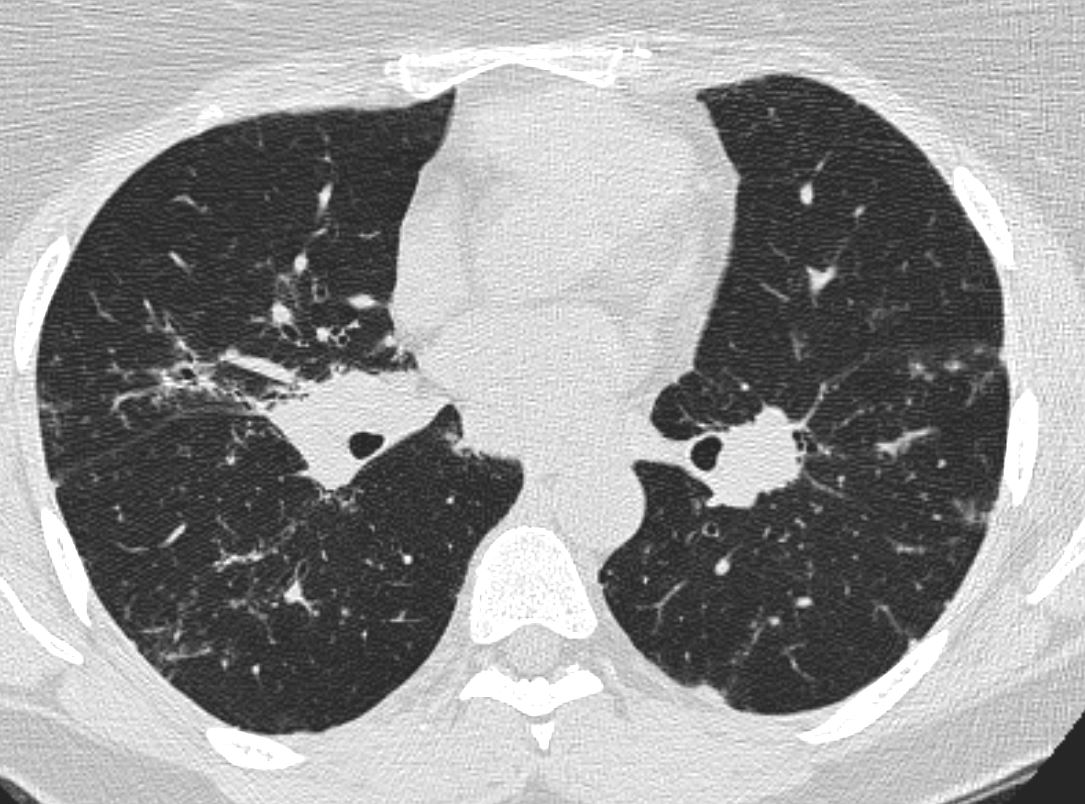
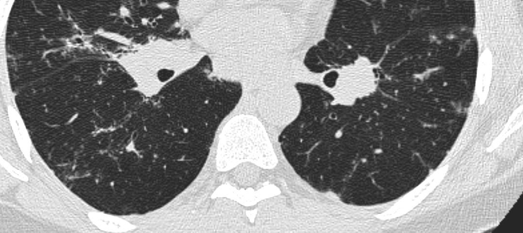
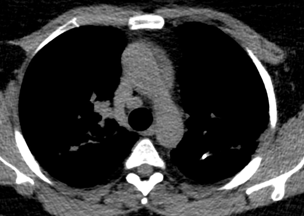
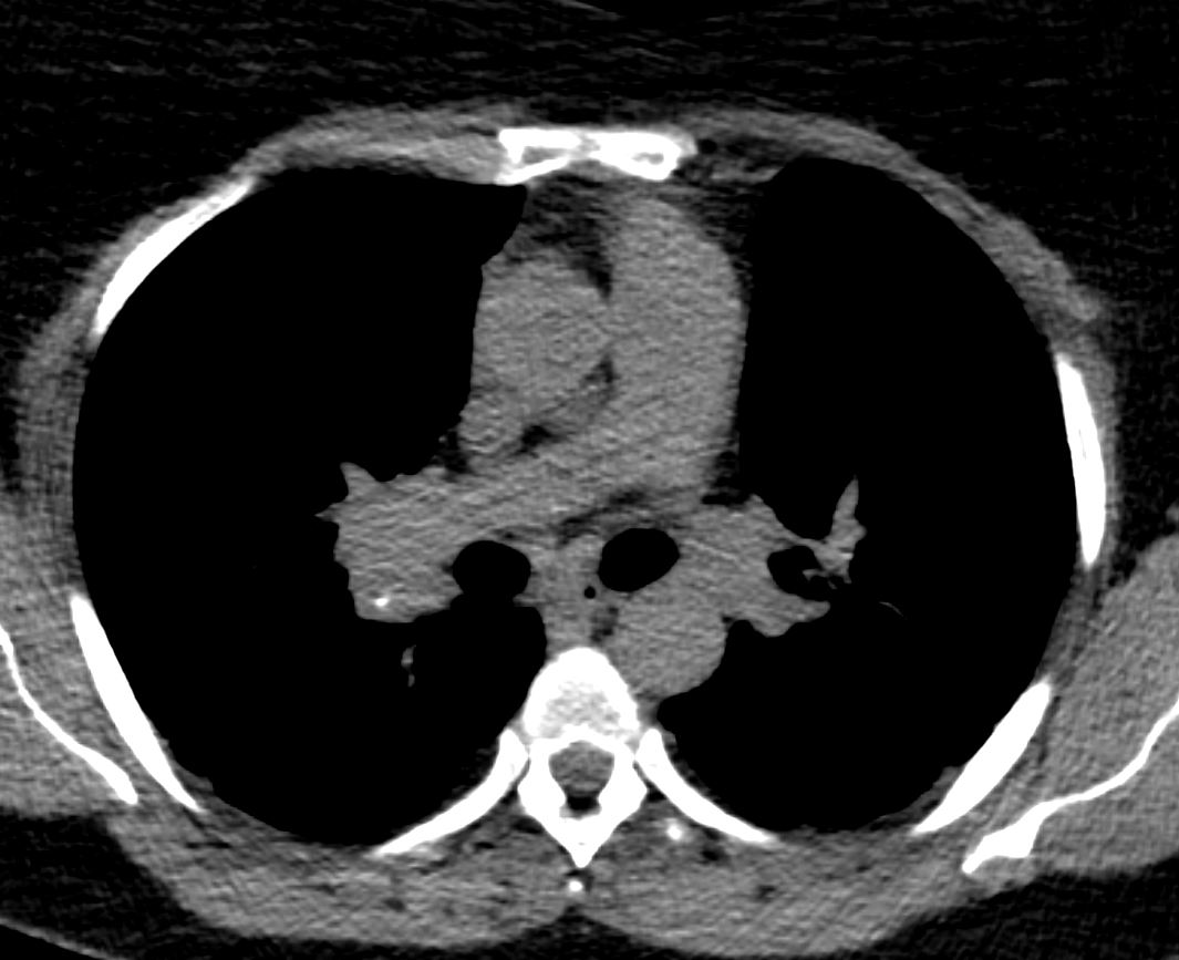
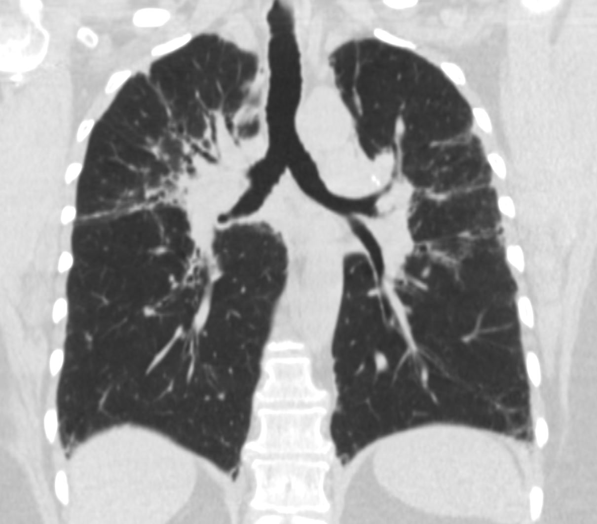
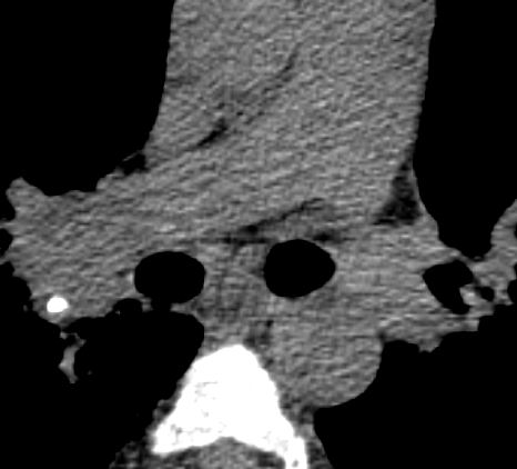
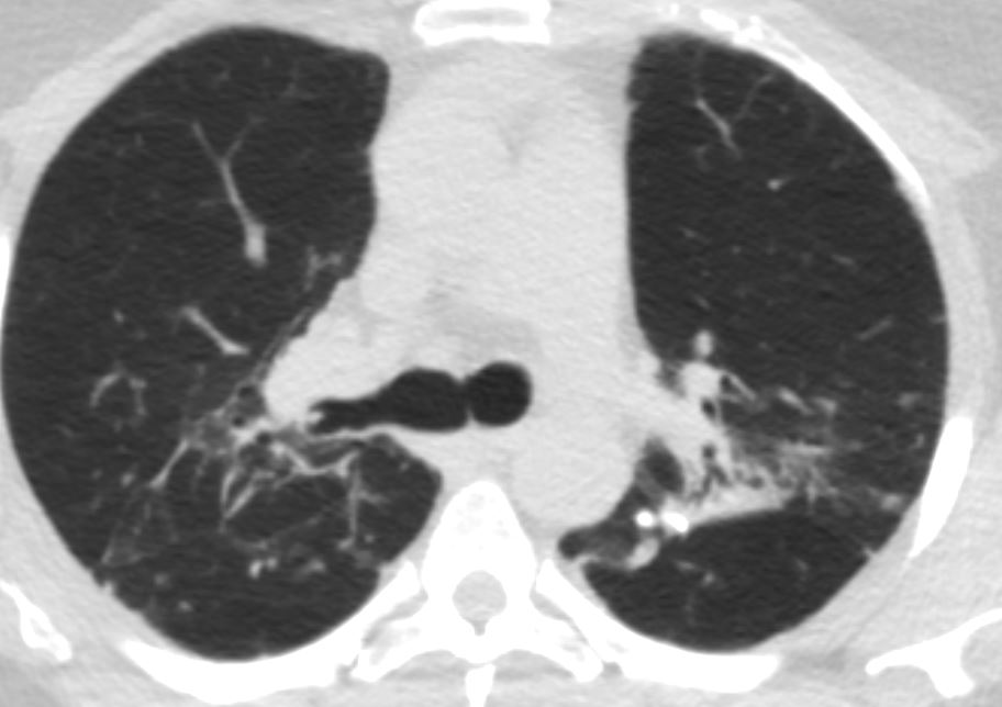
CT 10 years ago
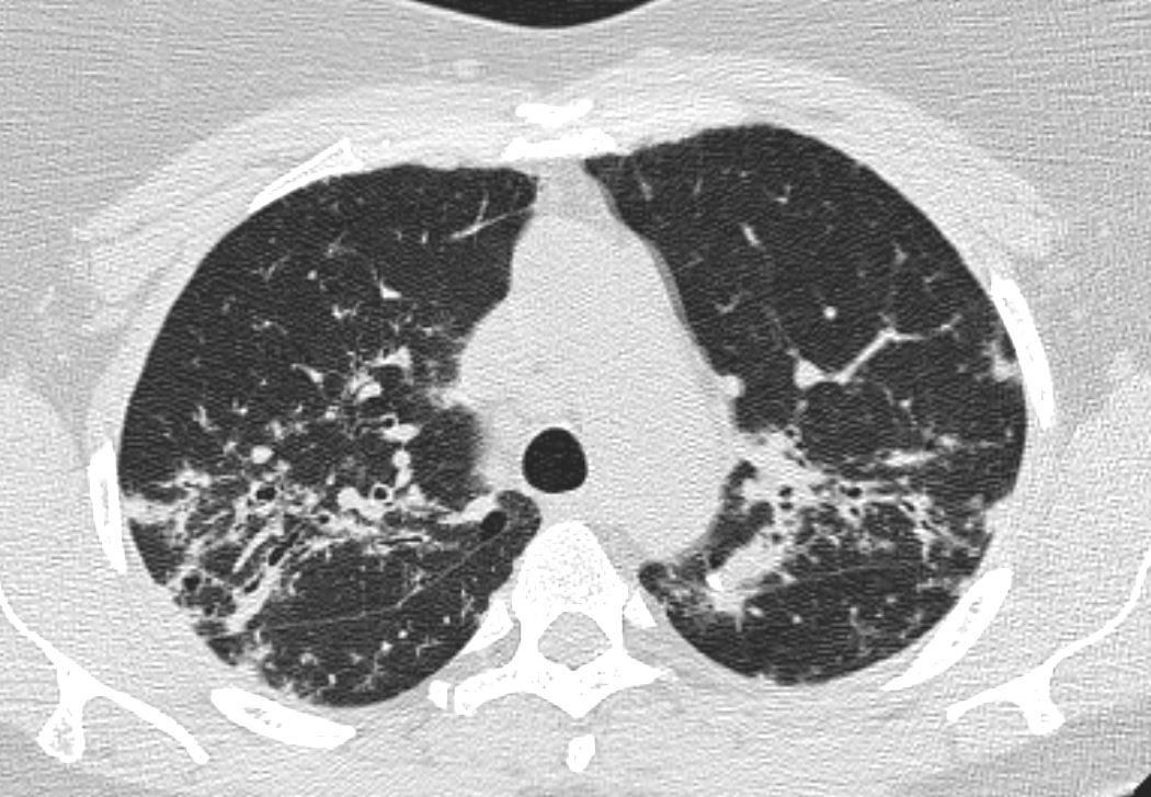
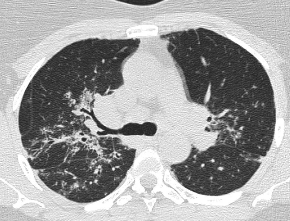
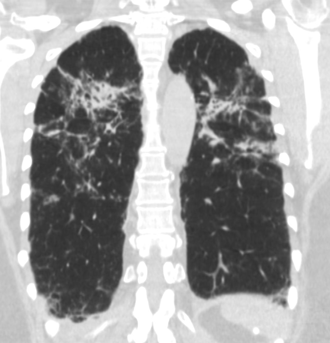
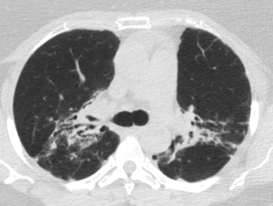
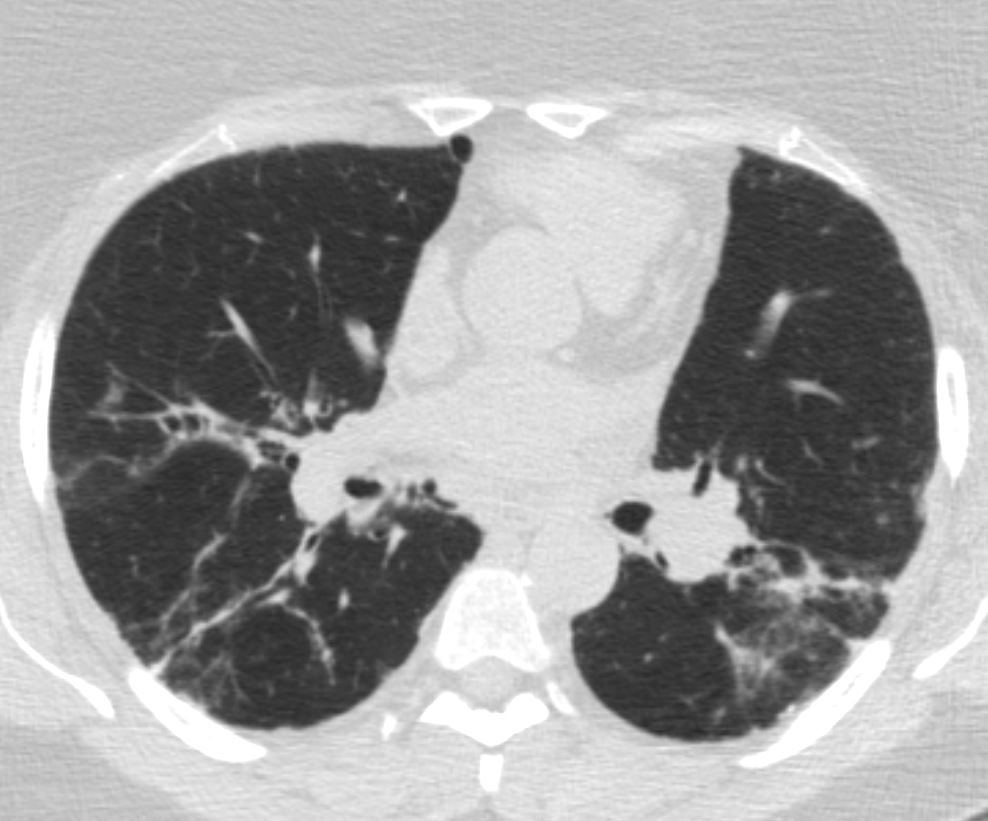
hilar adenopathy upper lobe reticular and nodular changes
bronchiectasis
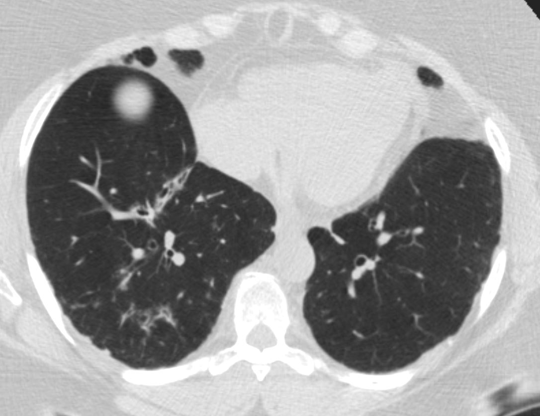
Lower Lobe Nodules
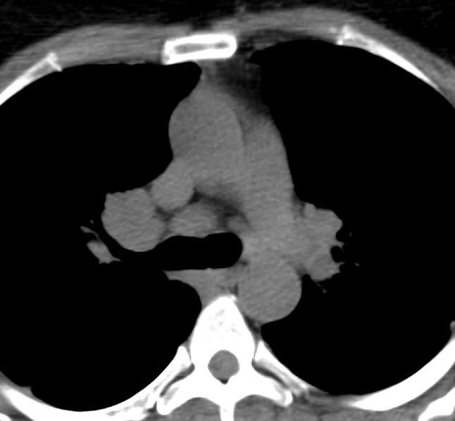
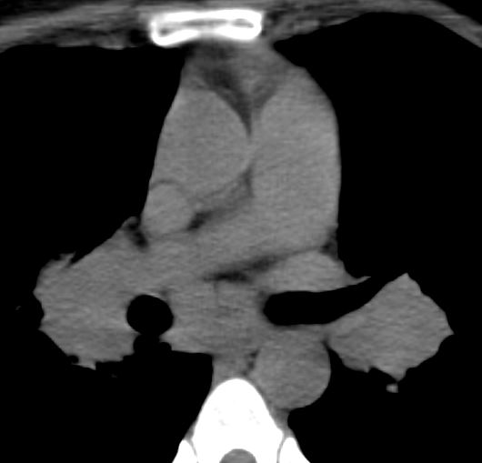
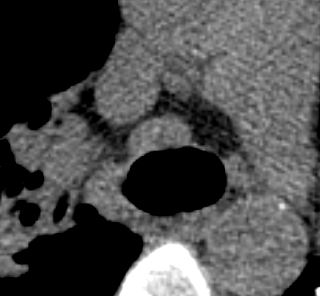
Mediastinal and Hilar Adenopathy 10 Years Ago
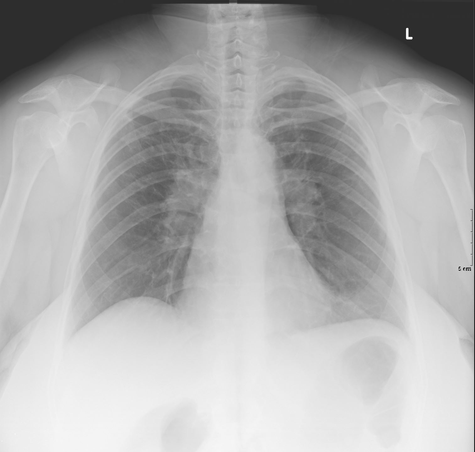
CXR hilar adenopathy upper lobe reticular changes 6years ago
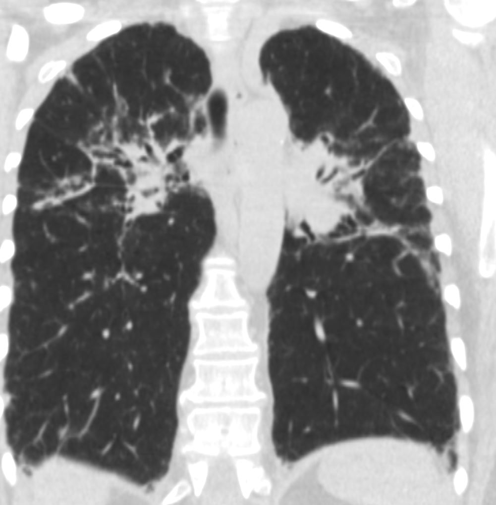
sarcoidosis stage 4 6 YearsAgo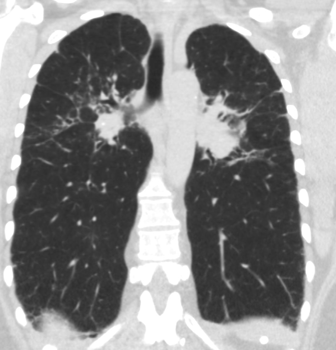
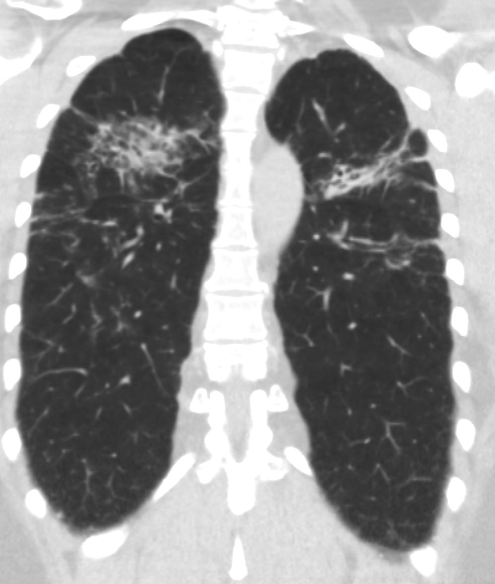
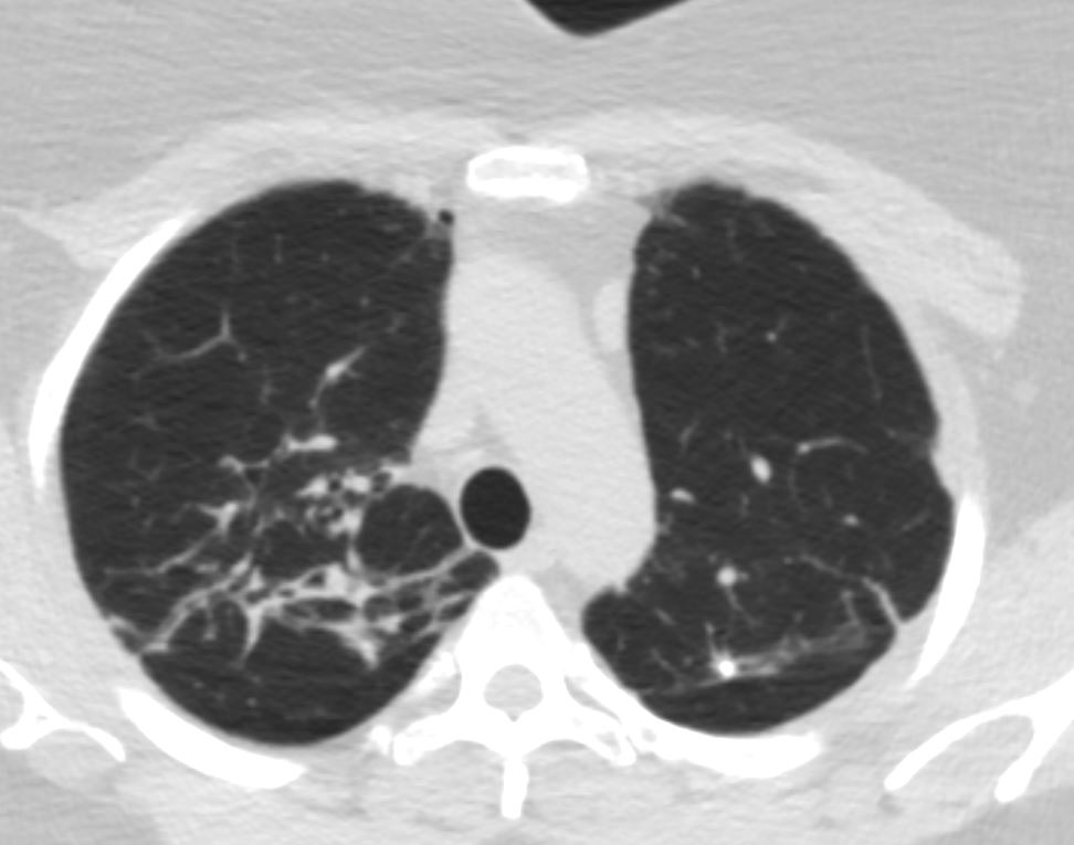
Lymph Nodes Decreasing 6 years ago
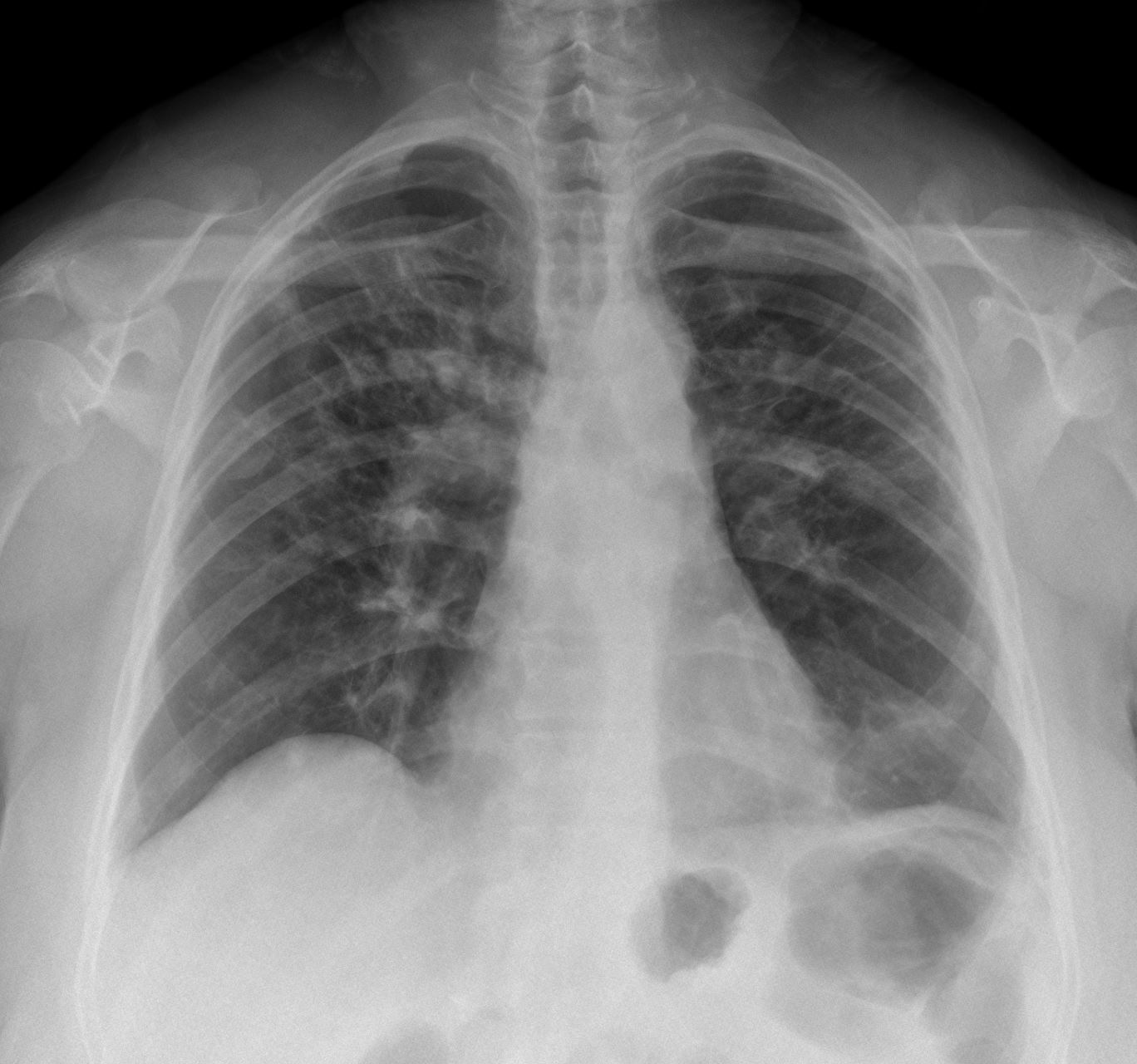
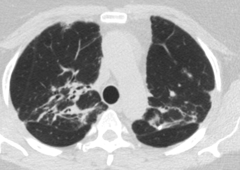
Current

