- 40-year-old gentleman with Type 1 DM
- presenting with several months of
- worsening weakness, falls,
- deconditioning brought in by EMS
- in DKA
CXR – Vague Mid and Lower Lung Infiltrate
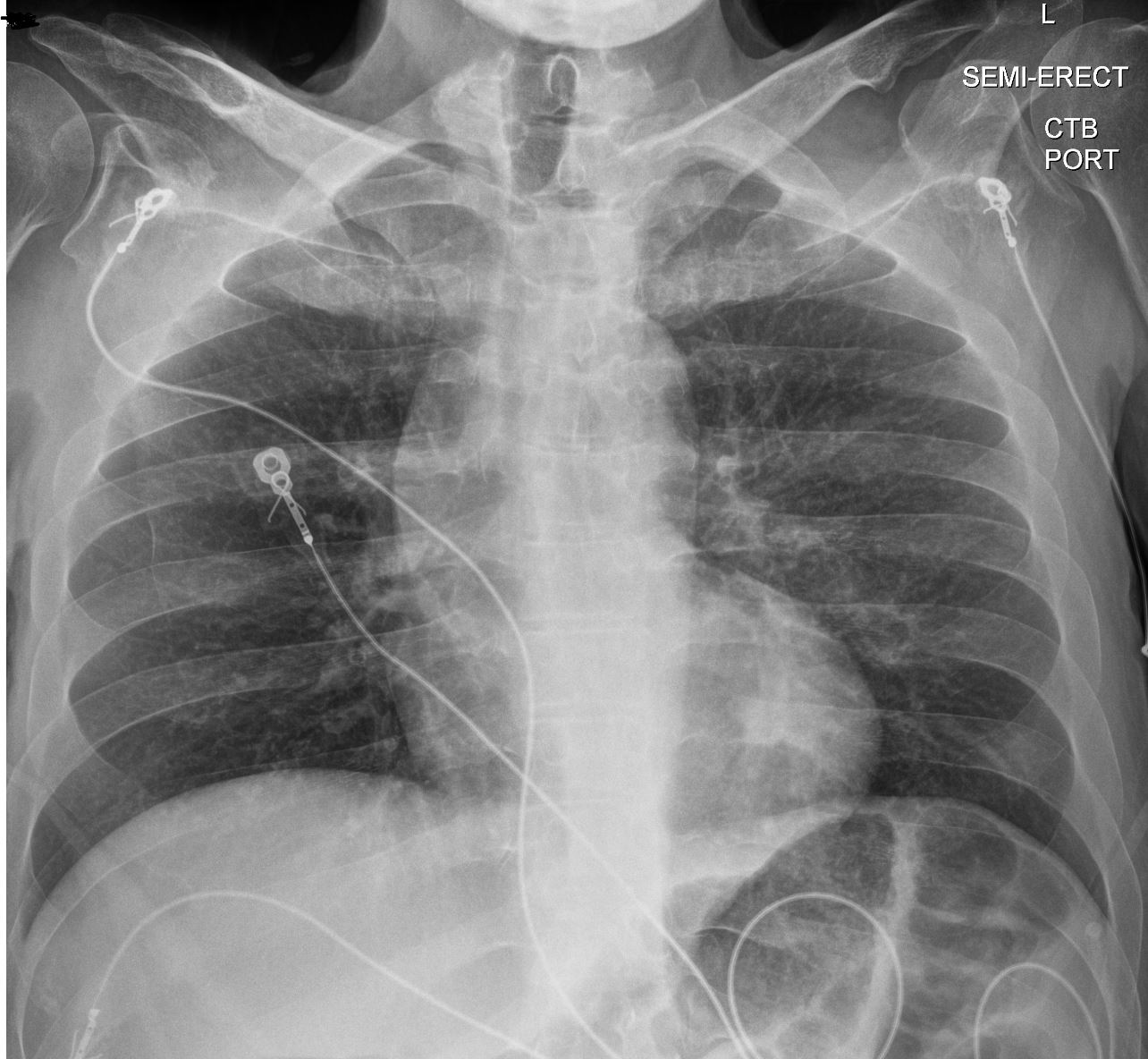
Ashley Davidoff
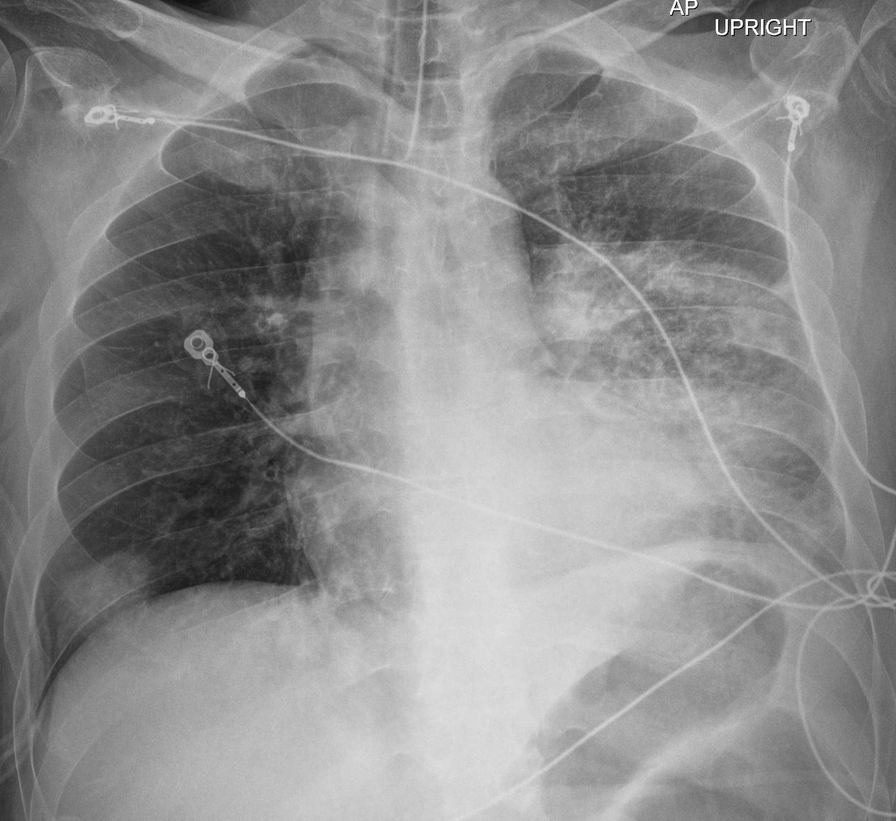
The Frontal CXR shows a rapidly progressive interstitial infiltrate in the left upper lobe with a focal subsegmental infiltrate in the right lower lobe Diagnosis Aspergillus Infection
Ashley Davidoff TheCommonVein.net
- Management
- initially managed for DKA with
- insulin drip,
- fluid management, and
- electrolyte management.
- DKA resolved but
- became acutely more ill and
- developed chest pain.
- Insulin was to transition back to an insulin drip
- initially managed for DKA with
CXR 2 days later


The Frontal CXR shows a rapidly progressive interstitial infiltrate in the left upper lobe with a focal subsegmental infiltrate in the right lower lobe Diagnosis Aspergillus Infection
Ashley Davidoff TheCommonVein.net
- Chest CT
-
- revealed bilateral consolidative opacities
- reversed halo sign (Atoll)
- concerning for fungal infection.
-
- Bronchoscopy was performed
-
- frank hyphae and
- fungal balls in the bronchus.
-
CT Scan Large Infiltrate with Halo Sign and Reversed Halo
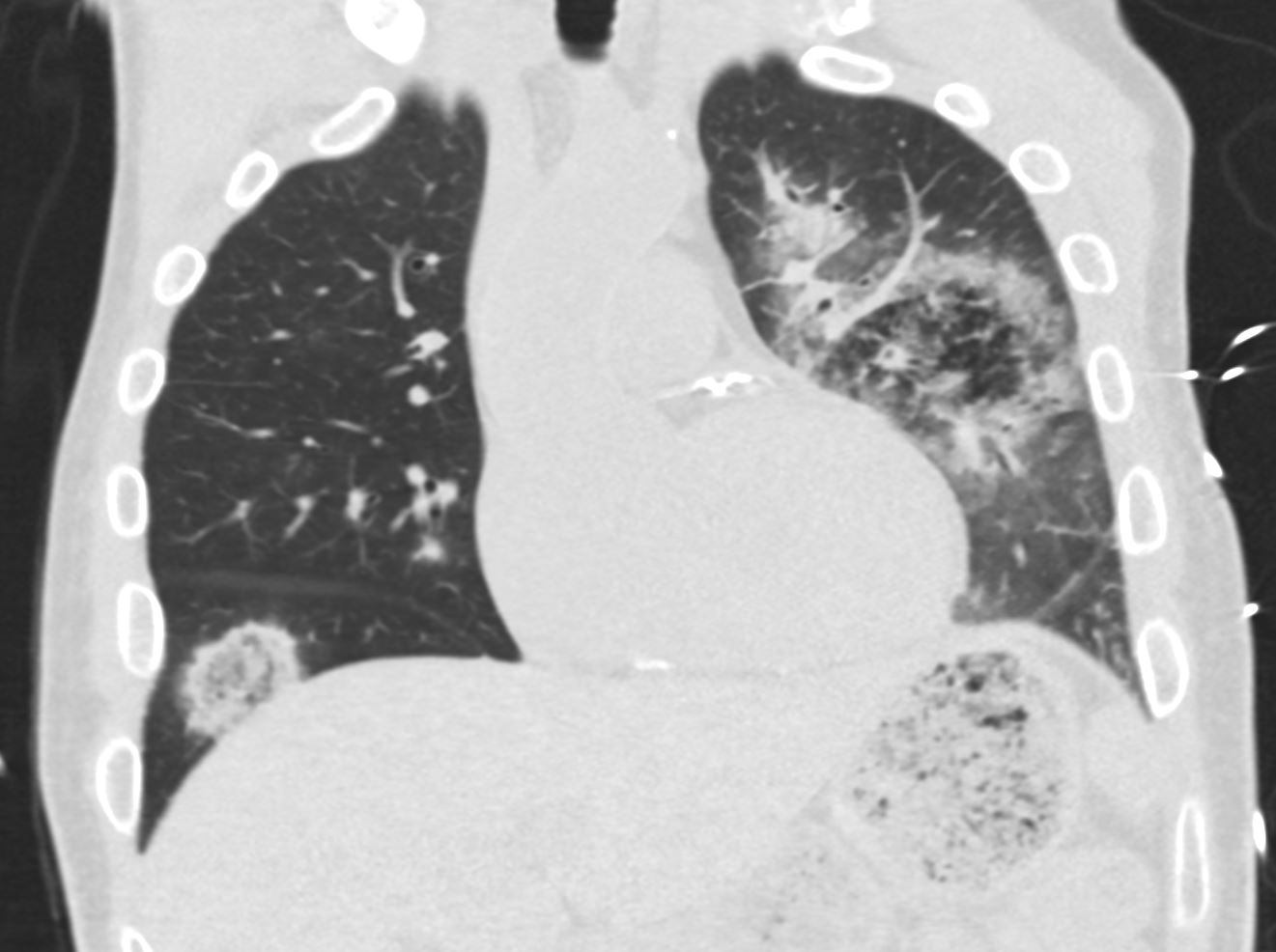

The coronal CT scan shows a rapidly progressive interstitial infiltrate in the left upper lobe (halo sign) with a focal subsegmental infiltrate in the right lower lobe, showing features of a reversed halo sign (Atoll sign) There is evidence of bronchial wall thickening.
Diagnosis Aspergillus Infection
Ashley Davidoff TheCommonVein.net 219Lu-004
Features of the Left Sided Infiltrate
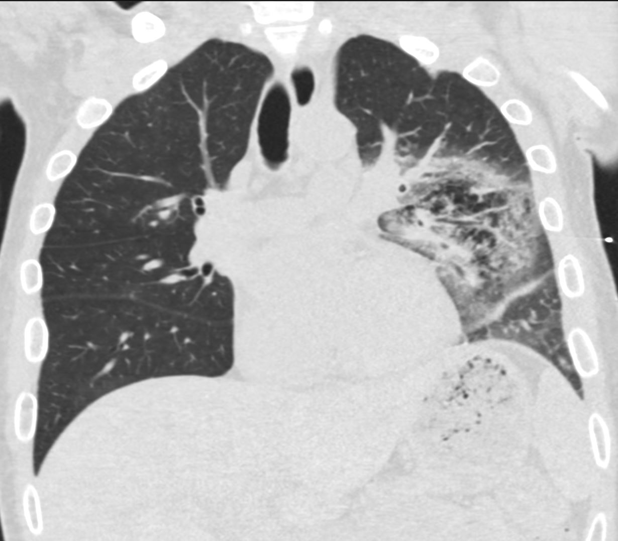

The coronal CT scan shows a rapidly progressive interstitial infiltrate in the left upper lobe showing features of a halo sign There is evidence of bronchial wall thickening.
Diagnosis Aspergillus Infection
Ashley Davidoff TheCommonVein.net 219Lu-002
Note Significant Bronchial Wall Thickening
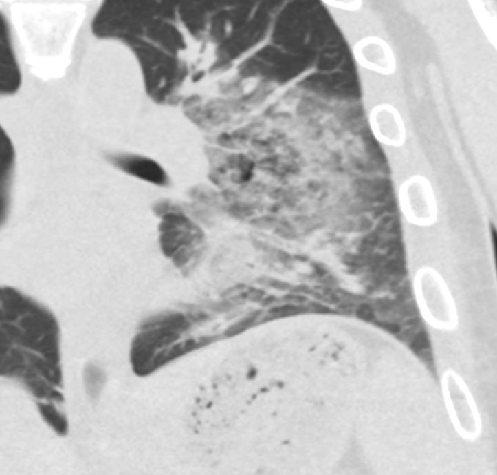

The coronal CT scan shows a rapidly progressive interstitial infiltrate in the left upper lobe with a focal subsegmental infiltrate in the right lower lobe, both showing features of a reversed halo sign (Atoll sign) There is evidence of bronchial wall thickening.
Diagnosis Aspergillus Infection
Ashley Davidoff TheCommonVein.net 219Lu-003
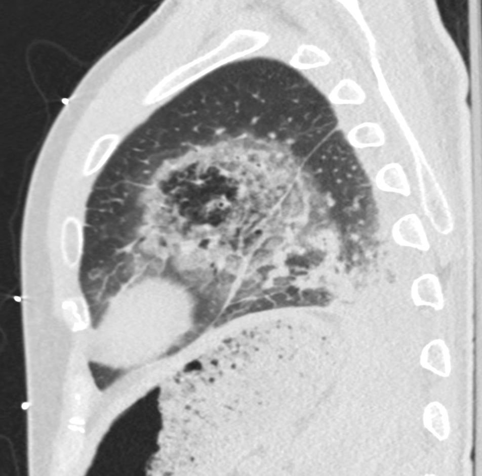

The coronal CT scan shows a rapidly progressive interstitial infiltrate in the left upper lobe showing features of a halo sign. There is evidence of bronchial wall thickening.
Diagnosis Aspergillus Infection
Ashley Davidoff TheCommonVein.net 219Lu-005

The coronal CT scan shows a rapidly progressive interstitial infiltrate in the left upper lobe showing features of a halo sign There is evidence of bronchial wall thickening.
Diagnosis Aspergillus Infection
Ashley Davidoff TheCommonVein.net 219Lu-008
Consolidative Changes in the Lung Bases with Obstruction of the Lower Lobe Airways and Finger in Glove Appearance
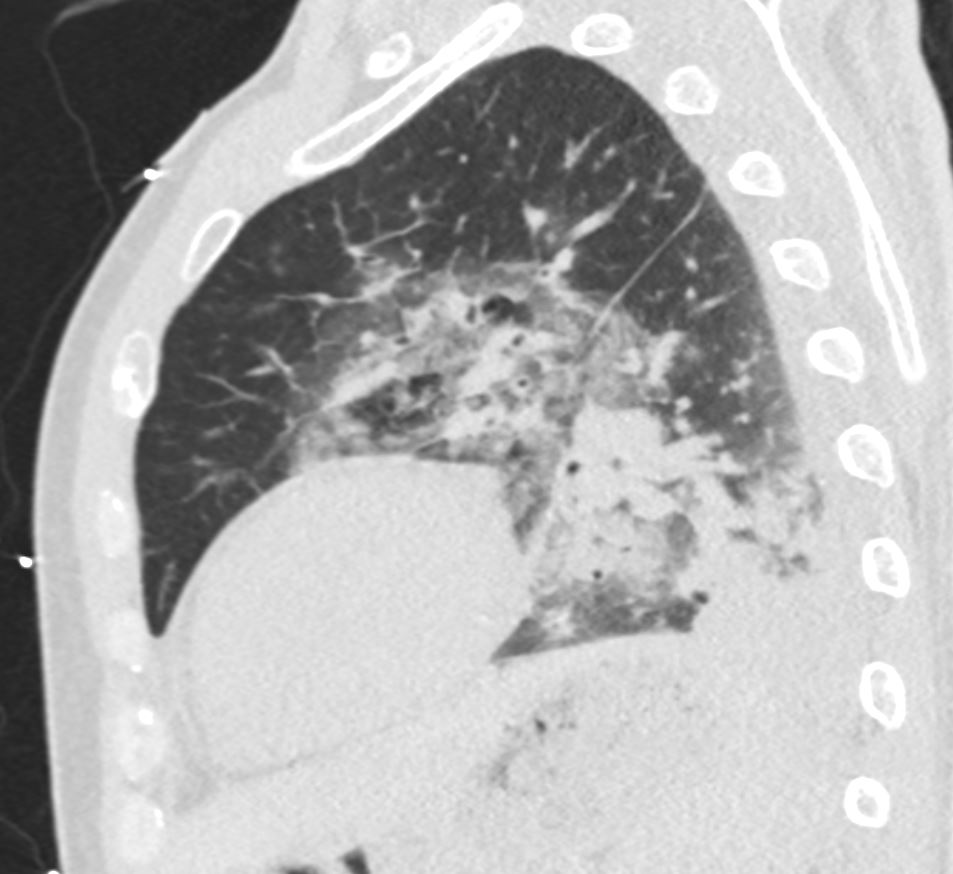

The sagittal CT scan shows a rapidly progressive interstitial infiltrate in the left upper and lower lobes showing features of a reversed halo sign (Atoll sign) There is evidence of basilar consolidation Extensive disease of the segmental and subsegmental airways are noted Fungal Hyphae were identified at bronchoscopy
Diagnosis Aspergillus Infection
Ashley Davidoff TheCommonVein.net 219Lu-006
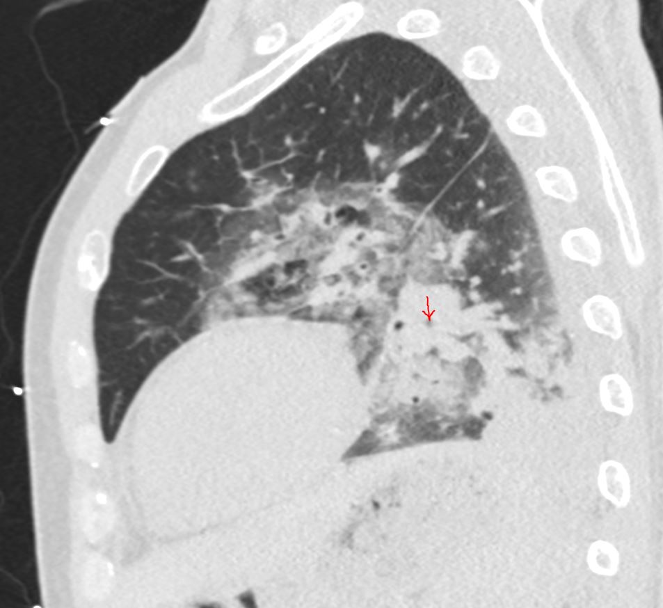

The sagittal CT scan shows a rapidly progressive interstitial infiltrate in the left upper and lower lobes showing features of a halo sign. In addition there is a left basilar consolidation Extensive disease of the segmental and subsegmental airways is noted (red arrow) Fungal Hyphae were identified at bronchoscopy
Diagnosis Aspergillus Infection
Ashley Davidoff TheCommonVein.net 219Lu-007
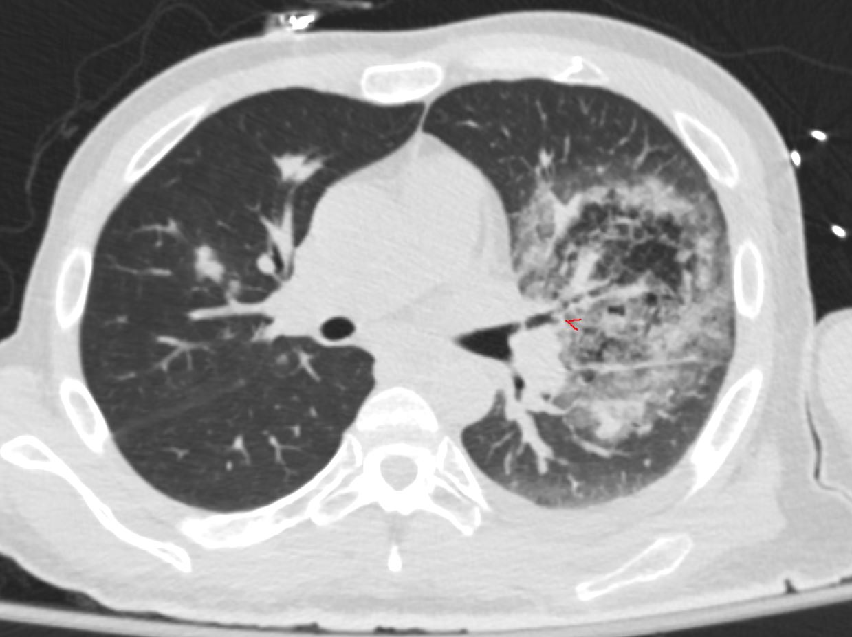
Suggestion of Finger in Glove of the Posterior and Latera Segmental Airways
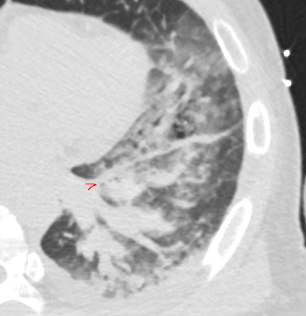

The axial CT scan shows interstitial infiltrate in the left upper and lower lobes There is evidence of basilar band like densities like due to impaction of dilated posterior and lateral segmental airways filled with hyphae of the fungus (bronchoscopic observation) This appearance is reminiscent of the finger in glove sign. The red arrowhead shows a diffusely diseased anterior segmental airway
Diagnosis Aspergillus Infection
Ashley Davidoff TheCommonVein.net 219Lu-009
Ashley Davidoff TheCommonVein.net
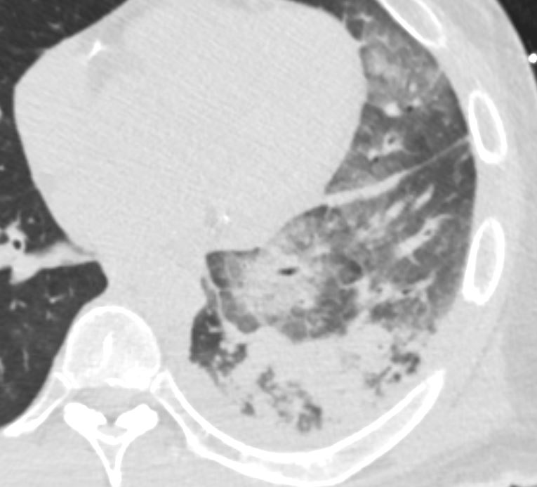

The axial CT scan shows multi-segmental consolidation involving the posterior and lateral segments of the LLL Fungal hyphae were identified at bronchoscopy
Diagnosis Aspergillus Infection
Ashley Davidoff TheCommonVein.net 219Lu-011
Atoll Sign in the Right Lower Lobe
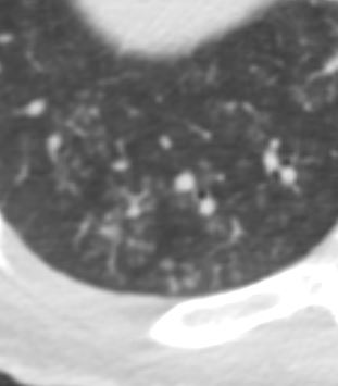


Axial Ct scan focused on the right lung base shows ground glass micronodules revealing a combination of centrilobular nodules and tree in bud sign indicating small airway disease
Diagnosis Aspergillus Infection
Ashley Davidoff TheCommonVein.net 219Lu-015
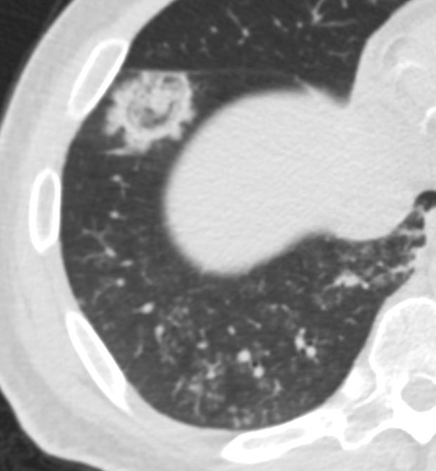

Diagnosis Aspergillus Infection
Ashley Davidoff TheCommonVein.net219Lu-014
Small Airway Disease Ground Glass Micronodules and Probable Tree in Bud Sign


Axial Ct scan focused on the right lung base shows ground glass micronodules revealing a combination of centrilobular nodules and tree in bud sign indicating small airway disease
Diagnosis Aspergillus Infection
Ashley Davidoff TheCommonVein.net 219Lu-015
-
-
- became acutely altered,
- unresponsive, with
- sluggish pupils and
- agonal breathing.
- intubated for
- airway protection and
- hypoxemic respiratory failure. P
- cardiac arrest
- PEA arrest
- repeat bronchoscopy
- redemonstration of his fungal infection
- cardiac arrest again
- was pronounced dead
-
