28-year-old female on OCP with
leg swelling, chest pain and dyspnea and positive DVT study.
CXR Acute Pulmonary Embolism (PE) Normal
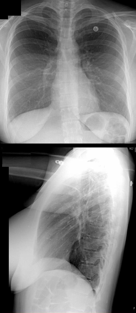
28-year-old female on OCP with leg swelling, chest pain and dyspnea.
CXR in frontal and lateral projection is normal
Ashley Davidoff MD TheCommonVein.net 274Lu 110060ac01
Mismatched Ventilation- Perfusion (V/Q) Scan
Multiple Bilateral Pulmonary Emboli
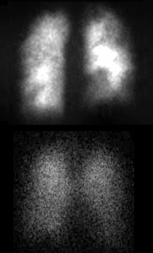
28-year-old female on OCP with leg swelling, chest pain and dyspnea.
Previously performed CXR was normal. Perfusion scan (above) shows multiple bilateral perfusion defects which are not matched on the ventilation scan (below). These findings are consistent with multiple pulmonary emboli
Ashley Davidoff MD TheCommonVein.net 274Lu 11006c02
Overview of Mismatched Ventilation- Perfusion (V/Q) Scan showing
Multiple Bilateral Pulmonary Emboli
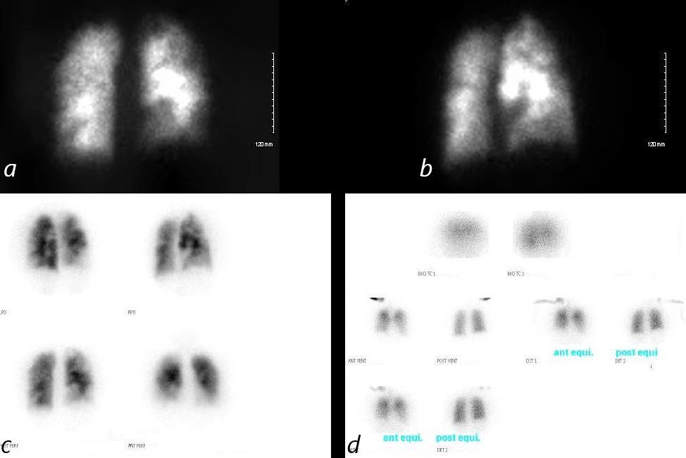
28-year-old female on OCP with leg swelling, chest pain and dyspnea.
Previously performed CXR was normal. DVT study was positive. Perfusion scan (a,b,c) shows multiple bilateral perfusion defects which are not matched on the ventilation scan (d). These findings are consistent with multiple pulmonary emboli
Ashley Davidoff MD TheCommonVein.net 274Lu 110060b01L
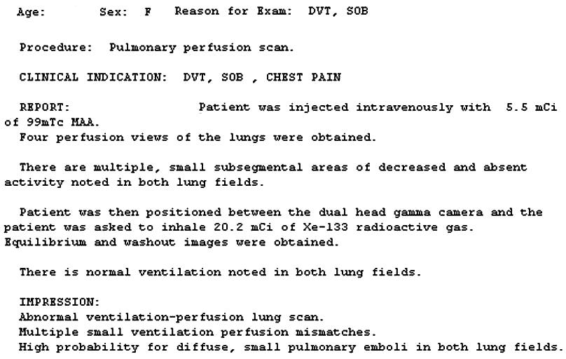
V-Q Scan Report with Technique
Ashley Davidoff MD TheCommonVein.net 274Lu 11006b02
Mismatched Ventilation- Perfusion (V/Q) Scan CT Multiple Bilateral Pulmonary Emboli
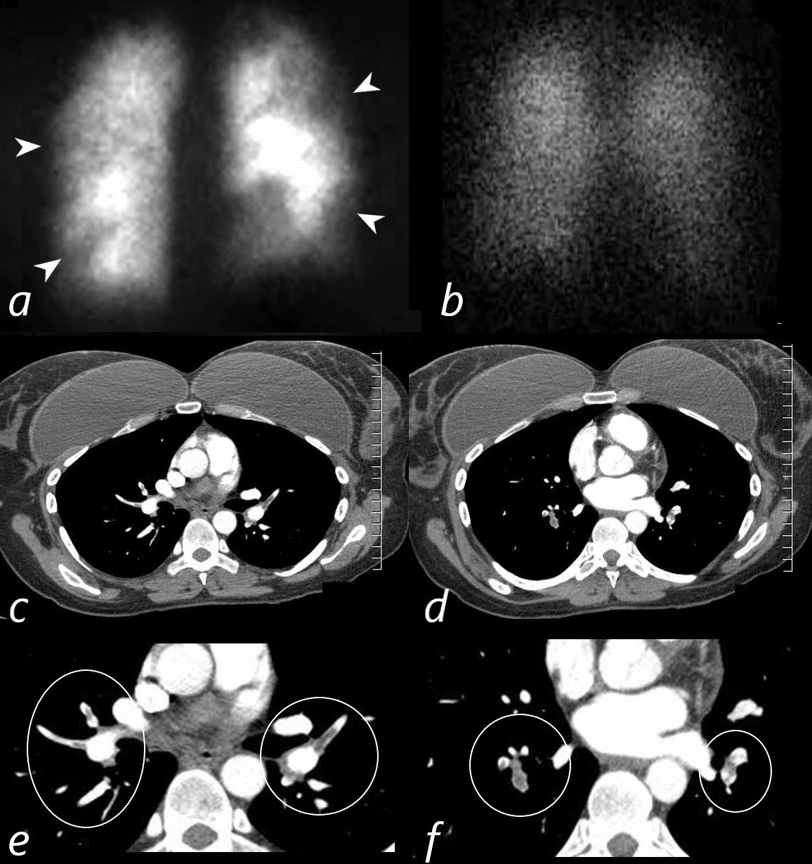
28-year-old female on OCP with leg swelling, chest pain and dyspnea.
Previously performed CXR was normal. Perfusion scan (a) shows multiple bilateral perfusion defects (white arrowheads) which are not matched on the normal ventilation scan (b). These findings are consistent with multiple pulmonary emboli. CT scan through the upper and mid portions of the chest (c,d) confirm the presence of multiple occlusive and non-occlusive pulmonary emboli magnified and ringed (e,f)
Ashley Davidoff MD TheCommonVein.net 274Lu 11006c03L
