75-year-old male with cardiomyopathy and treated for 3 months with amiodarone for atrial fibrillation and
now presents with dyspnea.
CXR Right Upper and Lower Lobe Infiltrates
RUL infiltrate without a fever, and thought to be related to amiodarone therapy.
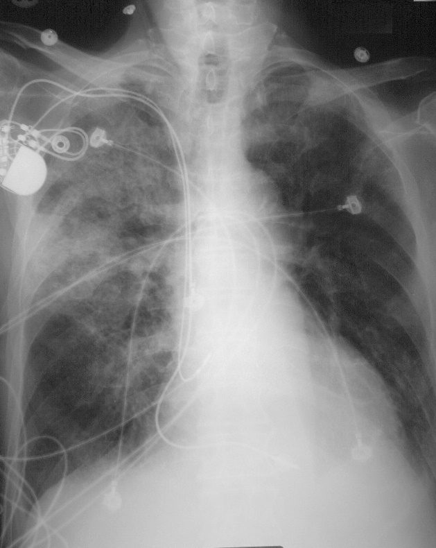
75-year-old male with cardiomyopathy and treated for 3 months with amiodarone for atrial fibrillation and now presents with dyspnea. CXR shows a prominent right upper lobe infiltrate with air bronchograms, a smaller RLL infiltrate, and no evidence of CHF
Ashley Davidoff MD TheCommonVein.278Lu 32475
75-year-old male with cardiomyopathy atrial fibrillation and treatment with amiodarone and a
There was no clinical evidence nor radiological evidence of heart failure.
CT 2 Days Later
Congestive Cardiomyopathy
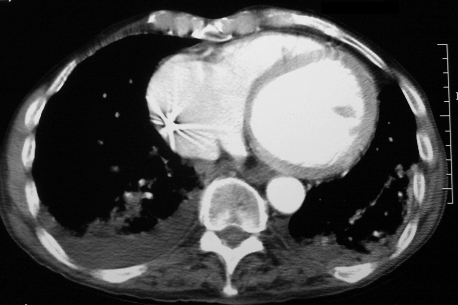
75-year-old male with congestive cardiomyopathy, and atrial fibrillation presents with dyspnea. Has been treated with amiodarone
CT scan shows a dilated left ventricle and right atrium with bibsilar infiltrates and a left pleural effusion. Pacing lead noted in the RA
Ashley Davidoff MD TheCommonVein.net
278 Lu 32473
Ground Glass Changes and Crazy Paving
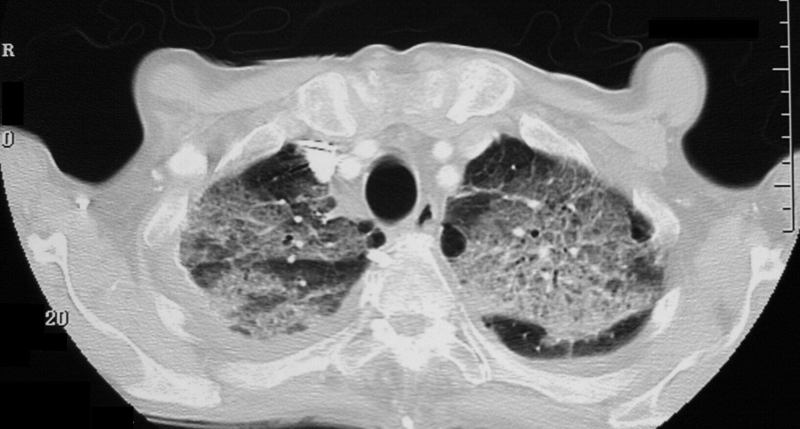
75-year-old male with cardiomyopathy atrial fibrillation and treatment with amiodarone and a RUL infiltrate thought to be related to amiodarone therapy.
There was no clinical evidence nor radiological evidence of heart failure.
CT scan shows ground glass changes with multicentric crazy paving appearance that was thought to be related to amiodarone toxicity.
Ashley Davidoff MD TheCommonVein.net
278 Lu 32470
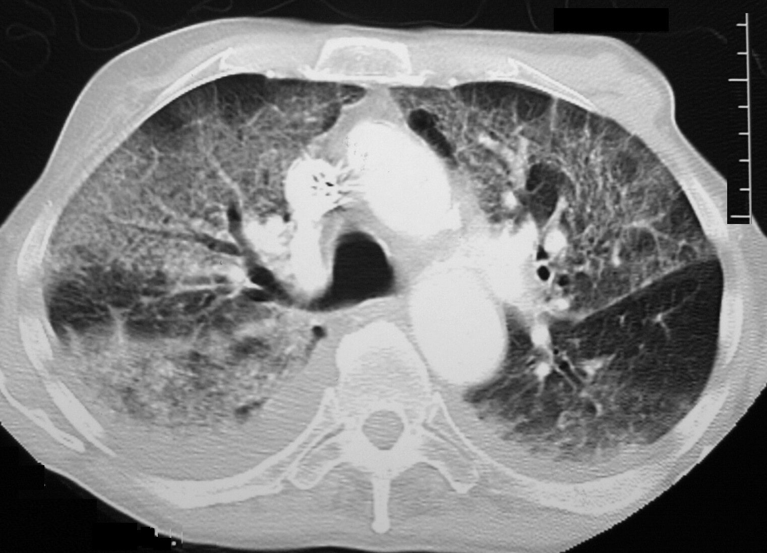
75-year-old male with cardiomyopathy atrial fibrillation and treatment with amiodarone and a RUL infiltrate thought to be related to amiodarone therapy. There was no clinical evidence nor radiological evidence of heart failure.
CT scan shows ground glass changes with multicentric crazy paving appearance that was thought to be related to amiodarone toxicity. bilateral small effusions are present, right greater than left. Following withdrawal of amiodarone and steroids administration he improved clinically and radiologically 3 months later confirming the probability of amiodarone toxicity
Ashley Davidoff MD TheCommonVein.278 Lu 32471
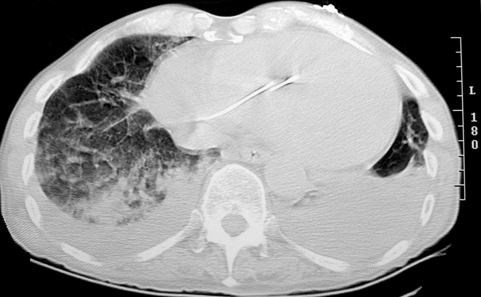
75-year-old male with cardiomyopathy atrial fibrillation and treatment with amiodarone
There was no clinical evidence heart failure or infection.
CT scan shows cardiomegaly with RV lead in place and bibasilar infiltrates and bilateral effusions
Ashley Davidoff MD TheCommonVein.net 278 Lu 32472
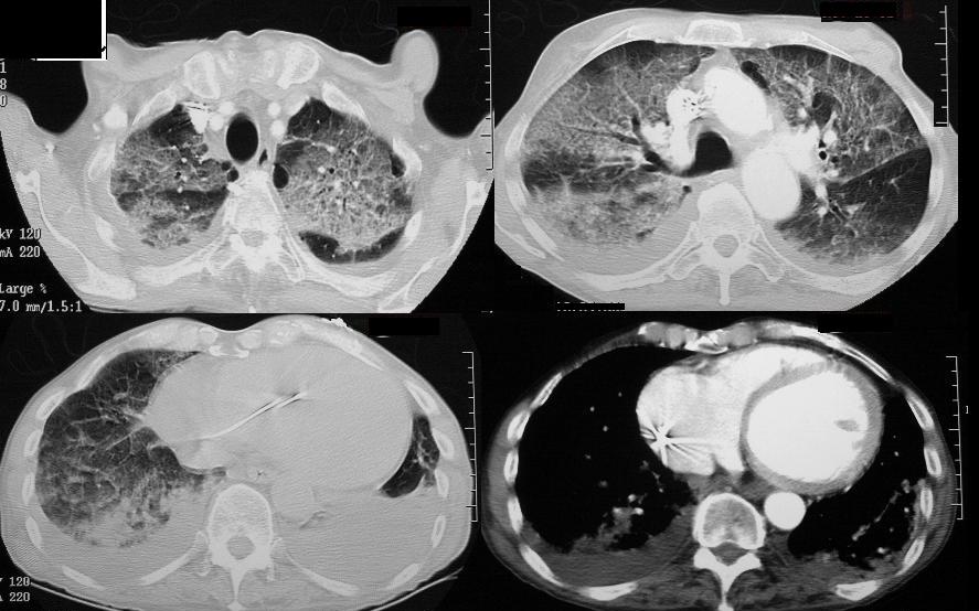
75-year-old male with cardiomyopathy atrial fibrillation and treatment with amiodarone
There was no clinical evidence heart failure or infection.
CT scan shows ground glass changes with multicentric crazy paving appearance in the upper lung fields, that was thought to be related to amiodarone toxicity (upper panels). There are bilateral effusions and thickened interlobular septa in the right lower lobe (left lower image) and evidence of a dilated LV and RA noted in the right lower panel.
Ashley Davidoff MD TheCommonVein.net 278 Lu 32469cd
CT 3 Later
After Amiodarone Withdrawn and Treatment with Steroids.
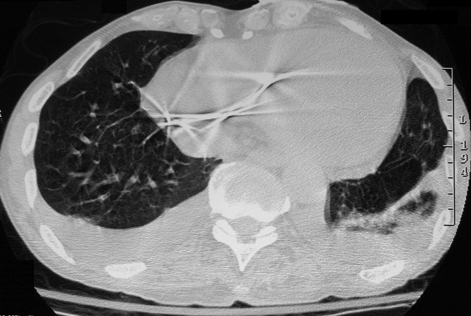
CT scan shows improvement of RLL changes and mild reduction in left lower effusion
Ashley Davidoff MD TheCommonVein.net 278Lu 32480
Following withdrawal of amiodarone and steroids administration he improved clinically and radiologically 6weeks later confirming the probability of amiodarone toxicity
CXR 6 Weeks Later After Amiodarone Withdrawn and Treatment with Steroids
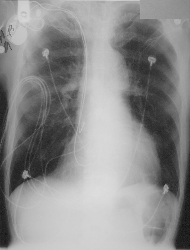
75-year-old male with cardiomyopathy atrial fibrillation and treatment with amiodarone and a RUL infiltrate thought to be related to amiodarone therapy.
CXR 6 weeks after withdrawal of amiodarone and treatment with steroids shows almost complete resolution of previously identified infiltrates. These findings are consistent with amiodarone toxicity
Ashley Davidoff MD TheCommonVein.net 278Lu 32479.8
Summary
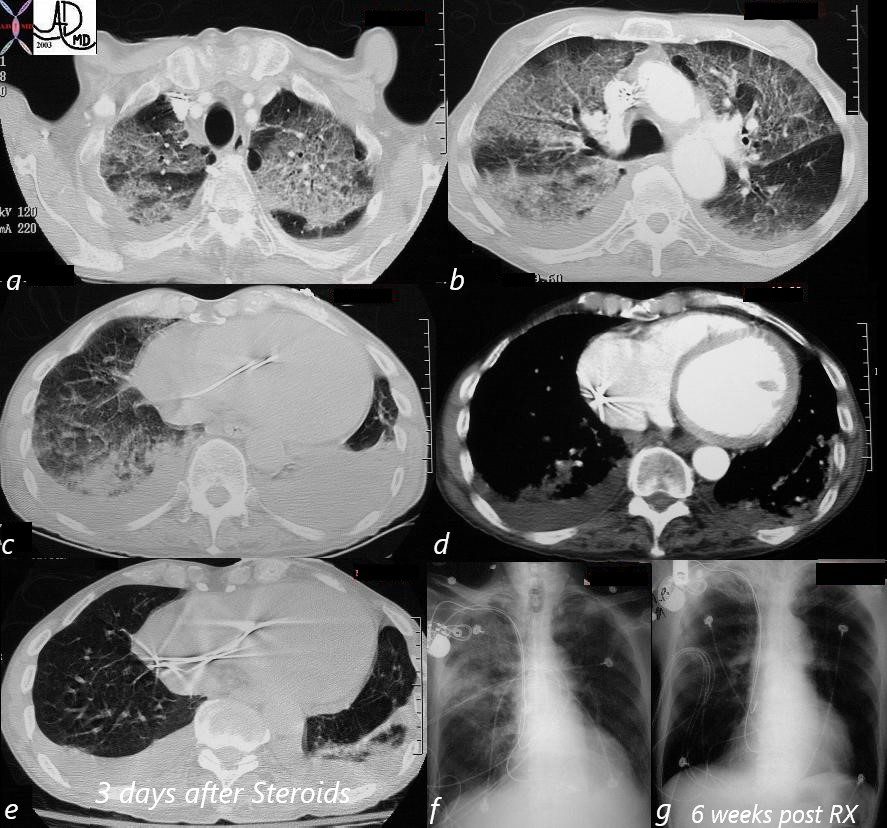
CT (a,b,c) shows diffuse ground glass pattern, dominating in the upper lobes, and global cardiomegaly (d). A diagnosis of amiodarone toxicity was entertained and 3 days after therapy the bases of the lungs showed marked improvement (e). The CXR on admission (f) still shows a dominant RUL infiltrate. 6weeks later sfter cessation of amiodarone and treatment with steroids the CXR (g) shows marked improvement.
Courtesy Ashley Davidoff MD. TheCommonVein.net 32469cLb01
key words lungs pulmonary fx infiltrate ground glass groundglass dx amiodarone toxicity cardiac heart fx enlarged imaging CXR chest X-ray plain film CTscan
