55-year old female presents with a chronic cough
CXR – Lingular Infiltrate
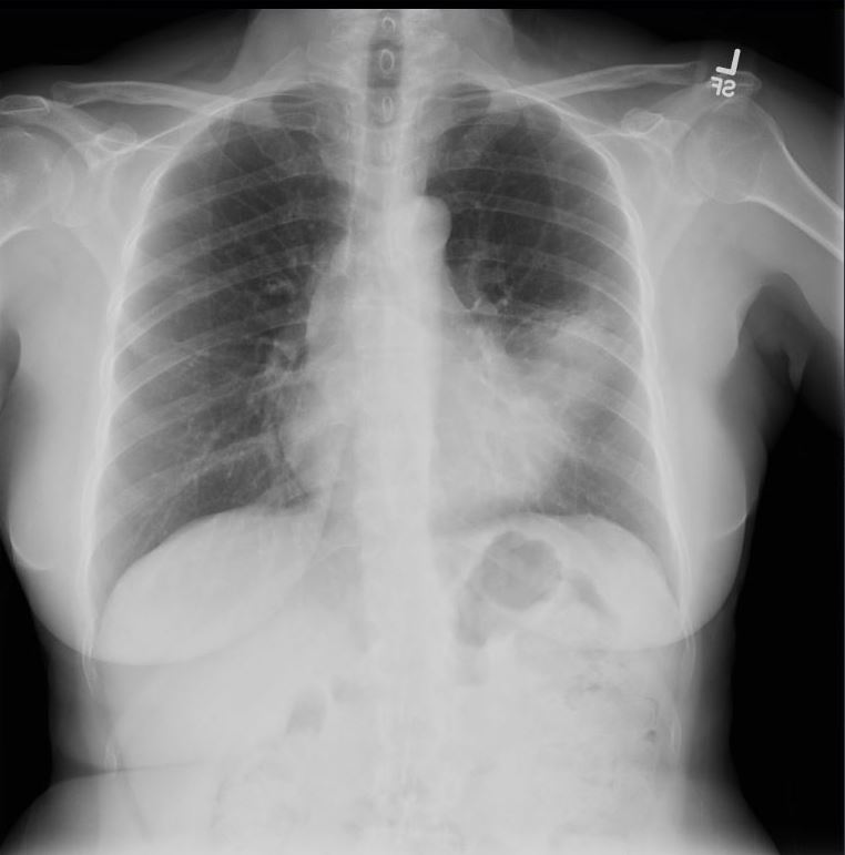
55-year-old female presents with a chronic cough
Frontal CXR shows an infiltrate involving the superior segment of the lingula, with partial silhouetting of the left heart border, and without associated secondary changes of volume loss
Final diagnosis was an obstructing hamartoma of the superior lingula bronchus
Courtesy Ashley Davidoff MD TheCommonVein.net 290 Lu 136563
Why an Infiltrate?
At this stage since there are no clinical or radiological findings to suggest whether it is pneumonia (no fever) and no radiological signs of volume loss (no shifting of the diaphragm or mediastinal structures ) itis reasonable to call the density in the LUL an infiltrate.
CXR and CT – Lingular Infiltrate
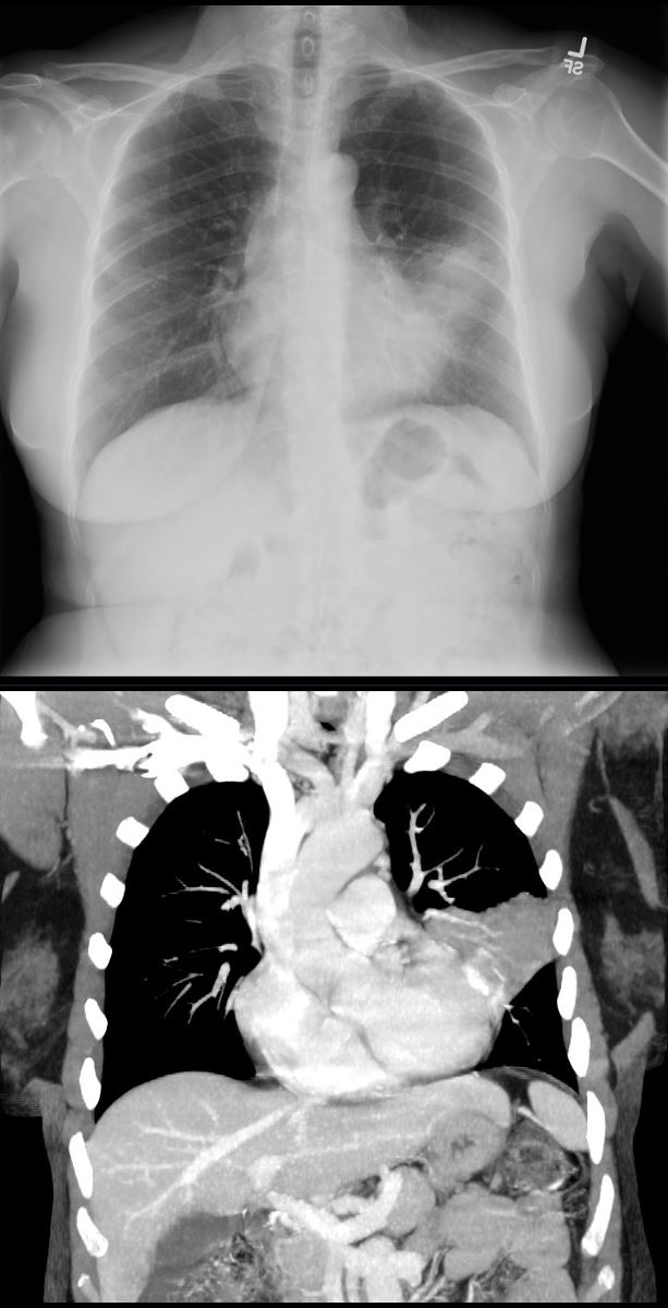
55-year-old female presents with a chronic cough
Frontal CXR and CT in the coronal plane shows an infiltrate involving the superior segment of the lingula, reflecting segmental post obstructive atelectasis and partial silhouetting of the superior aspect of the left heart border.
Final diagnosis was an obstructing hamartoma of the superior lingula bronchus
Courtesy Ashley Davidoff MD TheCommonVein.net 290 Lu 136564b
CT – Lingular Infiltrate with Obstructing Nodule
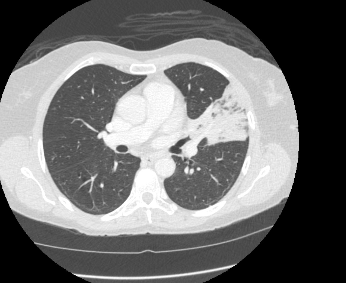
55-year-old female presents with a chronic cough
CT in the axial plane shows an infiltrate involving the superior segment of the lingula, reflecting segmental post obstructive atelectasis. A rounded soft tissue filling defect is noted in the subtending bronchus with downstream mucus accumulation
Final diagnosis was an obstructing hamartoma of the superior lingula bronchus
Courtesy Ashley Davidoff MD TheCommonVein.net 290 Lu 136565
Why Atelectasis?
At this stage since the obstructing nodule and downstream mucus support a diagnosis of atelectasis despite only minimal amount of volume loss
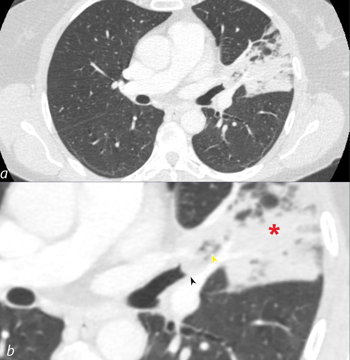
55-year-old female presents with a chronic cough
CT in the axial plane shows an infiltrate involving the superior segment of the lingula, reflecting segmental post obstructive atelectasis (b red asterisk). A rounded soft tissue filling defect is noted in the subtending bronchus (b, black arrowhead) with downstream mucus accumulation (b yellow arrowhead)
Final diagnosis was an obstructing hamartoma of the superior lingula bronchus
Courtesy Ashley Davidoff MD TheCommonVein.net 290 Lu 136566cL
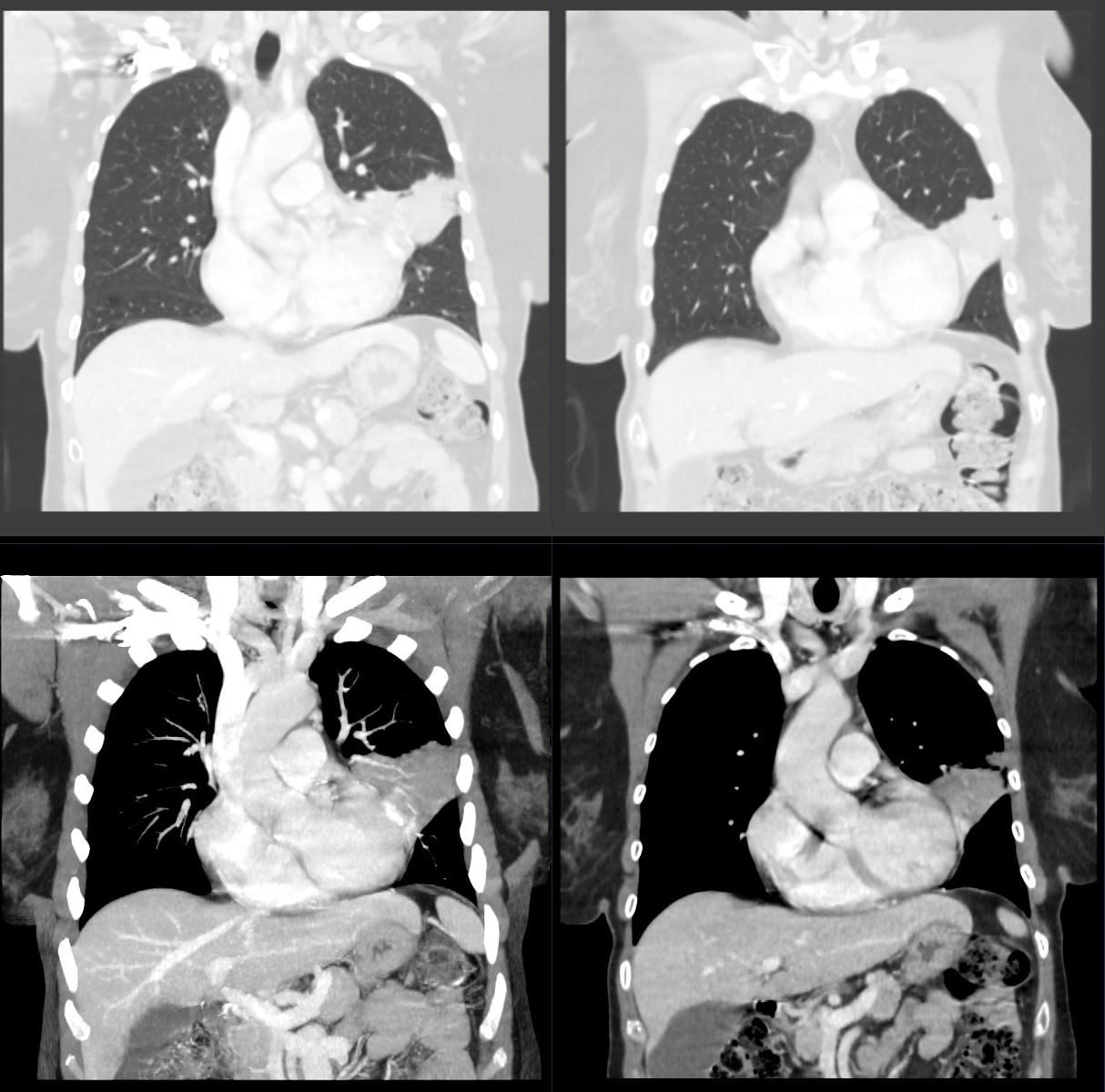
55-year-old female presents with a chronic cough
CT in the coronal plane shows an infiltrate involving the superior segment of the lingula, reflecting segmental post obstructive atelectasis and partial silhouetting of the superior aspect of the left heart border. Images above are in lung window settings and below are soft tissue/mediastinal settings
Final diagnosis was an obstructing hamartoma of the superior lingula bronchus
Courtesy Ashley Davidoff MD TheCommonVein.net 290 Lu 136567
