-
- The lung lobes are distinct, anatomically defined regions of the lungs that are separated by fissures. Each lung is divided into lobes, which are functional subdivisions of the lung tissue, facilitating efficient gas exchange.
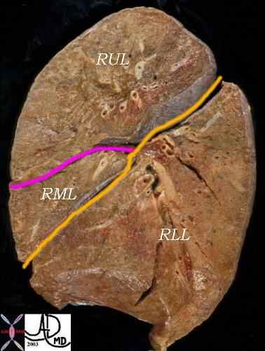
Right Lung – 3 Lobes

This lateral examination of the corresponding lung specimen in sagittal section demonstrates the major fissure in yellow/orange which divides the RLL from the RML and RUL. The minor fissure is in pink and it divides the RUL from the RML.
key words anatomy. normal
Ashley Davidoff MD. TheCommonVein.net 32159B06
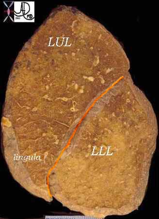
Left Lung – 2 Lobes
- Etymology:
The term “lobe” comes from the Latin “lobus,” meaning a rounded or spherical part, referring to the division of the lung into separate regions. - Parts:
- Right lung: The right lung is divided into three lobes:
- Upper lobe
- Middle lobe
- Lower lobe
- Left lung: The left lung is divided into two lobes:
- Upper lobe
- Lower lobe
- Fissures:
- Right lung: Separated by the horizontal fissure (between the upper and middle lobes) and the oblique fissure (between the middle and lower lobes).
- Left lung: Separated by the oblique fissure (between the upper and lower lobes). The left lung has no middle lobe, but a lingula, a small tongue-like projection of the upper lobe, is sometimes considered a part of the upper lobe.
- Right lung: The right lung is divided into three lobes:
- Size and shape:
- The lobes are irregular in shape, with the right lung being broader due to the position of the liver on the right side. The left lung is narrower, accommodating the heart on the left side.
- Each lobe’s size can vary slightly depending on individual anatomy but generally follows the same division patterns.
- Position:
- The right lung occupies the right hemithorax and is divided into three lobes (upper, middle, and lower), separated by the horizontal and oblique fissures.
- The left lung occupies the left hemithorax, divided into two lobes (upper and lower), with the oblique fissure separating them.
- Character:
- Each lobe is made up of pulmonary parenchyma, consisting of lobar bronchi dividing into bronchioles, alveoli, and their corresponding blood vessels. The lobes function independently, although they communicate through the bronchial tree.
Gas exchange occurs primarily in the alveolar sacs located within the lobes.
- Each lobe is made up of pulmonary parenchyma, consisting of lobar bronchi dividing into bronchioles, alveoli, and their corresponding blood vessels. The lobes function independently, although they communicate through the bronchial tree.
- Blood supply:
- Each lobe receives blood from the corresponding lobar artery. The bronchial arteries, arising from the aorta, provide oxygenated blood to the y the bronchi, bronchioles, and supporting structures.
- Venous drainage
Blood is drained from each lobe by the pulmonary veins, which empty into the left atrium of the heart. - Lymphatic drainage:
- Lymph from each lung lobe drains into the regional lymph nodes, including hilar, interlobar, and mediastinal nodes, and ultimately into the thoracic duct.
- Nerve supply:
- The lungs are innervated by the autonomic nervous system, with the parasympathetic fibers from the vagus nerve and sympathetic fibers from the sympathetic trunk. These fibers regulate bronchoconstriction and vasodilation.
- Embryology:
- Applied anatomy:
- The lobe divisions are important for surgical procedures, such as lobectomy or segmentectomy, in cases of lung cancer, infection, or trauma. These divisions also help in identifying lung pathology on imaging, such as pneumonia, tumors, or atelectasis, which may be localized to a specific lobe.
- Imaging Application:
- Chest X-ray: The lobes can be visualized as distinct areas of lung tissue on a chest X-ray, with the fissures acting as dividing lines.
- CT Scan: High-resolution CT provides detailed visualization of the lobes, including fissure lines and any pathology within the lobes, such as consolidation, tumors, or interstitial lung disease.
- MRI: MRI is less commonly used for lung imaging but can provide detailed imaging of lung lobes in specialized cases, such as in patients with tumors or vascular anomalies.

