- Lung Acinus

This diagram illustrates the acinus which consists of the respiratory bronchioles (rb 1, 2, 3) the alveolar duct (ad) the alveolar sac (as) and the alveoli. (a)
Courtesy Ashley Davidoff MD 42446b12 TheCommonVein.net
-
- Etymology
- Derived from the Latin word “acinus,” meaning berry or grape, referencing the grape-like clusters of alveoli.
- AKA and Abbreviation
- Also known as “primary lobule.”
- What is it?
- The acinus is the smallest functional unit of the lung responsible for gas exchange because it integrates multiple structures required for efficient oxygen-carbon dioxide exchange, including respiratory bronchioles, alveolar ducts, and alveoli. While the alveolus is the site of gas exchange, it lacks the additional structural and functional components needed for the coordinated delivery and removal of gases.
- It is called the “primary lobule” because it represents the first distinct functional subunit within the respiratory division of the lung, with all elements necessary for complete respiratory function.
- It includes all structures distal to the terminal bronchiole, comprising respiratory bronchioles, alveolar ducts, and alveoli.
- Principles
- Parts
- Terminal bronchiole: Precedes the acinus.
- Respiratory bronchioles: Initiate gas exchange.
- Alveolar ducts: Lead to alveolar sacs.
- Alveoli: Primary sites of oxygen and carbon dioxide exchange.
- Size
- Diameter: 6-10 mm.
- Each acinus contains 2000-5000 alveoli.
- Shape
- Grape-like clusters of alveoli surrounding alveolar ducts.
- Position
- Distal to the terminal bronchioles within secondary pulmonary lobules.
- Character
- Highly vascularized structures with thin-walled alveoli.
- Time
- Functional throughout life but susceptible to damage from environmental or pathological factors.
- Blood supply
- Supplied by the pulmonary arteries.
- Venous Drainage
- Drained by the pulmonary veins.
- Lymphatic drainage
- Lymphatic vessels drain into interlobular septa lymphatics.
- Nerve Supply
- Autonomic innervation from the pulmonary plexus.
- Embryology
- Development begins in the pseudoglandular phase (~16 weeks gestation) and matures through the alveolar phase (~36 weeks gestation to early childhood).
- Histology
- Lined by simple squamous epithelium.
- Supported by elastic fibers for recoil.
- Structurally integrated into the secondary pulmonary lobule, with each acinus contributing to the overall function of its lobule.
- Each secondary pulmonary lobule contains approximately 3 to 25 acini, with variation based on location within the lung (smaller near the pleura and larger near the hilum).
- Physiology and Pathophysiology
- Facilitates gas exchange through diffusion.
- Vulnerable to damage in diseases like emphysema, pneumonia, and interstitial lung diseases.
- Parts
- Applied Anatomy to Radiology
- CXR
- The acinus itself is not directly visible but contributes to imaging patterns.
- Acinar Shadows: Suggest filling of multiple acini within a secondary pulmonary lobule, appearing as ill-defined, nodular, or confluent opacities (1-2 cm) caused by fluid, pus, blood, or cells, commonly seen in conditions like pneumonia or pulmonary edema.
- CT
- The acinus itself is rarely imaged directly unless it is diseased.
- Observations on CT often represent changes at the secondary pulmonary lobule level:
- Acinar Shadows: Correlate with areas of consolidation or ground-glass opacity where groups of acini are filled or obliterated by pathological processes.
- CXR
- Etymology
A structural unit of the lung which is distal to the terminal bronchiole and contains respiratory bronchioles, alveolar ducts, and alveoli, which are all involved in gas exchange.
The Acinus
Artistic Rendering of an Acinus
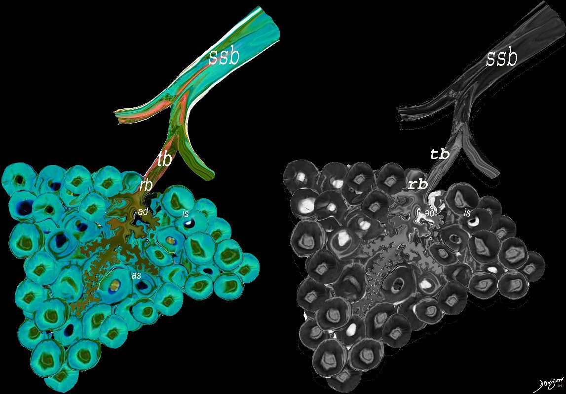
The subsegmental medium sized airways give rise to the terminal bronchiole (tb) which gives rise to the membranous airways. These include in order, the respiratory bronchiole (rb), alveolar duct (ad) and alveolar sac (as)
Ashley Davidoff Medical Art
TheCommonvein.net lungs-00680
Acinus Subtended by a Single First Order Terminal Respiratory Bronchiole
3-30 Acini (usually 20-30) per Secondary Lobule Measures 2-5mm
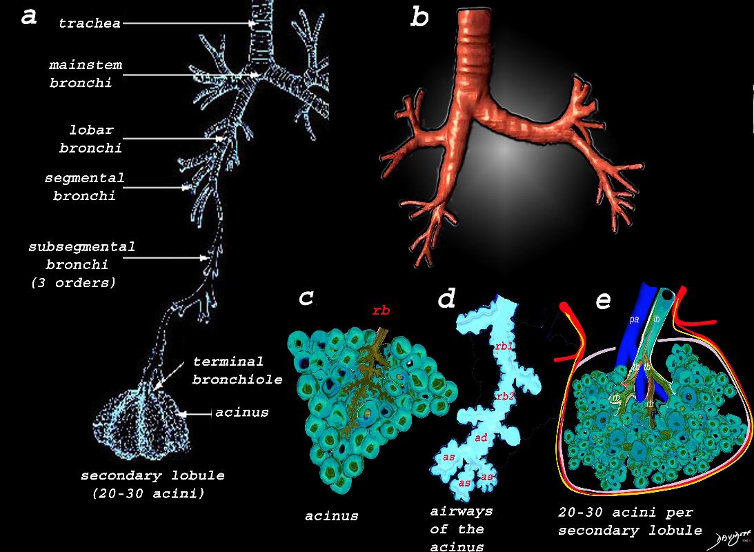
Image a shows the airways starting in the trachea and continuing to the mainstem bronchi, lobar bronchi, segmental bronchi, and subsegmental bronchi,. The subsegmental bronchi have 3 subsequent generations until the bronchiole is reached. The terminal bronchiole is the last of the transporting airways and is considered the most proximal small airway with a diameter of 2mm or less, and it gives rise to the respiratory bronchiole which is the feeding airway for the acinus . The acinus is the functional unit of the lung.
Image b is a 3D reconstruction of a CT scan showing the proximal airways from the trachea to the segmental airways.
Image c shows the structures that make up the acinus and the other parts of the small airways, starting with the respiratory bronchiole (rb) . The diagram in d, shows the detail of the small airways that participate in gas exchange, including the respiratory bronchiole, (rb) alveolar duct, (ad) and alveolar sac (as)
Image e shows the secondary lobule made from about 20-30 acini, arising from a single lobular bronchiole accompanied by a single pulmonary arteriole (pa).. Structure that surround and enclose the secondary lobule include the pulmonary venule, (red) lymphatics,(yellow) and a fibrous septum (pink).
Ashley Davidoff MD Medical Art TheCommonVein.net
lungs-0739
Normal acini are not visible on imaging, whereas abnormal acini may be visualized as small, rounded opacities referred to as “air space nodules”. (Etesami)
- The Acinus
- is a functional unit of the lung which is involved in gas exchange
- It is
- distal to the terminal bronchiole and
- contains
- respiratory bronchioles,
- alveolar ducts, and
- alveoli,
- which are
- all involved in gas exchange.
- Normal acini are not visible on imaging,
- whereas
- abnormal acini may be visualized as small, rounded opacities referred to as
- “air space nodules”.
- abnormal acini may be visualized as small, rounded opacities referred to as
- The acinus is also defined as the portion of lung that is
- distal to the terminal bronchiole and is
- supplied by the
- respiratory bronchiole and
- usually measures about
- 2-5mms in diameter.
It is the functional unit of the lung
The terminal bronchiole precedes the acinus. (Webb)
The bronchi proceed from the mainstem bronchus via 16 to 23 divisions into the terminal bronchioles. Thereafter sac-like protrusions develop in the system, which allow gas exchange to start taking place. The first branch that is able to perform this gas exchange is called the respiratory bronchiole. After three divisions the respiratory bronchioles become alveolar ducts and after further division become alveolar sacs. Finally, at the terminal end of this pathway, are the alveoli. The system that starts at the respiratory bronchiole and terminates at the alveoli is called an acinus, and it is functionally characterized by having the ability to both conduct air as well as enable gas exchange.
The acinus averages about 7mm (4-8mm)in diameter. When it is filled with fluid, it can be visualized on a CXR as a 6mm nodular density called an “acinar shadow.” or “air space nodules” As a structural entity it has little diagnostic utility. We will expand on the pulmonary lobule in the next section which has greater implications for the imaging of the lung. Functionally, however, the acinus can be considered as the unit of gas exchange in the lung.
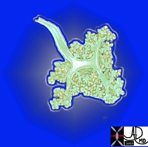
The acinus with its arborizations is shaped more like a bunch of grapes.
Courtesy of: Ashley Davidoff, M.D 42650
TheCommonVein.net
Respiratory System Anatomy Samuel Chen
Imaging
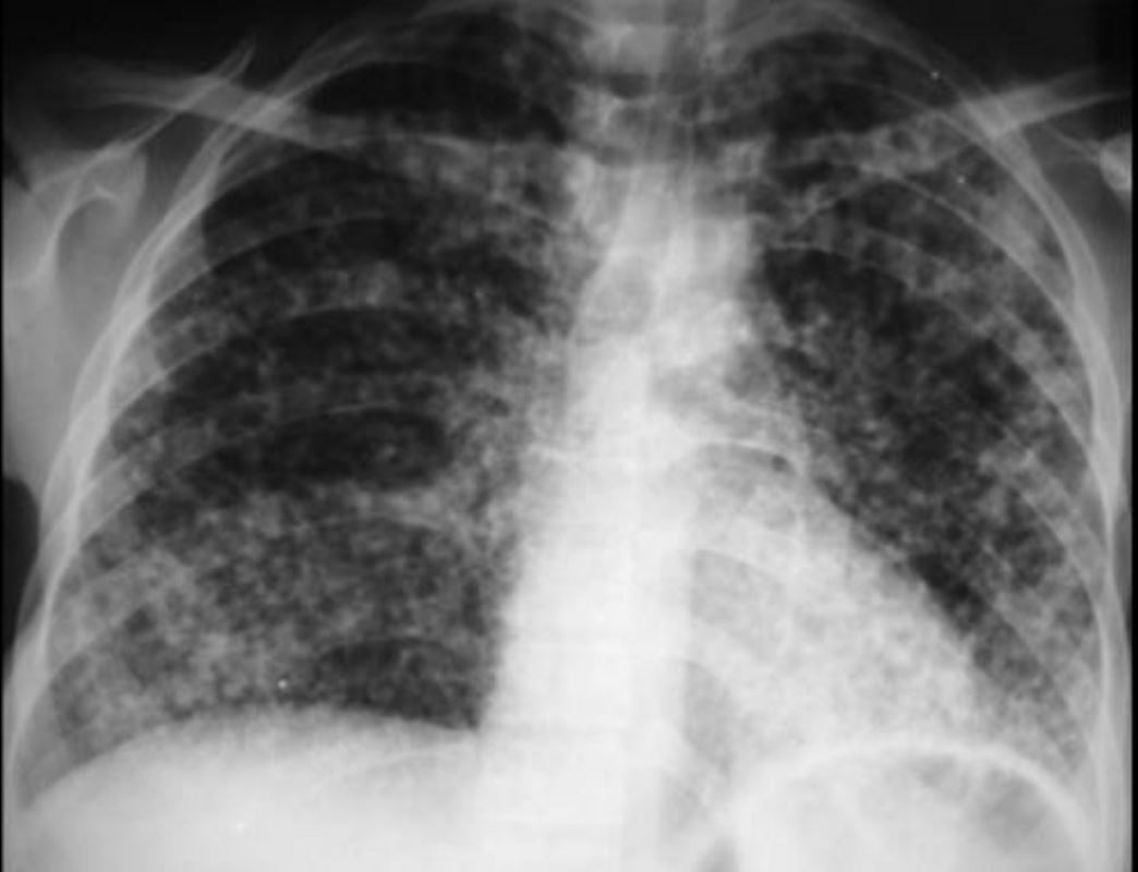
Acinar Shadows Pulmonary Hemorrhage GPA aka Wegener’s Granulomatosis
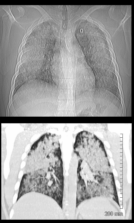
19 year old male previously well with history of hemoptysis, sweating, fevers, myalgias, arthritis over 3 weeks.
CT scan scout (above ) shows diffuse bilateral lobar infiltrates sparing the hila regions.
Coronal CT shows bilateral symmetrical lobar infiltrates involving the upper and lower lobes with acinar pattern. The upper lobes are more affected than the lower lobes.
Lab shows ANCA positivity, acute renal failure (creatinine 6) and renal biopsy showing crescentic glomerulonephritis.
Treated with cyclophosphamide
Ashley Davidoff MD TheCommonVein.net 139195c
Courtesy https://www.slideshare.net/
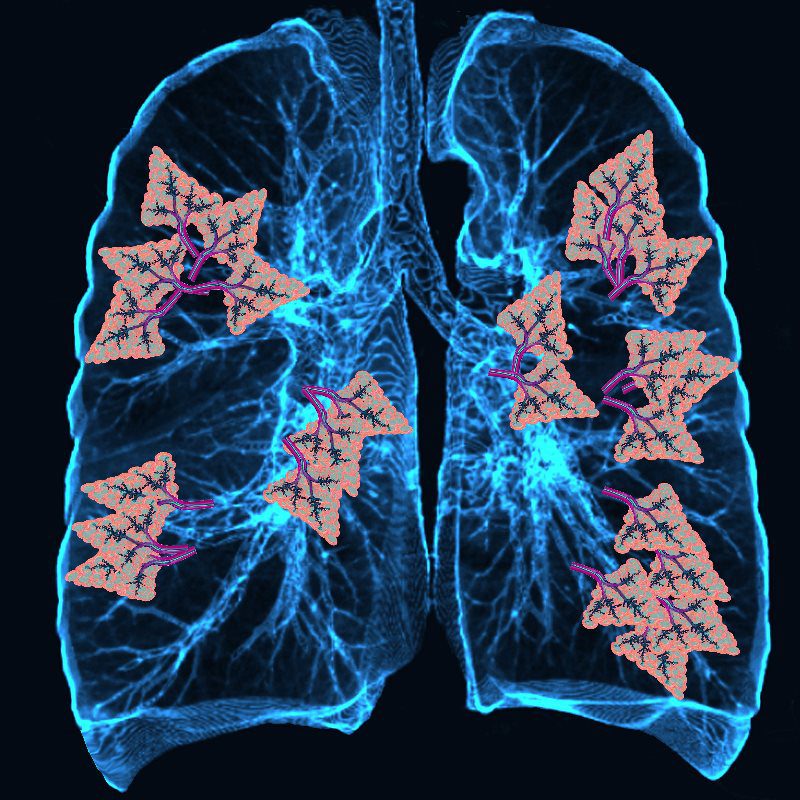
Other Artistic Examples of Normal Acini in the Lung
Ashley Davidoff TheCommonVein.net
The Acinus in Health and in Emphysema
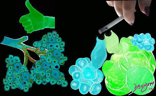
Image on the left shows normal size and appearance of terminal bronchioles and alveoli. On the right the image shows the effects on the respiratory bronchioles and when severe, on the alveoli as well. The respiratory bronchioles, are the earliest and most significantly affected. As the disease progresses, the destruction and enlargement of airspaces can extend distally to involve other portions of the acinus, such as the alveolar ducts and alveoli.
Ashley Davidoff MD TheCommonVein.net
Links and References
Fleischner Society
acinus
Anatomy.—The acinus is a structural unit of the lung distal to a terminal bronchiole and is supplied by first-order respiratory bronchioles; it contains alveolar ducts and alveoli. It is the largest unit in which all airways participate in gas exchange and is approximately 6–10 mm in diameter. One secondary pulmonary lobule contains between three and 25 acini (,4).
Radiographs and CT scans.—Individual normal acini are not visible, but acinar arteries can occasionally be identified on thin-section CT scans. Accumulation of pathologic material in acini may be seen as poorly defined nodular opacities on chest radiographs and thin-section CT images. (See also nodules.)
