Air crescent sign
- The air crescent sign is a radiological finding
- characterized by a
-
- crescent-shaped radiolucency that
- forms between
- a mass or nodule and the surrounding lung tissue
-
-
Etymology
- Derived from the Latin “aer” (air) and “crescere” (to grow), reflecting the crescent-shaped radiolucency seen on imaging.
AKA
- Mycetoma (when caused by fungal infection)
- Crescent sign
- Air cap sign
What is it?
- The air crescent sign is a radiologic finding characterized by a crescent-shaped radiolucency of air surrounding a central mass or nodule.
- It is typically seen in cavitary lesions and signifies the separation of necrotic tissue or a fungal ball from the cavity wall.
- Commonly associated with fungal infections, most frequently caused by Aspergillus spp. but can also involve other fungi.
Characterized by
- A crescent-shaped air collection outlining a central opacity (e.g., fungal ball or necrotic material).
- Represents the dynamic interaction between a lesion and its surrounding air.
- Seen predominantly in cavitary pulmonary lesions.
Anatomically affecting
- Typically involves the lungs, within cavitary lesions of varying etiology.
- Most commonly associated with the upper lobes due to the high prevalence of cavitary lesions in these regions.
Pathophysiology
- The sign reflects the formation of a cavity within the lung parenchyma due to necrosis or infection.
- Air enters the cavity, displacing necrotic material or fungal debris to create the crescent-shaped interface.
- Fungal Causes: Most commonly Aspergillus species, but also includes:
- Zygomycetes (e.g., Mucor and Rhizopus in mucormycosis).
- Candida spp.
- Cryptococcus spp.
- Histoplasma capsulatum (endemic mycoses).
- Coccidioides spp.
- Blastomyces dermatitidis.
How does it appear on each relevant imaging modality?
Principles
- Parts: Crescent-shaped air surrounding a dense central lesion.
- Size: Varies based on the size of the cavity and the lesion.
- Shape: Crescentic or semi-lunar.
- Position: Typically within the upper lobes or regions prone to cavitary changes.
- Character: Radiolucent air surrounding an opacity.
- Time: Appears during the late stages of cavitation or fungal ball formation.
CXR
- Appears as a crescent-shaped lucency adjacent to a denser central opacity.
- Best seen in upright or lateral decubitus views to highlight air-fluid levels.
- May also reveal associated findings such as cavitation or parenchymal scarring.
CT
- Provides superior visualization, clearly delineating:
- The cavity wall—thickness and irregularity can indicate underlying pathology (e.g., malignancy, infection).
- The air crescent—its sharp margins and configuration.
- The central opacity or debris (e.g., fungal ball, necrotic material).
- High-resolution CT (HRCT) is particularly useful for small or subtle air crescents not visible on CXR.
- Multiplanar reconstructions allow precise localization and characterization of the lesion and adjacent structures.
- Mobility of the fungal ball: The fungal ball typically changes position with changes in the patient’s posture, distinguishing it from fixed lesions such as malignancies.
MRI
- Rarely used for this finding but can show low signal intensity in the central mass and high signal from the surrounding air on T2-weighted images.
- Gadolinium-enhanced sequences may help identify vascularized cavity walls or underlying malignancies.
PET-CT
- May show increased metabolic activity in the cavity wall if associated with active infection or malignancy.
- Central necrotic or fungal material typically shows low metabolic activity.
- Useful for distinguishing benign from malignant cavitary lesions.
Other Modalities
- Ultrasound: Limited utility but may visualize fluid motion within cavities during real-time imaging.
- Fluoroscopy: Historically used in dynamic studies to evaluate cavity motion and air-fluid levels but now largely replaced by CT.
Differential Diagnosis
- Aspergilloma (most common association).
- Tuberculosis with cavitary lesions.
- Lung abscess (organizing necrotic debris).
- Wegener’s granulomatosis (cavitating granulomas).
- Lung cancer (necrotic cavitary tumor).
- Hydatid cyst rupture with air ingress.
Recommendations
- Further imaging:
- Contrast-enhanced CT to better evaluate the cavity wall and surrounding parenchyma.
- High-resolution CT for detailed structural analysis and subtle findings.
- Laboratory correlation:
- Sputum or bronchoalveolar lavage (BAL) for fungal cultures, bacterial cultures, or cytology.
- Biopsy: Consider if malignancy or uncertain etiology is suspected.
- Clinical correlation:
- Assess for symptoms like fever, hemoptysis, or weight loss, which may suggest infection or malignancy.
Key Points and Pearls
- The air crescent sign is most classically associated with aspergilloma but can occur in a variety of cavitary diseases.
- It is usually a late finding, appearing after cavity formation and partial resolution of the inflammatory process.
- The sign helps localize and define cavitary lesions, guiding diagnostic and therapeutic interventions.
- On CT, the air crescent provides detailed insights into cavity dynamics and underlying pathology.
- The mobility of the fungal ball on CT imaging with positional changes is a hallmark feature, aiding differentiation from fixed lesions.
- Multimodal imaging, including PET-CT, can refine diagnosis and management strategies.
CXR Air Crescent Sign in the Right Upper Lobe
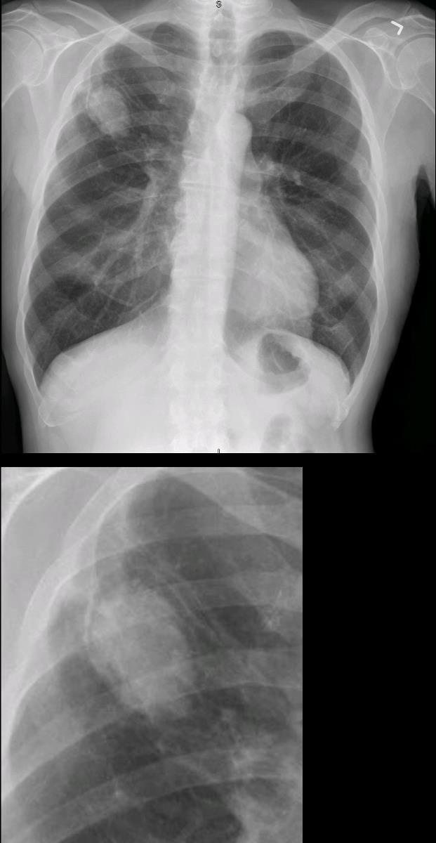
66- year-old malnourished, immunodeficient male presents with a chronic cough.
CXR in the frontal view shows a right upper lobe nodule with a subtle rim of a crescentic accumulation of air on the lateral border of the nodule (yellow arrowheads). CT confirmed an air -crescent sign consistent with a diagnosis of an aspergilloma
Ashley Davidoff TheCommonVein.net 293Lu 113533c
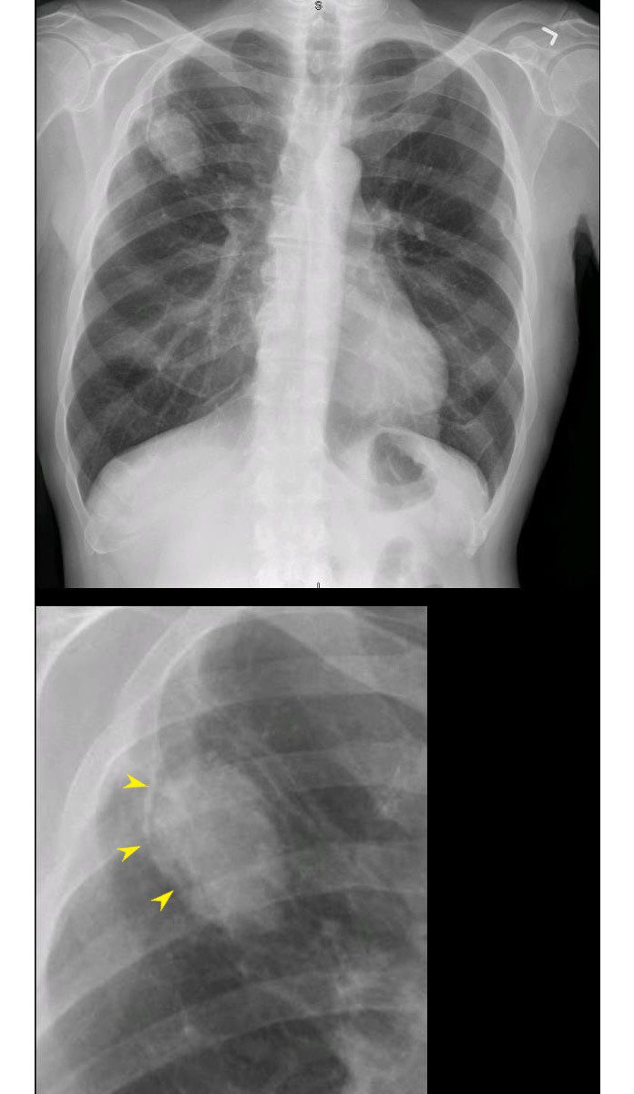
66- year-old malnourished, immunodeficient male presents with a chronic cough.
CXR in the frontal view shows a right upper lobe nodule with a subtle rim of a crscentic accumulation of air on the lateral border of the nodule (yellow arrowheads). CT confirmed an air -crescent sign consistent with a diagnosis of an aspergilloma
Ashley Davidoff TheCommonVein.net 293Lu 113533cL
CT Air Crescent Sign in the Right Upper Lobe
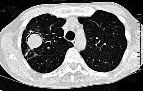
66 year malnourished immunodeficient male with right upper lobe aspergilloma in the lung
CT scan in the axial plain shows an air-crescent sign with right Upper lobe mass consistent with aspergilloma
Ashley Davidoff TheCommonVein.net 113530b01
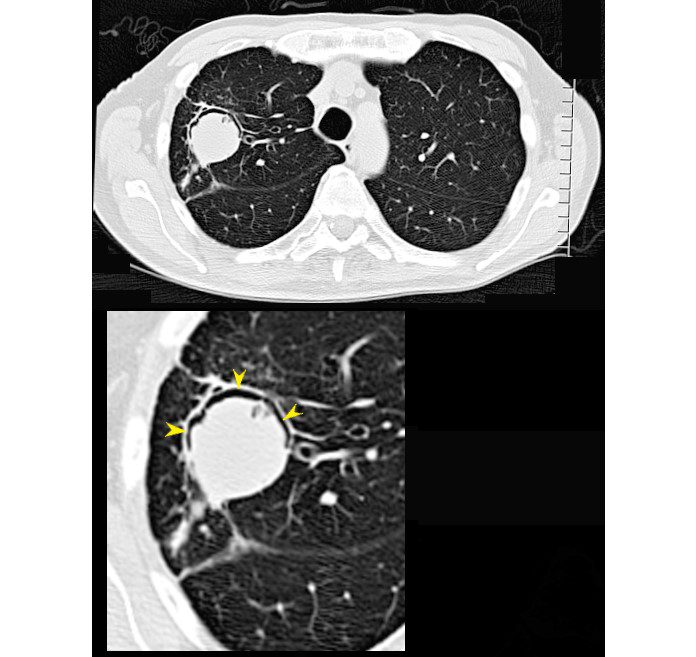


66- year-old malnourished, immunodeficient male presents with a chronic cough.
CT in the axial plane shows a 3.2cms right upper lobe mass with a rim of a crescentic accumulation of air in the dependent portion of the mass (magnified in the lower image- yellow arrowheads) while the aspergilloma “sinks” to the most dependent portion of the cavity . This finding reflects an air -crescent sign and is consistent with a diagnosis of an aspergilloma
Ashley Davidoff TheCommonVein.net 293Lu 113530cL
-
-
- It is often associated with
- infectious or inflammatory conditions like
- invasive aspergillosis,
- especially in immunocompromised patients, where damaged lung and necrotic tissue
- begins to retract,
- leaving a space filled with
- air (cavitation).
- invasive aspergillosis,
- infectious or inflammatory conditions like
- Structurally, the air crescent sign indicates
- cavitary lesions or areas of necrosis,
- Clinically
- presents with symptoms like
- cough and hemoptysis.
- presents with symptoms like
- Diagnosis is made through
- clinical symptoms,
- imaging (primarily CT), and
- laboratory tests, including
- cultures or
- serology for infectious agents like
Aspergillus. (Etesami)
- It is often associated with
-


66 year malnourished immunodeficient male with right upper lobe aspergilloma in the lung
CT scan in the axial plain shows an air-crescent sign with right Upper lobe mass consistent with aspergilloma
Ashley Davidoff TheCommonVein.net 113530b01
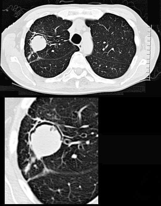

66- year-old malnourished, immunodeficient male presents with a chronic cough.
CT in the axial plane shows a 3.2cms right upper lobe mass with a rim of a crescentic accumulation of air in the dependant portionof the mass (magnified in the lower image) while the aspergilloma “sinks” to the most dependent portion of the cavity . This finding reflects an air -crescent sign and is consistent with a diagnosis of an aspergilloma
Ashley Davidoff TheCommonVein.net 293Lu 113528c


66- year-old malnourished, immunodeficient male presents with a chronic cough.
CT in the axial plane shows a 3.2cms right upper lobe mass with a rim of a crescentic accumulation of air in the dependant portionof the mass (magnified in the lower image- yellow arrowheads) while the aspergilloma “sinks” to the most dependent portion of the cavity . This finding reflects an air -crescent sign and is consistent with a diagnosis of an aspergilloma
Ashley Davidoff TheCommonVein.net 293Lu 113528cL


Source
Signs in Thoracic Imaging
Journal of Thoracic Imaging21(1):76-90, March 2006
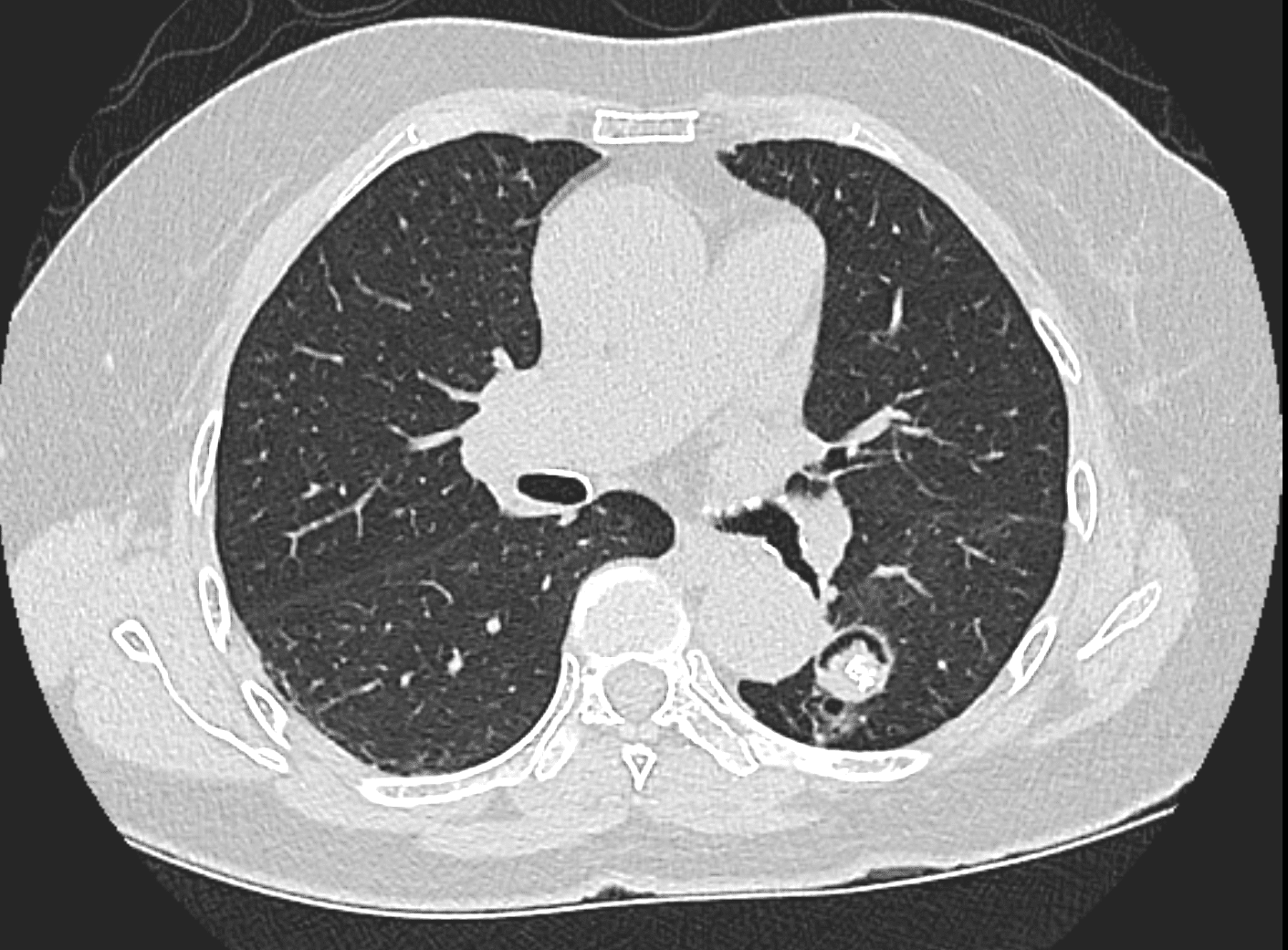
The air crescent sign appears as a variably sized, peripheral crescentic collection of air surrounding a necrotic central focus of infection on thoracic radiographs (Fig. 1A) and CT (Fig. 1B).2–4 It is often seen in neutropenic patients who have undergone bone marrow or organ transplantation and is most characteristic of infection with invasive pulmonary aspergillosis. The fungus invades the pulmonary vasculature, causing hemorrhage, thrombosis, and infarction. With time, the peripheral necrotic tissue is reabsorbed by leukocytes and air fills the space left peripherally between the devitalized central necrotic tissue and normal lung parenchyma.5 Thus, the presence of the air-crescent sign heralds recovery of granulocytic function.4 Other causes of the air crescent include cavitating neoplasms, bacterial lung abscesses, and infections such as tuberculosis or nocardiosis.6


Ashley Davidoff MD


Ashley Davidoff MD
Links and References
Fleischner Society
air crescent
Radiographs and CT scans.—An air crescent is a collection of air in a crescentic shape that separates the wall of a cavity from an inner mass (,Fig 3). The air crescent sign is often considered characteristic of either Aspergillus colonization of preexisting cavities or retraction of infarcted lung in angioinvasive aspergillosis (,9,,10). However, the air crescent sign has also been reported in other conditions, including tuberculosis, Wegener granulomatosis, intracavitary hemorrhage, and lung cancer. (See also mycetoma.)
-
Parallels with Human Endeavors
- The air crescent sign mirrors adaptive structural dynamics seen in engineering, where voids and separations create new patterns, such as spaces forming around central objects in erosion processes.
- It also reflects the concept of containment and isolation, akin to protective barriers forming around compromised systems to prevent further damage.
- The crescent has symbolic meanings across cultures, representing growth, renewal, and protection—qualities reflected in the body’s adaptive response to isolate necrotic material or infection within a cavity.
- Country Flags with Crescents: The crescent appears on the flags of at least 11 nations, including Turkey, Pakistan, Malaysia, and Algeria, symbolizing Islam, progress, and unity.
- Cultural Significance: The crescent moon has historically represented transition, renewal, and guidance, aligning with the medical interpretation of the air crescent as a marker of healing and separation from pathology.
-
Waxing crescent
:max_bytes(150000):strip_icc():focal(749x0:751x2):format(webp)/waxing-crescent-moon-060624-24705a8e86d44039b9aec43c3cccdada.jpg)
Waxing crescent.Darwin Fan/Getty A symbol of hope and promise of things to come, the first glimpse of moonlight encourages us to take the initial steps toward our goals, harnessing the energy of growth and transformation to manifest our dreams into reality.
Waning crescent
:max_bytes(150000):strip_icc():focal(749x0:751x2):format(webp)/waning-crescent-moon-060624-a1f7a98fab6c4d0ca7a41c3f1955fe70.jpg)
Waning Crescent.Yaorusheng/Getty As the moonlight fades into the darkness, the waning crescent moon represents surrender, rest and renewal. This lunar phase invites us to surrender to the natural rhythms of life while encouraging us to trust the process of death and rebirth that follows the completion of every lunar cycle.
