Rounded Atelectasis
Etymology
- Derived from the Latin word rotundus, meaning “round,” and the Greek word atelectasis, meaning “incomplete expansion.” The term refers to a localized, round-like collapse of lung tissue associated with adjacent pleural disease.
AKA
- Folded lung syndrome
- Blesovsky syndrome
Definition
What is it?
- Rounded atelectasis is a localized form of lung collapse that appears as a round or oval mass-like opacity on imaging, typically associated with pleural thickening or fibrosis. It is benign and often related to chronic pleural diseases, such as asbestos exposure.
Caused by
- Chronic pleural inflammation or fibrosis, commonly due to:
- Asbestos-related pleural disease
- Tuberculosis
- Empyema
- Radiation therapy
- Prior hemothorax
- Pleural effusions leading to folding or invagination of adjacent lung tissue
Resulting in
- A rounded or oval mass-like opacity on imaging
- Traction and invagination of lung parenchyma into the pleural thickening
- Associated volume loss in the affected area
Structural Changes
- Folding or invagination of lung parenchyma
- Thickened pleura adjacent to the affected area
- Traction bronchiectasis and vascular distortion
Pathophysiology
- Rounded atelectasis develops due to chronic pleural disease that causes localized scarring and contraction of the pleura. This leads to folding or invagination of the underlying lung tissue, which appears as a round or oval opacity on imaging. Traction from pleural fibrosis causes distortion of bronchi and blood vessels, creating the characteristic “comet-tail sign.”
Pathology
- Collapsed alveoli with folded lung parenchyma
- Associated pleural thickening or fibrosis
- Evidence of chronic inflammation and fibroblastic activity in the pleura
Radiology in Detail
CXR
Findings
- Round or oval mass-like opacity, often in the peripheral lung adjacent to pleural thickening
- Associated volume loss and possible displacement of adjacent structures
Associated Findings
- Pleural thickening or calcifications
- No mediastinal shift unless associated with significant volume loss
CT
Parts
- Lung parenchyma adjacent to thickened pleura
Size
- Varies but typically ranges from 2 to 5 cm in diameter
Shape
- Round or oval opacity with smooth or slightly irregular margins
- “Comet-tail sign” caused by distorted bronchi and vessels converging toward the mass
Position
- Commonly located in the lower lobes, particularly adjacent to areas of pleural thickening or effusion
Character
- “Comet-tail sign” caused by distorted bronchi and vessels converging toward the mass
- Adjacent pleural thickening or calcifications
Time
- Chronic process developing over weeks to months in the context of pleural disease
Associated Findings
- Possible subpleural atelectasis or pleural plaques
Other Imaging Modalities
MRI/PET CT/NM/US/Angio
- MRI: Rarely used but may confirm tissue characteristics and rule out malignancy
- PET-CT: Typically demonstrates low metabolic activity, helping differentiate from malignancy
- Ultrasound: Useful for detecting pleural effusion and guiding interventions
Key Points and Pearls
- Rounded atelectasis is a benign condition often associated with chronic pleural diseases, especially asbestos exposure.
- The “comet-tail sign” on CT is a hallmark feature, representing distorted bronchi and vessels.
- It is essential to differentiate rounded atelectasis from malignancy, particularly in patients with a history of asbestos exposure or smoking.
- Treatment typically focuses on addressing the underlying pleural condition if symptomatic; the lesion itself usually requires no intervention.
- Recognizing the imaging characteristics can prevent unnecessary biopsies or surgeries.
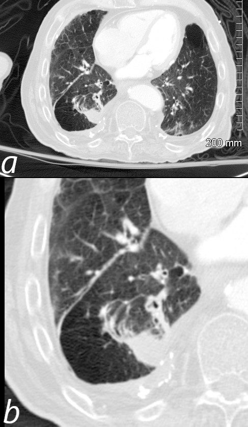
72-year-old male with a history of asbestos exposure presents with a cough. Axial CTscan using lung windows, shows a pleural based nodule with a subtending curvilinear comet tail formed by a trailing bronchovascular bundle. There is associated pleural thickening and pleural based calcification reminiscent of asbestos related disease.
Ashley Davidoff MD TheCommonVein.net 118433c 240Lu
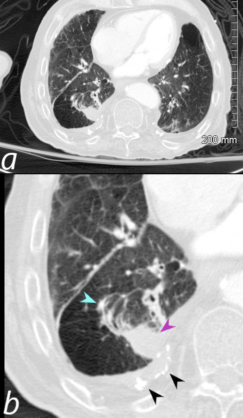
72-year-old male with a history of asbestos exposure presents with a cough. Axial CTscan using lung windows, shows a pleural based nodule (purple arrowhead b) with a subtending curvilinear comet tail formed by a trailing bronchovascular bundle (teal blue b). There is associated pleural thickening and pleural based calcification (black arrowheads ,b), reminiscent of asbestos related disease.
Ashley Davidoff MD TheCommonVein.net 118433cL 240Lu
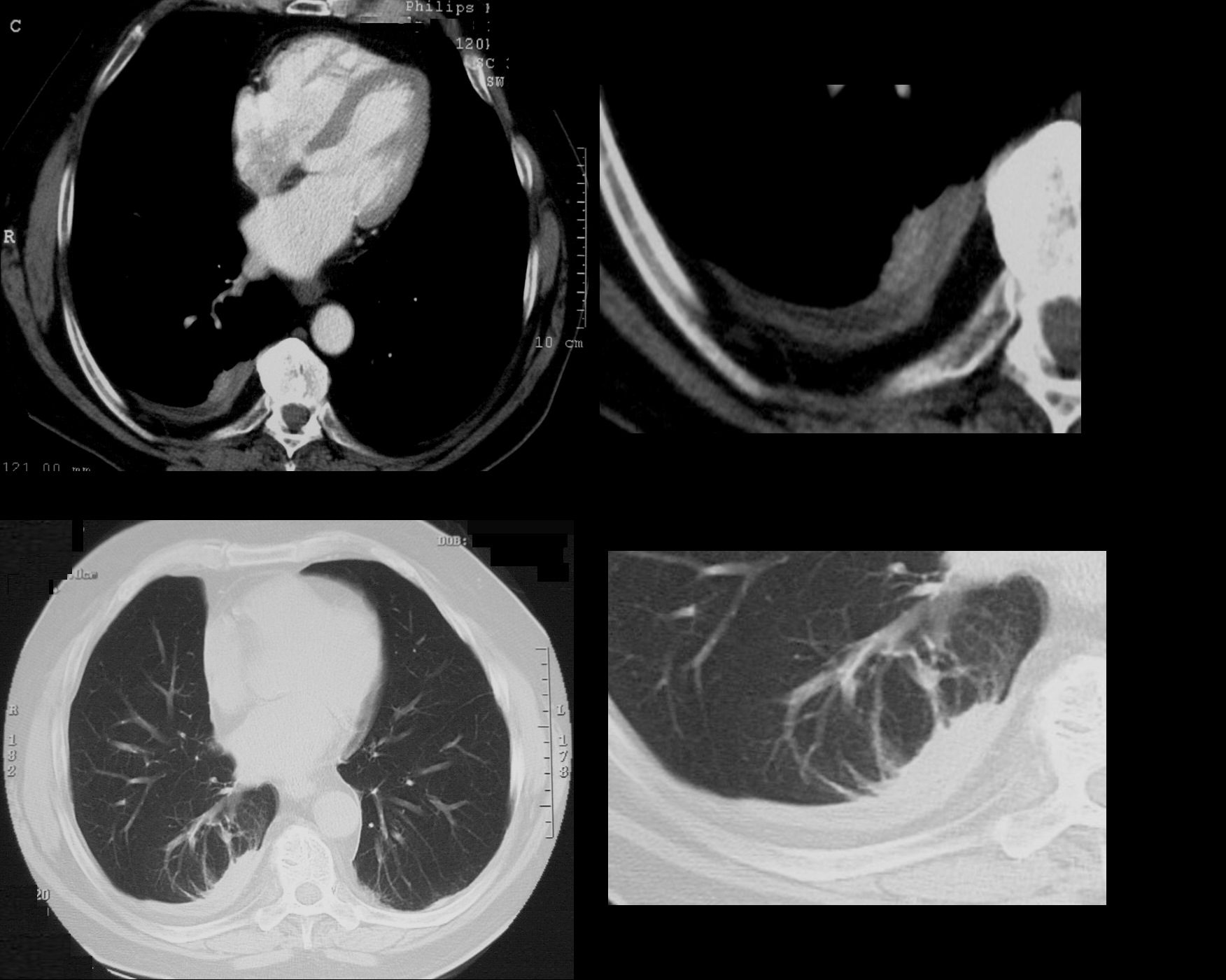
74 year old male with a cough.
CT shows split pleura sign with thickened visceral and parietal pleura with regions of early spiraling of an atelectatic process in the right lower lobe consistent with early rounded atelectasis
Ashley Davidoff MD TheCommonVein.net
31563c
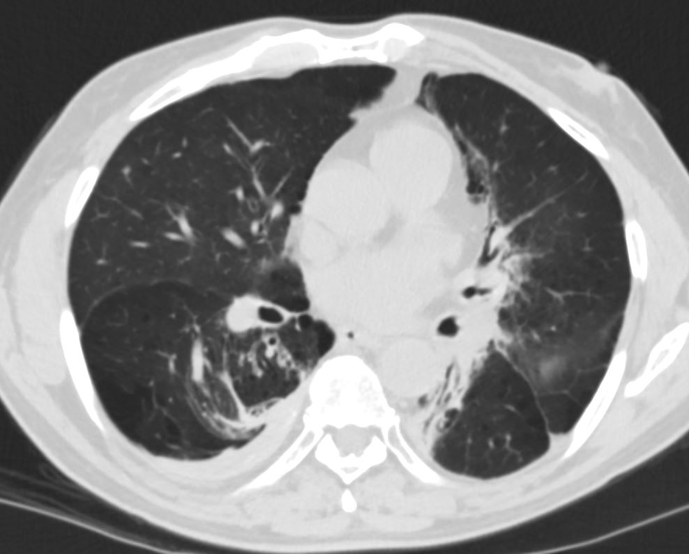
CT shows focal region of pleural thickening with calcification
Also note hyperlucent right lower lobe
Ashley Davidoff MD TheCommonVein.net
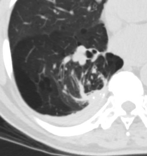
CT shows focal region of pleural thickening with calcification
Also note hyperlucent right lower lobe
Ashley Davidoff MD TheCommonVein.net
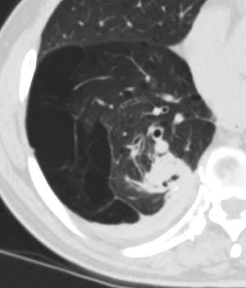
CT shows focal region of pleural thickening with calcification
Also note hyperlucent right lower lobe
Ashley Davidoff MD TheCommonVein.net
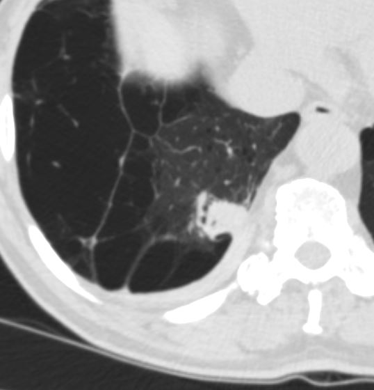
CT shows focal region of pleural thickening with calcification
Also note hyperlucent right lower lobe
Ashley Davidoff MD TheCommonVein.net
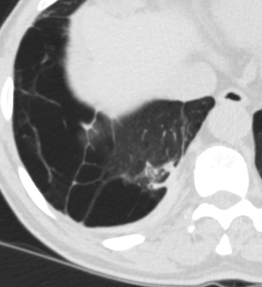
CT shows focal region of pleural thickening with calcification
Also note hyperlucent right lower lobe
Ashley Davidoff MD TheCommonVein.net
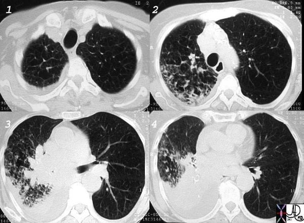
Courtesy Ashley Davidoff MD.
TheCommonVein.net
32426_03cl keywords
lung bronchus lyphatic infiltrate mass obstruction atelectasis thickening interlobular septa neoplasm malignant primary malignancy small cell carcinoma imaging radiology CTscan
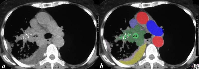
This CT scan is from a middle aged female with poorly differentiated small cell carcinoma, with extensive mediastinal and hilar involvement (dark green in b) extending into and almost occluding the right main stem bronchus light green with black air in the centre) and occluding the smaller airways (light green surrounded by white bronchial cartilage). A complex right effusion (yellow) and atelectasis is seen (light pink). The relationship to the SVC (light blue ) is noted without mass effect at this time, with aorta in red and left pulmonary artery (royal blue). Parenchymal disease is suspected based on the increased soft tissue noted in the mediastinal windows in the right lower lobe.
Courtesy Ashley Davidoff MD
TheCommonVein.net
32429bc03.8s
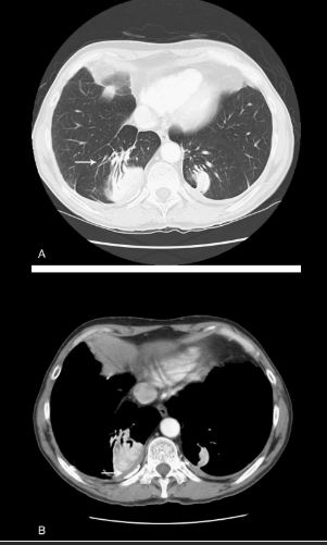
Source
Signs in Thoracic Imaging
Journal of Thoracic Imaging 21(1):76-90, March 2006.
