Introduction
In this section we deal with the back and forth battle of the malignant process with the bodies defence mechanism with the structural and functional result that follows.
Initiating Event
The initiating event is presumably the effect of a carcinogen on the genetic makeup of a cell or group of cells that survive the insult and continue to grow and thrive at the expense of the host tissue and organ. The oncogenes become dominant and there tends to be a reduced effect of recessive tumor-suppressor genes. 10 to 20 genetic mutations of the aberrant cells may occur by the time the tumor is clinically apparent. The lack of contact inhibition allows the local spread of the disease.
Growth
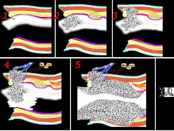
An area in the epithelium of in situ cytological atypia presumably forms resulting a small space occupying lesion on the mucosa.. With progression, the conglomerate of malignant cells elevates the mucosa which may eventually ulcerate.

This is a collage showing the evolution of a malignant cell (1) into a nodule restricted to the epithelium (2) which with time penetrates the basement membrane and progressively extends into the submucosa and muscularis (3). Subsequent extension into local lymph nodes and blood vessels occurs (4) as well as growth into the lumen. As it grows circumferentially, narrowing and eventually obstruction of the lumen complicates the process (5) Space in tubular systems is limited and malignant growth has no respect for this space nor for boundaries. By definition malignant disease is space occupying.
Ashley Davidoff
TheCommonVein.net
32336c
The mass grows along lines of least resistance but can overcome natural planes. Therefore it may grow into the lumen, or through subsequent layers of basement membrane, muscularis and adventitia or through the lung parenchyma.
The invasion of lymphatics, venous structures and pleura is inevitable with time.
The frequency of nodal involvement varies slightly with the histological pattern but averages greater than 50%. The adrenals are involved almost 50% while the liver (30 to 50%), brain (20%), and bone (20%) are commonly involved.
Response
The body is able to respond to this aggressor, but it has to recognize the cell or group of cells as foreign. Most the time it is able. There are presumably many instances when such activity is identified as clandestine and is treated effectively by the immune system… However there are instances where the malignant cells get away with murder, perhaps under the continued stimulus of a carcinogen in a background of genetic predisposition, and find the loophole in the immune system that allows the malignant cells to survive.
Result
The result is space occupying disease and depending on the location and degree of structural change symptomatology clinical and radiological findings will differ. Partial obstruction may cause focal emphysema while a total obstruction may lead to segmental or subsegmental atelectasis. Secondary infection may result in bronchitic, and or pneumonic change and subsequently even abscess formation
Venous structures can also be compressed the most dramatic of which is the involvement of the SVC and resulting SVC syndrome with total obstruction of the SVC, and head and brain swelling.
