
Rossi, SE et al Tree-in-Bud Pattern at Thin-Section CT of the Lungs: Radiologic-Pathologic Overview RadioGraphics Vol. 25, No. 3 2005
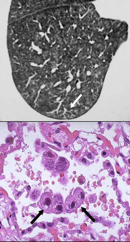
Cytomegalovirus pneumonia. CT scans in a 31-year-old woman with a history of type 1 diabetes mellitus complicated by end-stage renal disease. The patient had previously undergone kidney and pancreas transplant and presented with 2 days of right lower quadrant abdominal pain associated with nausea and vomiting. Upon further work-up, patient was found to have cytomegalovirus viremia. (above) Axial and (be;low) coronal CT images demonstrate diffuse randomly distributed small pulmonary nodules (arrows), many of which are ill-defined and distributed in the secondary pulmonary lobules and perilymphatic regions.
Parekh, M et al Review of the Chest CT Differential Diagnosis of Ground-Glass Opacities in the COVID Era Radiology Vol. 297, No. 3 July 2020