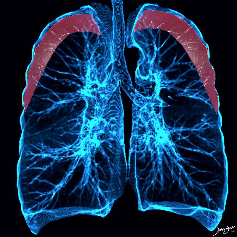
Chronic eosinophilia is characterised by alveolar filling with eosinophils and inflammatory exudates(a) and interalveolar interstitial thickening, (overlaid in red in b). The infiltrates are classically peripherally positioned, usually upper lobes, more commonly bilateral but can be unilateral, and manifest as consolidation and or ground glass opacities. The CT shows bilateral peripheral consolidations in the upper lobes
Ashley Davidoff MD The CommonVein.net lungs-0765
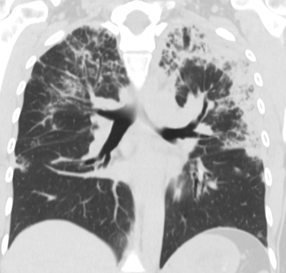
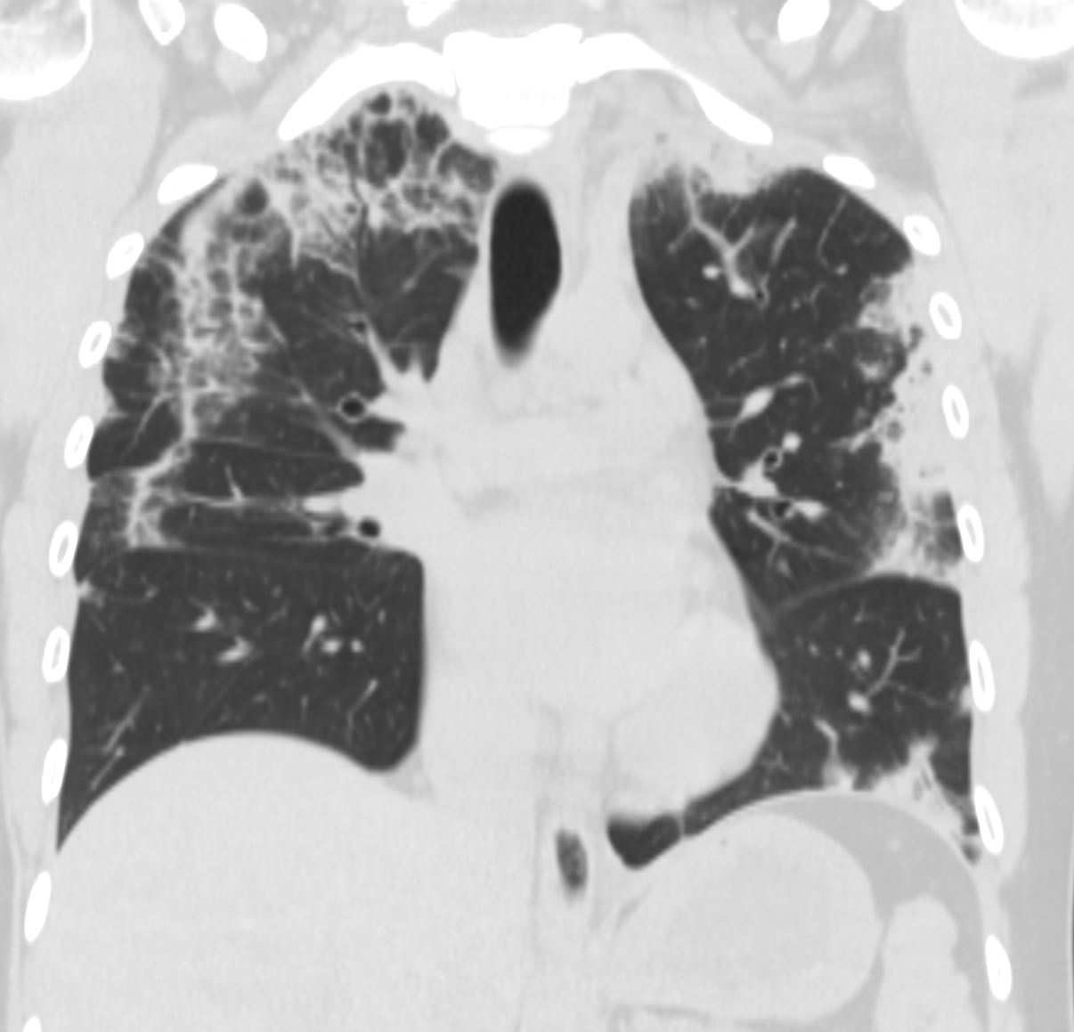
CT scan in the coronal performed 6 months ago at the time of clinical presentation shows upper lobe predominant peripheral infiltrates with small left lower lobe peripheral infiltrate Subsequent diagnosis by BAL of chronic eosinophilic pneumonia (CEP)
Ashley Davidoff TheCommonVein.net
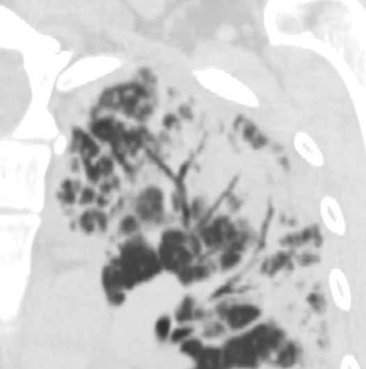
CT scan in the coronal performed 6 months ago at the time of clinical presentation shows upper lobe predominant peripheral infiltrates more prominent in the left upper lobe. Subsequent diagnosis by BAL of chronic eosinophilic pneumonia (CEP) was made
Ashley Davidoff TheCommonVein.net
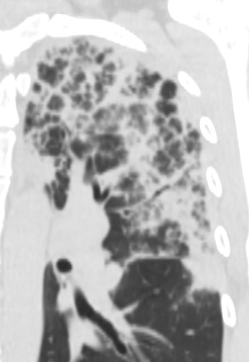
CT scan in the coronal performed 6 months ago at the time of clinical presentation shows upper lobe predominant peripheral infiltrates more prominent in the left upper lobe. Subsequent diagnosis by BAL of chronic eosinophilic pneumonia (CEP) was made
Ashley Davidoff TheCommonVein.net
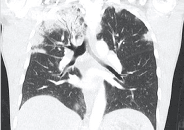
Crowe M et al Therapeutics and Clinical Risk Management Volume 15:397-403 March 2019
https://www.researchgate.net/publication/331691288_Chronic_eosinophilic_pneumonia_clinical_perspectives
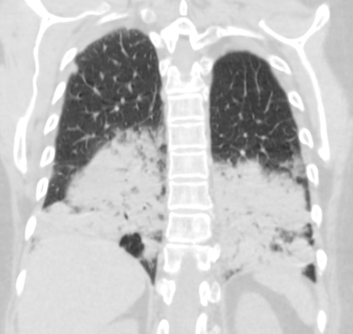
Ashley Davidoff MD TheCommonVein.net
eosinophilic-pneumonia-011
