Atelectasis and Bronchiectasis with Volume Loss

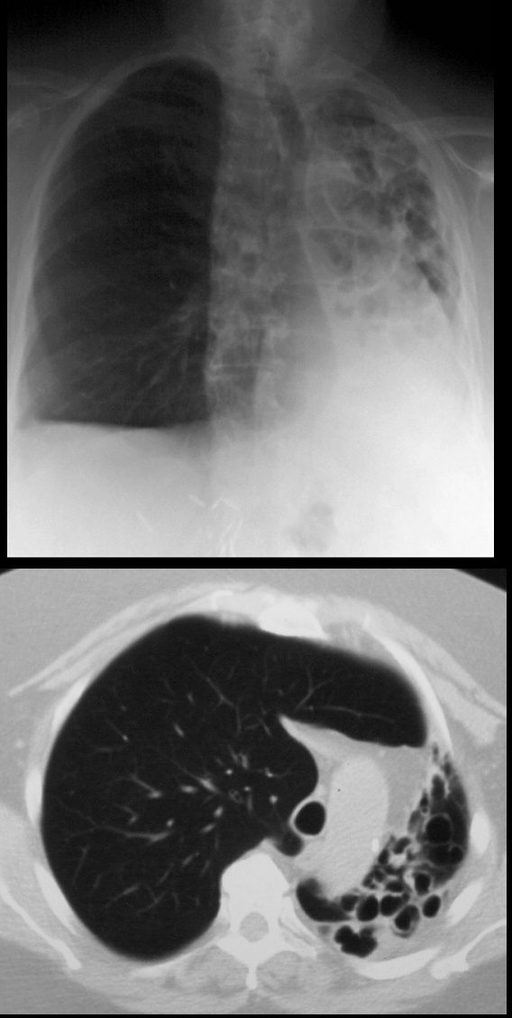
68year old male presents with chronic cough. Chest Xray reveals evidence of tram tracking and bronchiectasis in the left lung with volume loss of the entire right lung with secondary hyperinflation of the right lung, and mediastinal shift. CT scan shows severe volume loss and hyperinflation of the right lung.
Ashley Davidoff MD TheCommonVein.net
Combination of Cicatrization and Obstruction

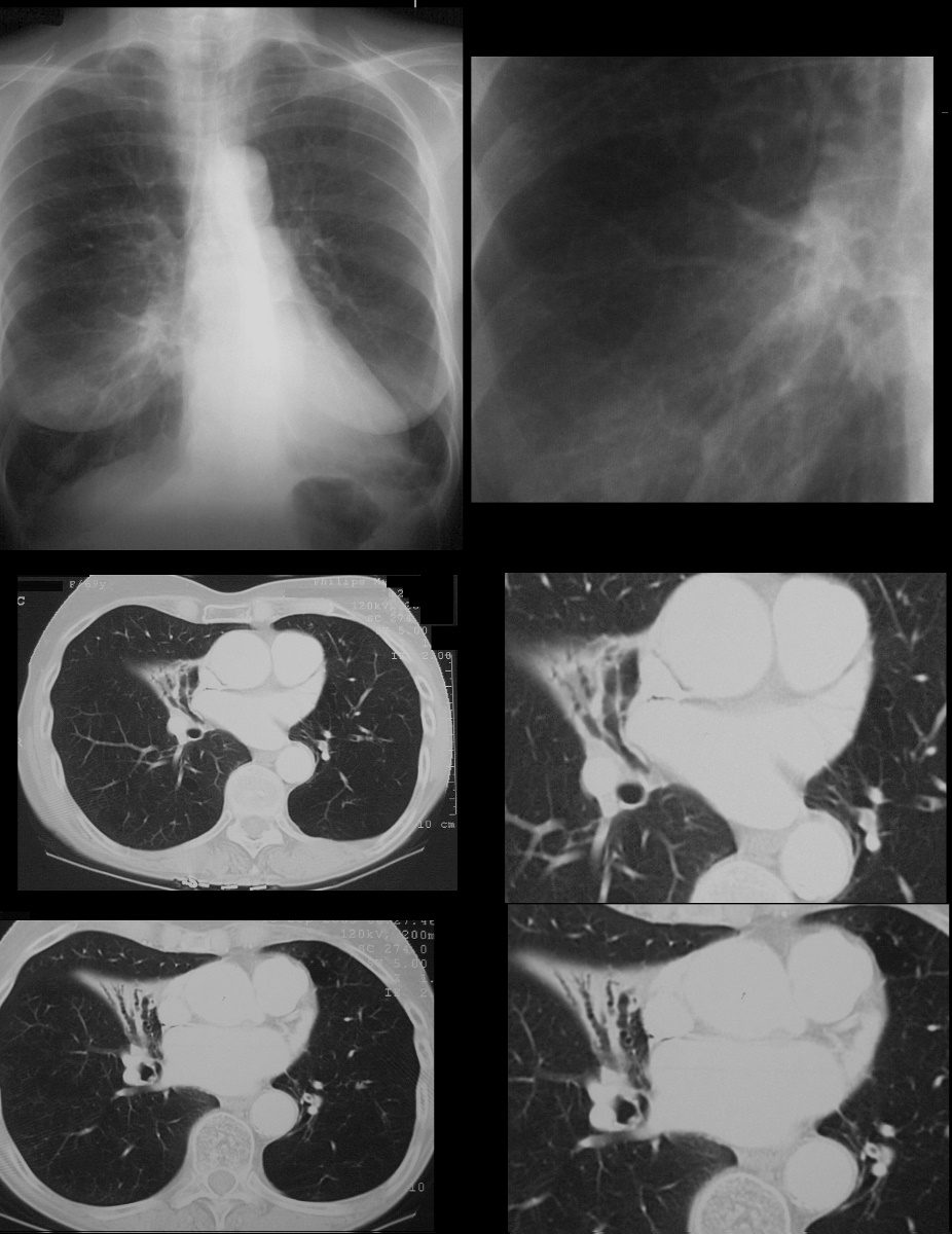
69year old female with varicose bronchiectasis atelectasis and mucus plugging. The CXR suggests a hilar process with an indistinct margin of the right heart border possibly relating to a middle lobe process. CT scans confirm atelectasis of the middle lobe, varicose bronchiectasis, and the presence of mucoid impaction best revealed in the left lower bronchi
Ashley Davidoff MD TheCommonVein.net
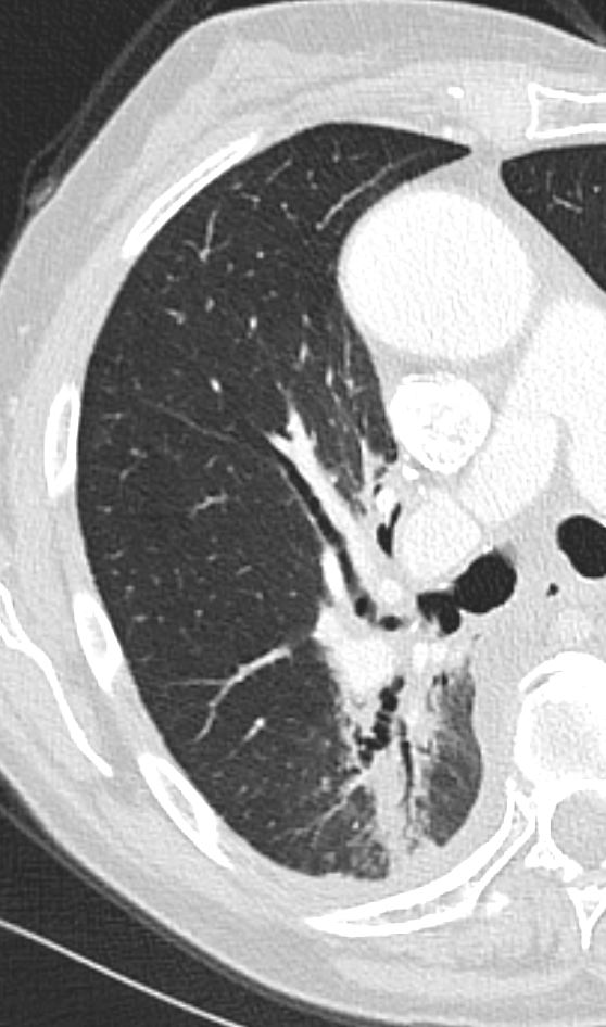
Radiation and Atelectasis

Ashley Davidoff MD TheCommonVein.net post XRT 001
Rounded Atelectasis

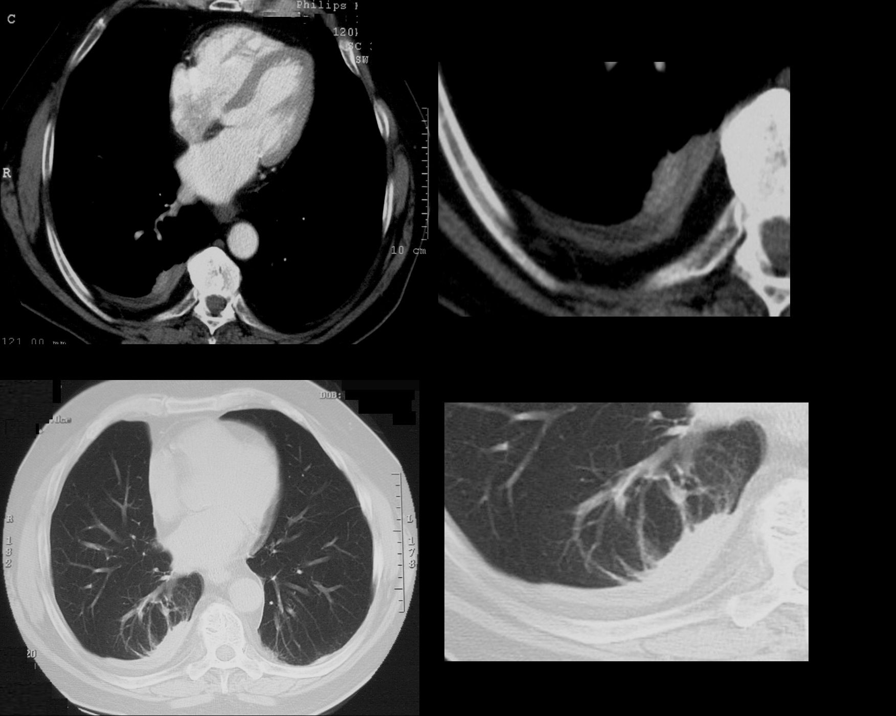
74 year old male with a cough.
CT shows split pleura sign with thickened visceral and parietal pleura with regions of early spiraling of an atelectatic process in the right lower lobe consistent with early rounded atelectasis
Ashley Davidoff MD TheCommonVein.net
31563c
