Parts
Size
Variably Sized
Chronic Pulmonary Langerhans Cell Histiocytosis (PLCH)
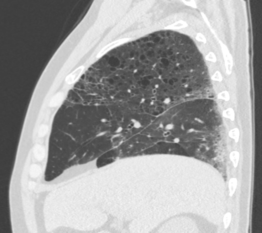
54 year old smoker presents with a chronic cough. CT in the sagittal plane shows multiple thin walled cysts of varying size with upper lung predominance.
Findings are consistent with chronic pulmonary Langerhans cell histiocytosis (PLCH)
Ashley Davidoff MD TheCommonvein.net 279Lu 136475
Shape
In a Wedge Shape Accumulation
Septic Emboli
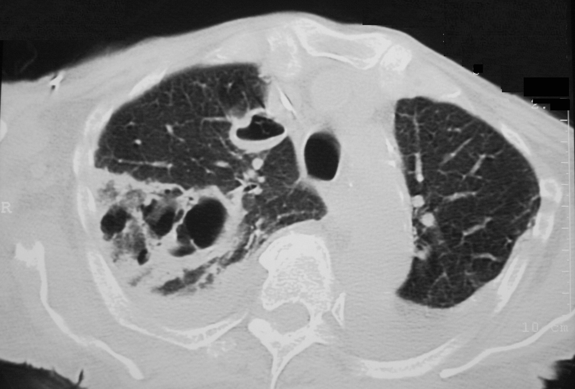
Axial CT reveals a large wedge shaped thick walled complex multicystic lesion associated with a feeding bronchovascular bundle (feeding vessel sign) in the right apex consistent with a cavitating infarction (cavitating Hampton’s hump). In addition there is a second smaller unilocular thick-walled cyst with a small air fluid level suggesting infection. There are pleural effusions. Echo showed tricuspid valve vegetations. Diagnosis is consistent with cavitating septic emboli
Ashley Davidoff TheCommonVein.net 33012 307Lu
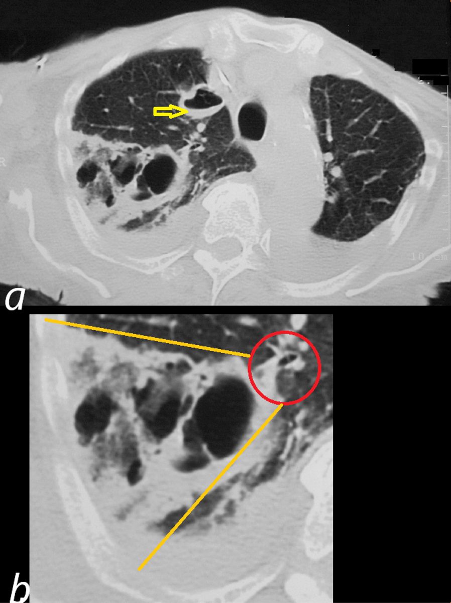
Axial CT reveals a large wedge shaped thick walled complex multicystic lesion ( bordered by orange lines in b) associated with a feeding bronchovascular bundle (red ring b -feeding vessel sign) in the right apex consistent with a cavitating infarction (cavitating Hampton’s hump). In addition there is a second smaller unilocular thick-walled cyst with a small air fluid level (yellow arrow, a) suggesting additional purulence in this clinical context. There are bilateral pleural effusions. Echo showed tricuspid valve vegetations. Diagnosis is consistent with cavitating septic emboli with pulmonary infarction.
Ashley Davidoff TheCommonVein.net 33012 307Lu
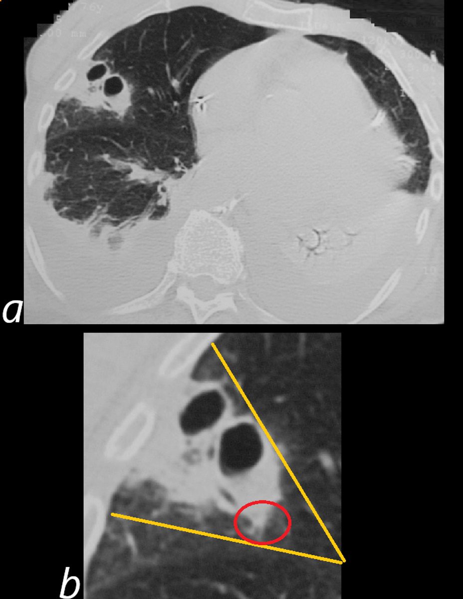

Axial CT reveals a thick walled wedge shaped bilocular cystic lesion in a region of subsegmental consolidation associated with a feeding bronchovascular bundle (feeding vessel sign). There are large bilateral pleural effusions associated with compressive atelectasis. Echo showed tricuspid valve vegetations. Diagnosis is consistent with cavitating septic emboli
Ashley Davidoff TheCommonVein.net 33015cL 307Lu
Position
Upper Lung Zones
Chronic Pulmonary Langerhans Cell Histiocytosis (PLCH)
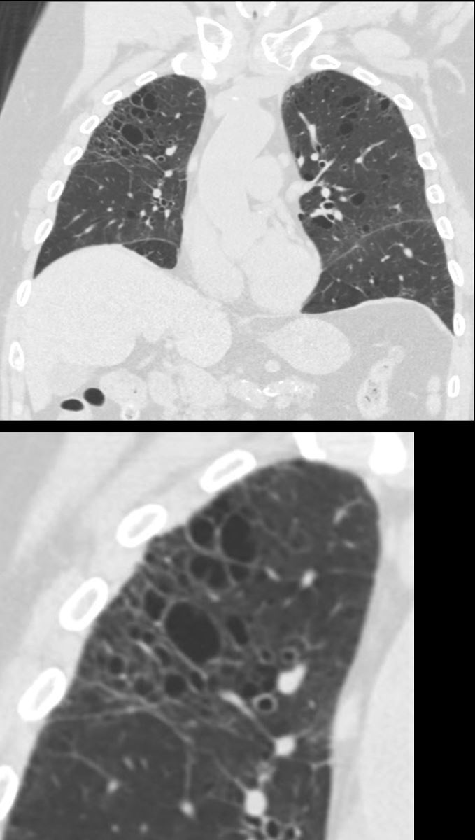

54 year old smoker presents with a chronic cough. CT in the coronal plane shows multiple thin walled cysts of varying size with upper lung predominance
Findings are consistent with chronic pulmonary Langerhans cell histiocytosis (PLCH)
Ashley Davidoff MD TheCommonvein.net 279Lu 136470c
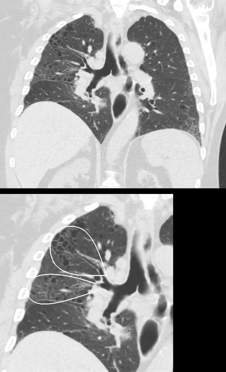

54 year old smoker presents with a chronic cough. CT in the coronal plane shows multiple thin walled cysts of varying size with upper lung predominance. The cysts appear to be aligned with a normal sized subsegmental airway with minimal wall thickening (ringed in white lower image)
Findings are consistent with chronic pulmonary Langerhans cell histiocytosis (PLCH)
Ashley Davidoff MD TheCommonvein.net 279Lu 136472cL

54 year old smoker presents with a chronic cough. CT in the sagittal plane shows multiple thin walled cysts of varying size with upper lung predominance.
Findings are consistent with chronic pulmonary Langerhans cell histiocytosis (PLCH)
Ashley Davidoff MD TheCommonvein.net 279Lu 136475
Character
Thick Walled Cavitating Septic Emboli


Axial CT reveals a large wedge shaped thick walled complex multicystic lesion ( bordered by orange lines in b) associated with a feeding bronchovascular bundle (red ring b -feeding vessel sign) in the right apex consistent with a cavitating infarction (cavitating Hampton’s hump). In addition there is a second smaller unilocular thick-walled cyst with a small air fluid level (yellow arrow, a) suggesting additional purulence in this clinical context. There are bilateral pleural effusions. Echo showed tricuspid valve vegetations. Diagnosis is consistent with cavitating septic emboli with pulmonary infarction.
Ashley Davidoff TheCommonVein.net 33012 307Lu
Time Associated Findings
Infection
Inflammation
Smoking Related
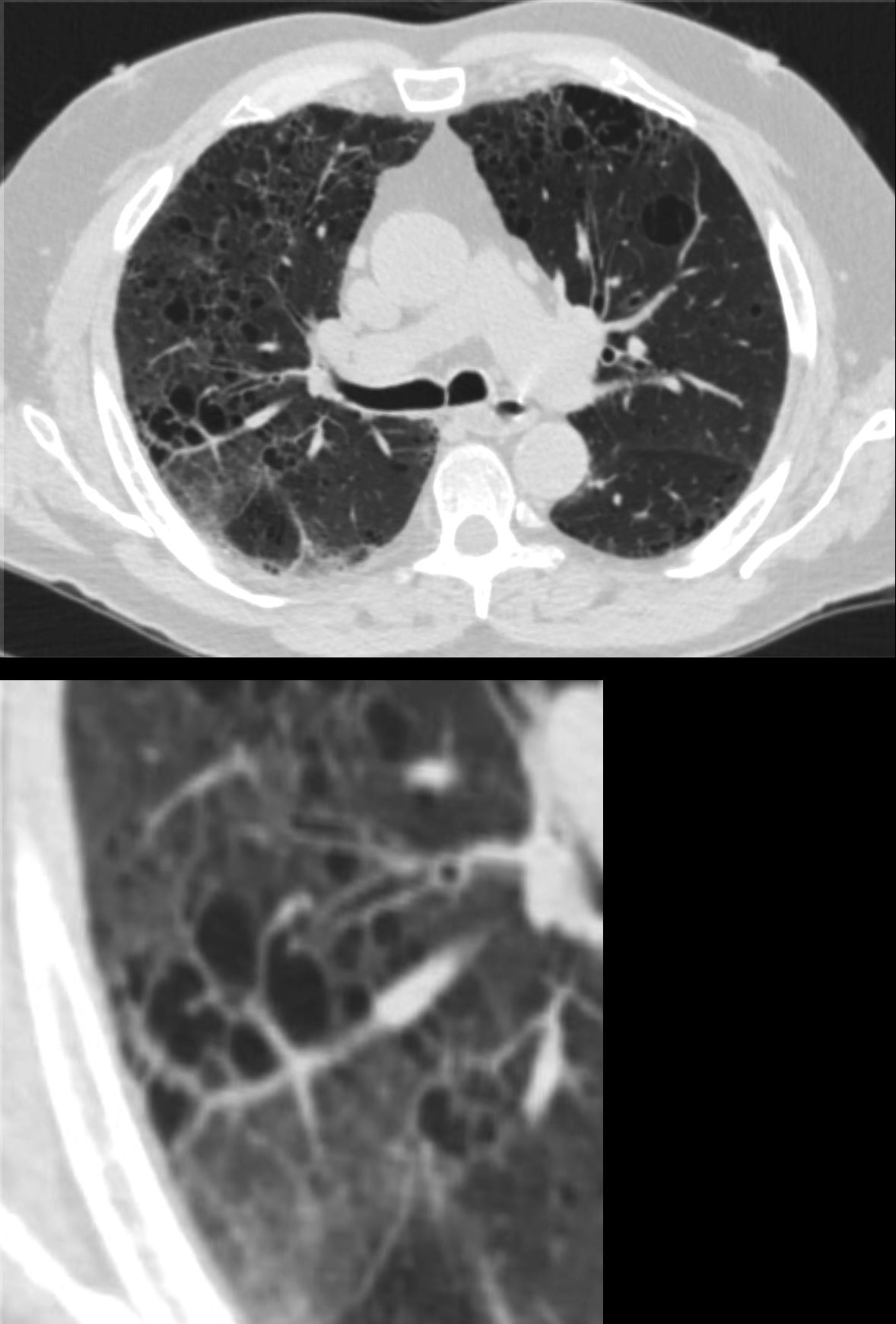

54 year old smoker presents with a chronic cough. CT in the axial plane plane shows multiple thin walled cysts of varying size with upper lung predominance. The cysts appear to be aligned with a normal sized subsegmental airway with minimal wall thickening (lower image)
Findings are consistent with chronic pulmonary Langerhans cell histiocytosis (PLCH)
Ashley Davidoff MD TheCommonvein.net 279Lu 136478c
Malignancy
CT Chest – Cavitating Metastatic Trophoblastic Tumor
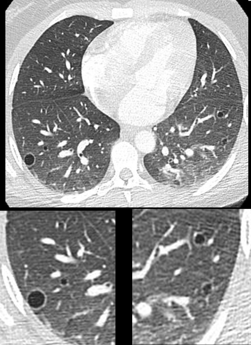

28year old female presents with vaginal bleeding for 3 days s/p ablation of a vascular molar pregnancy. CT of the chest shows multiple cystic lesions in the lungs bilaterally with slightly thickened walls. Wedge biopsy confirmed a diagnosis of placental site trophoblastic tumor
Ashley Davidoff MD TheCommonVein.net
280Lu 136464.
Thick Walled Cavitating Squamous Cell Carcinoma
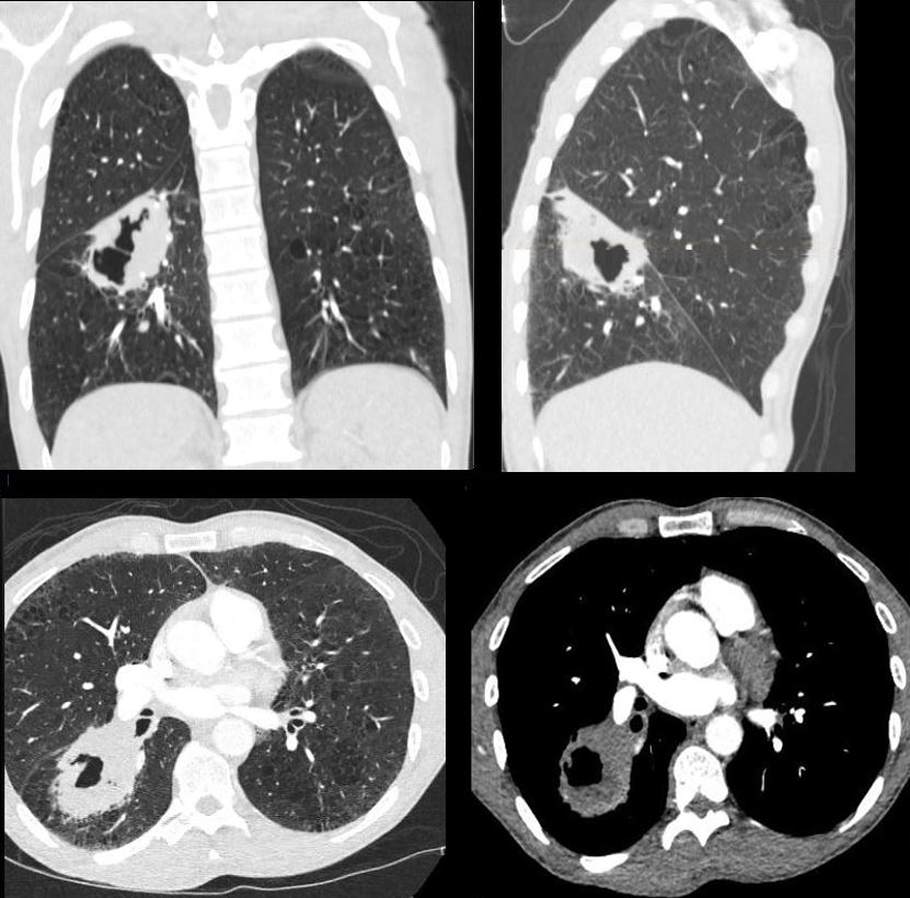

50 year old male with cough and weight loss
Coronal and sagittal CT reconstructions show a cavitating mass in the superior segment of the right lower lobe (upper images) correlated with axial images (lower panel)
Ashley Davidoff MD TheCommonVein.net 176Lu 136737
Mechanical
Atelectasis
Trauma
Metabolic
Circulatory-
Hemorrhage
Immune Infiltrative Idiopathic Iatrogenic
Inherited
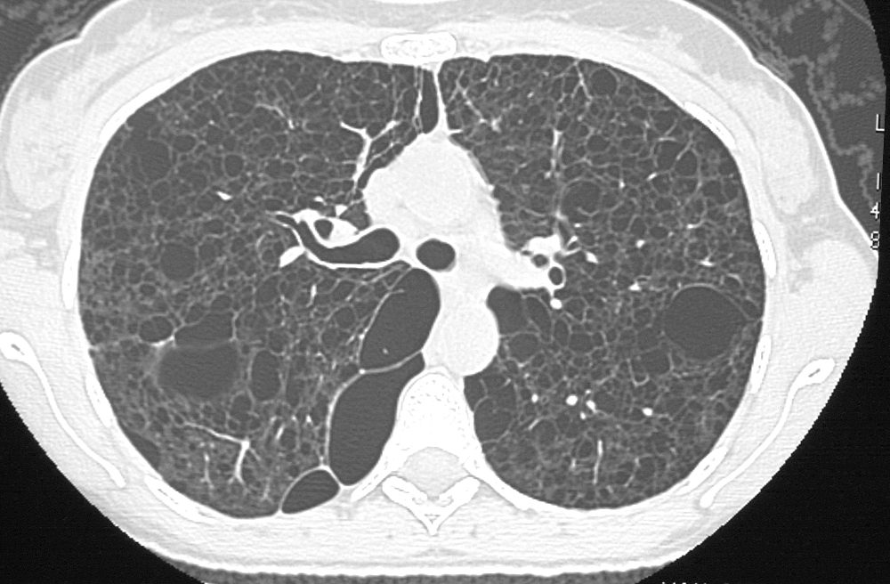

38-year-old patient with progressive dyspnea and cough
CXR (scout for CT) shows hyperinflated lungs with increased lung volumes with bilateral and extensive thin-walled cysts surrounded by very little normal lung parenchyma. The cysts are round and thin-walled except for air filled large irregular pocket in the right apex (image 27628/29) . Some of the cysts do not have walls at all and others have an irregular configuration.
In the abdomen multiple low density lymphangioleiomyomas are present that are due to lymphatic obstruction.
Ashley Davidoff MD TheCommonvein.net
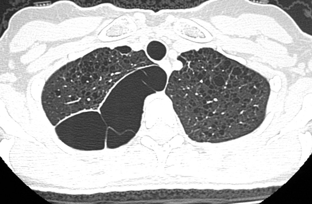

38-year-old patient with progressive dyspnea and cough
CXR (scout for CT) shows hyperinflated lungs with increased lung volumes with bilateral and extensive thin-walled cysts surrounded by very little normal lung parenchyma. The cysts are round and thin-walled except for air filled large irregular pocket in the right apex (image 27628/29) . Some of the cysts do not have walls at all and others have an irregular configuration.
In the abdomen multiple low density lymphangioleiomyomas are present that are due to lymphatic obstruction.
Ashley Davidoff MD
