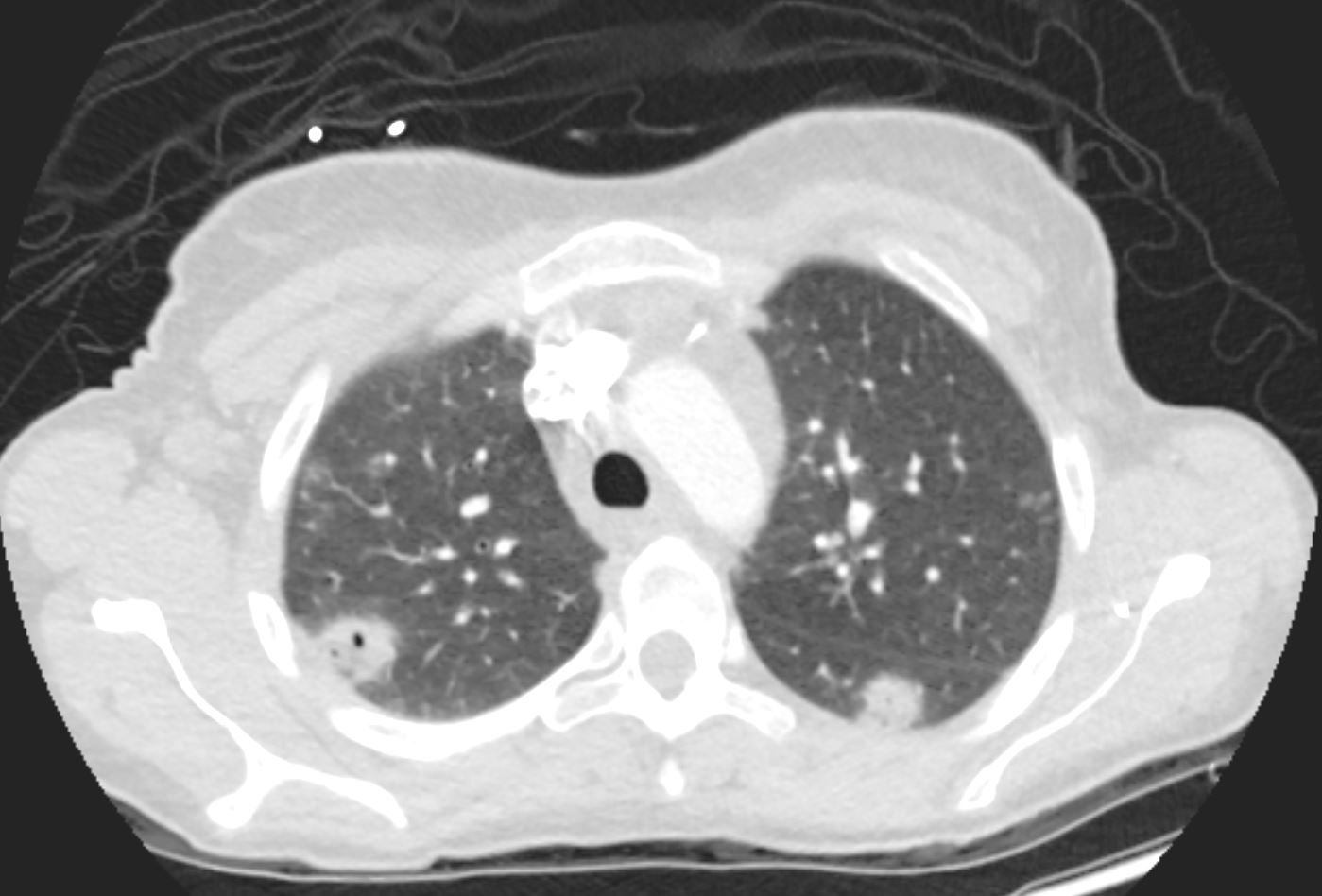
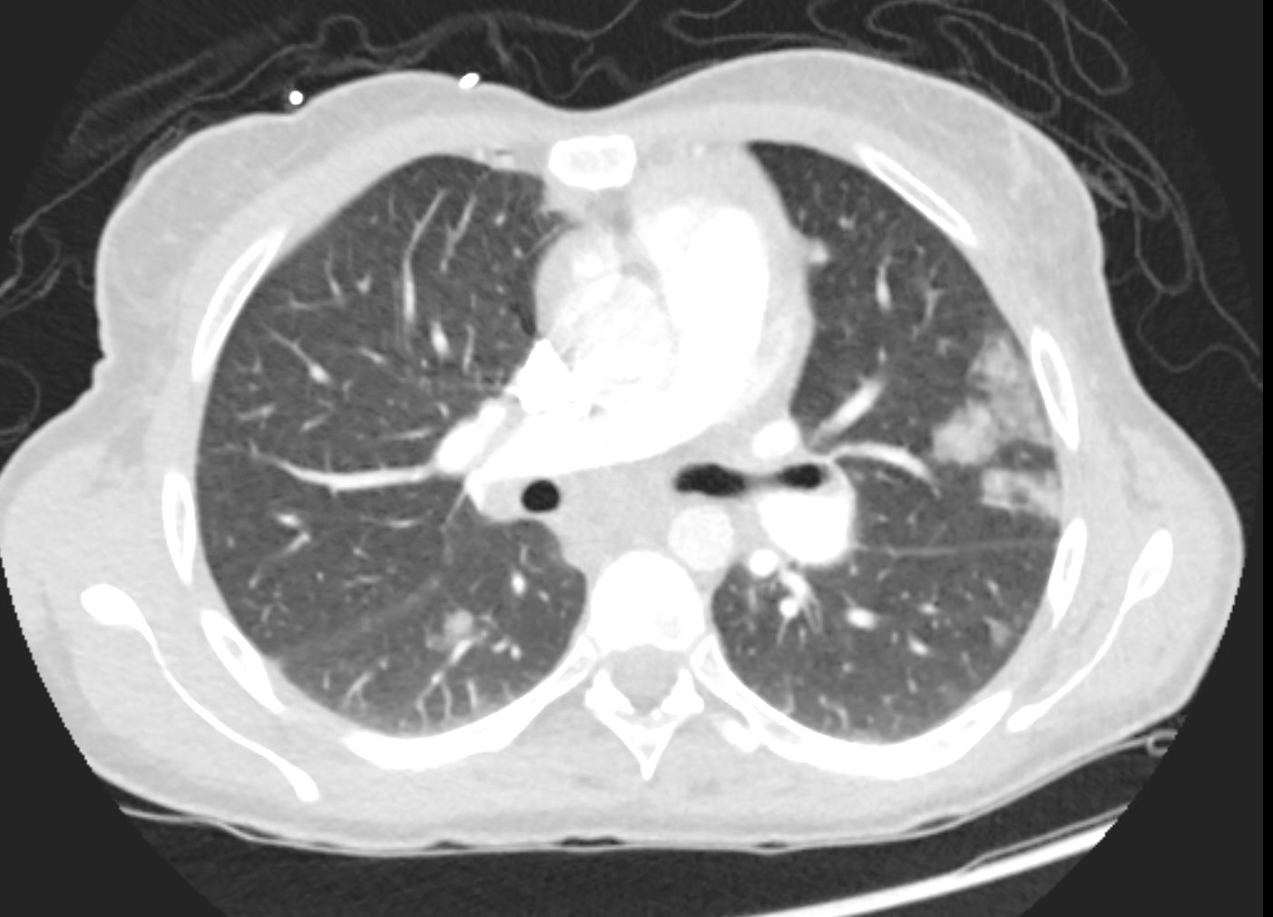
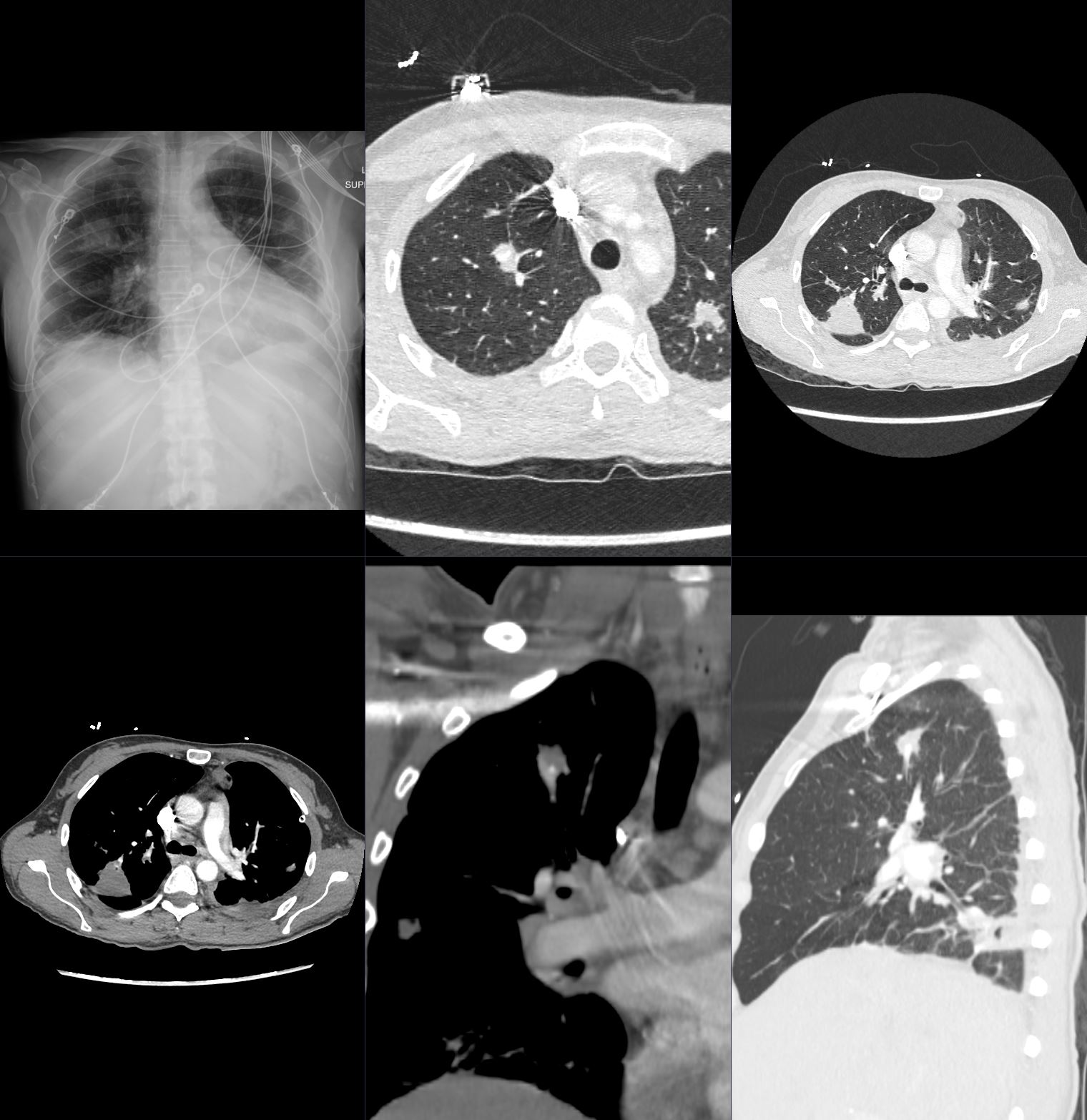
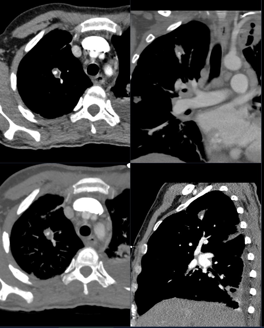
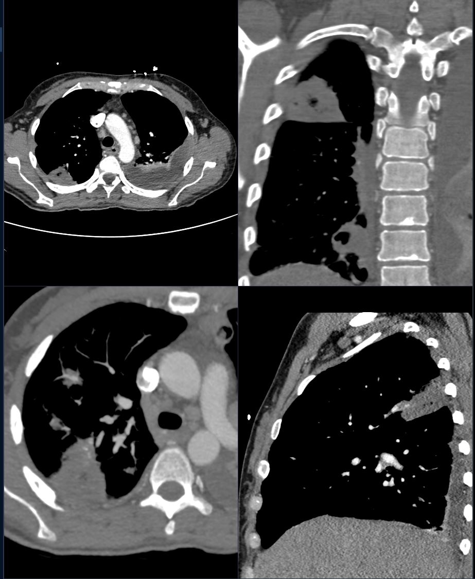
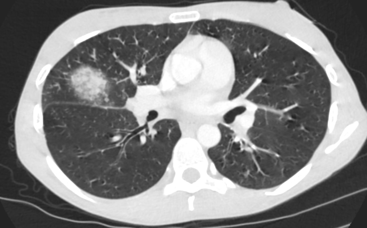
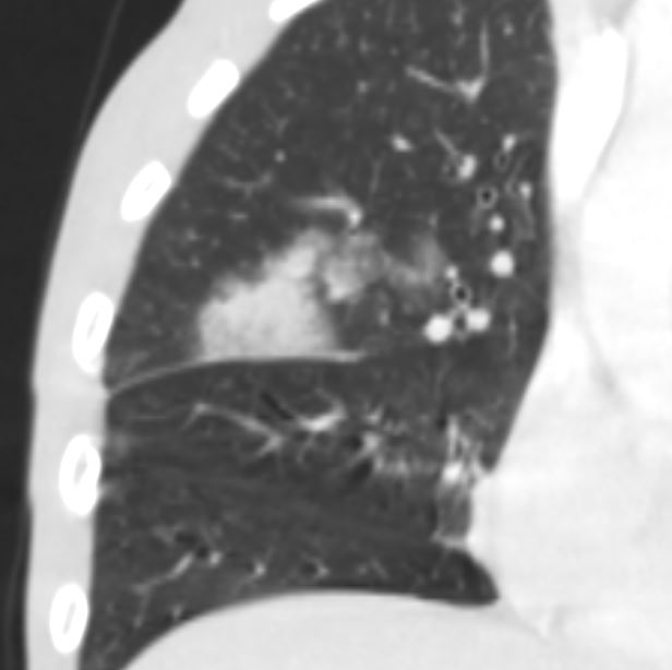
Faces of Endocarditis Cavitating Nodules in Bacterial Endocarditis 24 year old female with bacterial endocarditis with multiple pulmonary emboli, some cavitating. The CT shows multiple foci of consolidation with a foci of consolidations in the posterior segments of the upper lobes bilaterally The focal consolidation in the right upper lobe is cavitatingAshley Davidoff MD TheCommonvein.net 24f PE Hampton’s hump 002 24 year old female with bacterial endocarditis with multiple pulmonary emboli, some cavitating. The CT scan shows a wedge shaped conglomerate region of consolidation in the posterior segment of the left upper lobe consistent with a Hamptons hump caused by an embolic infarctionAshley Davidoff MD TheCommonvein.net 24f PE Hampton’s hump 003 24 year old female with bacterial endocarditis with multiple pulmonary emboli, some cavitating. The CT scan shows a wedge shaped conglomerate region of consolidation in the posterior segment of the left upper lobe consistent with a Hamptons hump caused by an embolic infarctionAshley Davidoff MD TheCommonvein.net 24f PE Hampton’s hump 003 Cavitating Nodules and Mycotic Pseudoaneurysms PseudoaneurysmBacterial Endocarditis (BE) and Mycotic Pseudoaneurysm Lung Abscess 39FAshley Davidoff TheCommonVein.net b11422b02 Feeding vessel SignBacterial Endocarditis (BE) and Mycotic Pseudoaneurysm Lung Abscess 39FAshley Davidoff TheCommonVein.net b11422b02 PseudoaneurysmBacterial Endocarditis (BE) and Mycotic Pseudoaneurysm Lung Abscess 39FAshley Davidoff TheCommonVein.net b11422b02 28year old female with Infective Endocarditis CT scan shows an Atoll sign or reversed halo sign characterised a rim of more dense consolidation surrounding a ground glass matrixAshley Davidoff TheCommonVein.net 28year old female with Infective Endocarditis CT scan shows an Atoll sign or reversed halo sign characterised a rim of more dense consolidation surrounding a ground glass matrixAshley Davidoff TheCommonVein.net