Multicentric Pulmonary Hemorrhage and Pneumonia with ANCA-associated Vasculitis
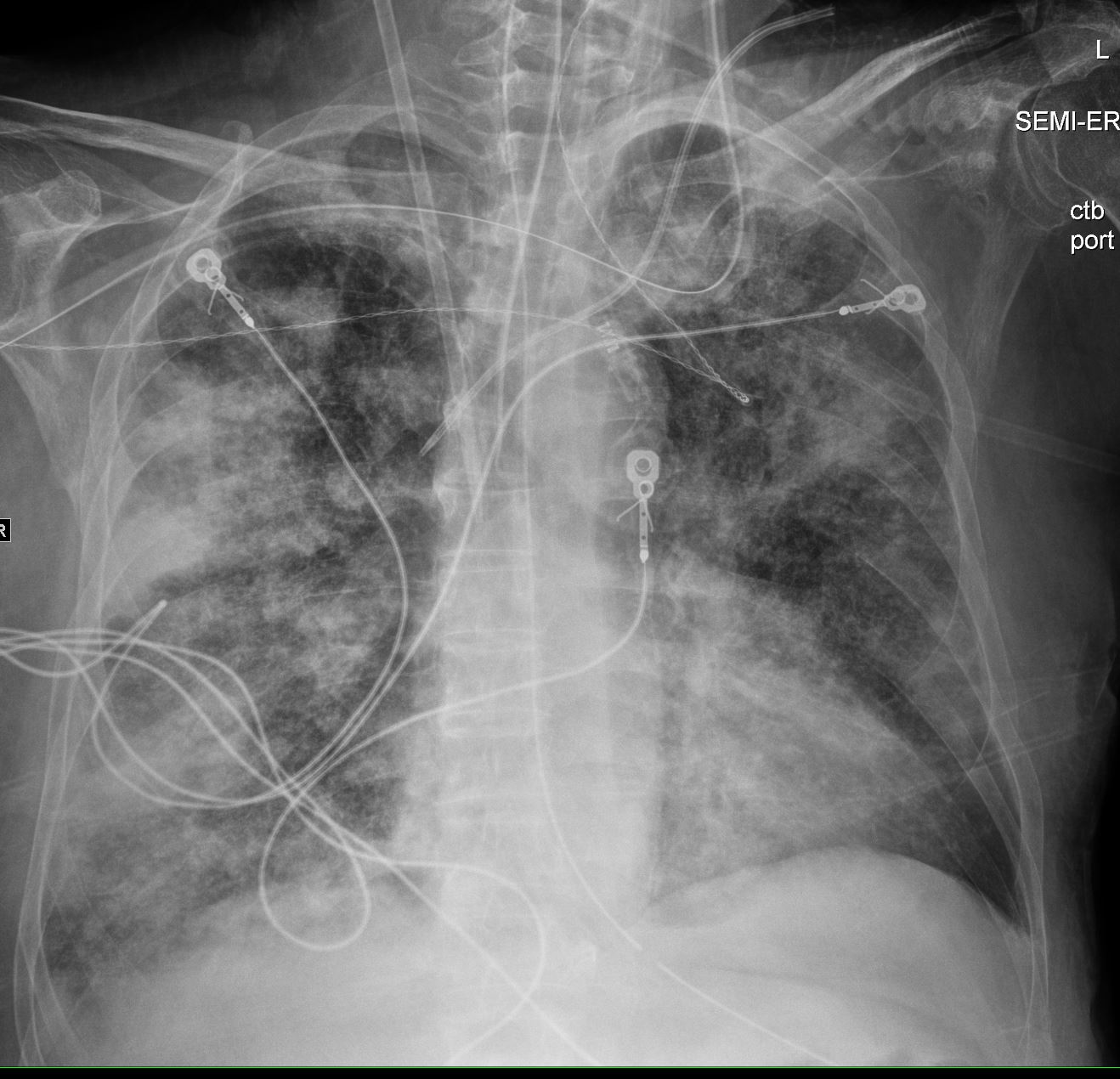
71 yo m w/ a hx of recently diagnosed ANCA-associated vasculitis (diagnosed by kidney biopsy), presents with acute respiratory failure. Bronchoscopic findings were consistent with diffuse alveolar hemorrhage associated with MSSA (Methicillin Sensitive Staph Aureus ) pneumonia/bacteremia
CXR shows multicentric consolidations likely a combination of alveolar hemorrhage and pneumonia
Ashley Davidoff TheCommonVein.net
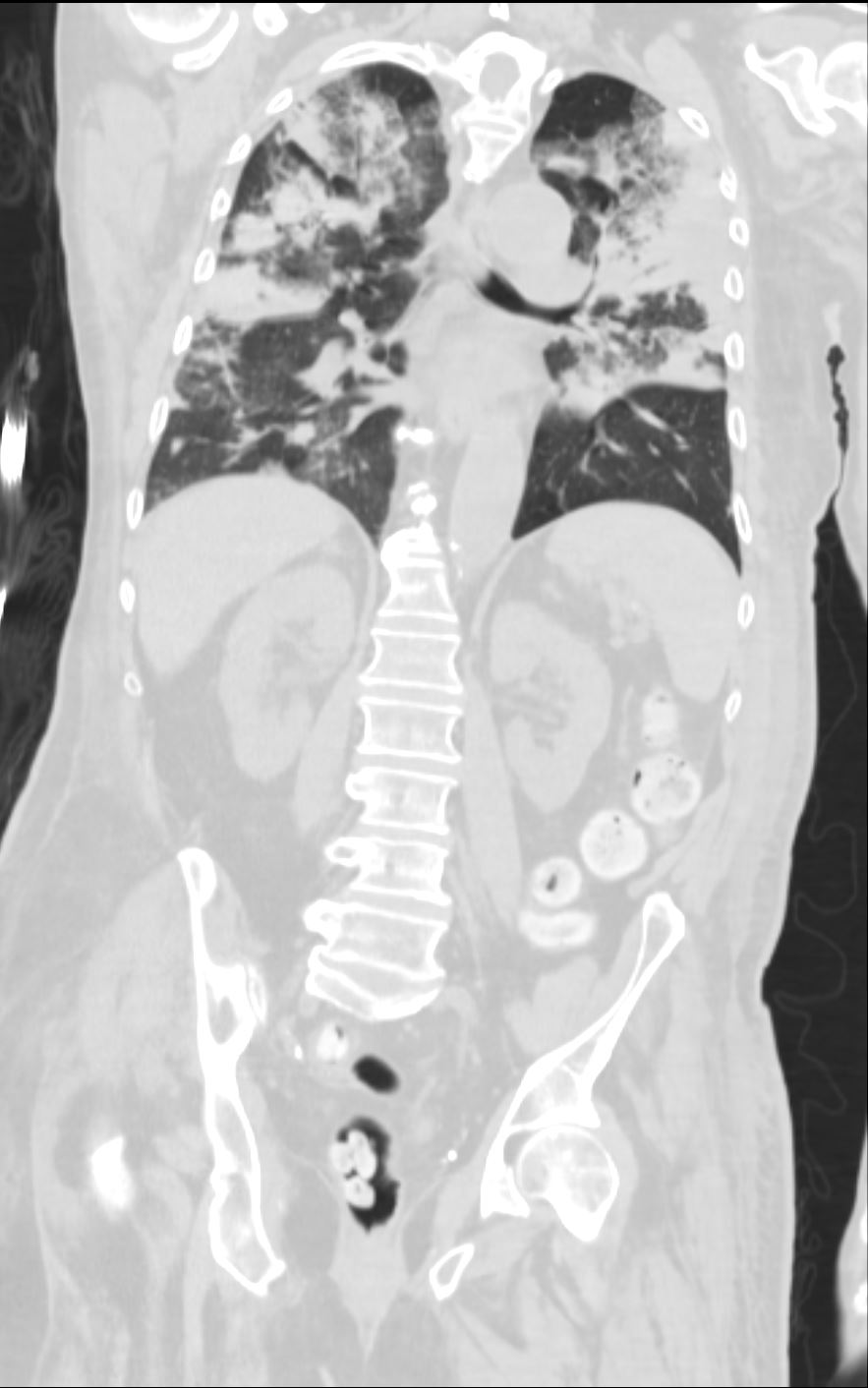
71 yo m w/ a hx of recently diagnosed ANCA-associated vasculitis (diagnosed by kidney biopsy), presents with acute respiratory failure. Bronchoscopic findings were consistent with diffuse alveolar hemorrhage associated with MSSA (Methicillin Sensitive Staph Aureus ) pneumonia/bacteremia. Note Consolidation surrounded by ground glass opacity, the latter likely reflecting a hemorrhagic component
CTscan shows multicentric consolidations likely a combination of alveolar hemorrhage and pneumonia
Ashley Davidoff TheCommonVein.net
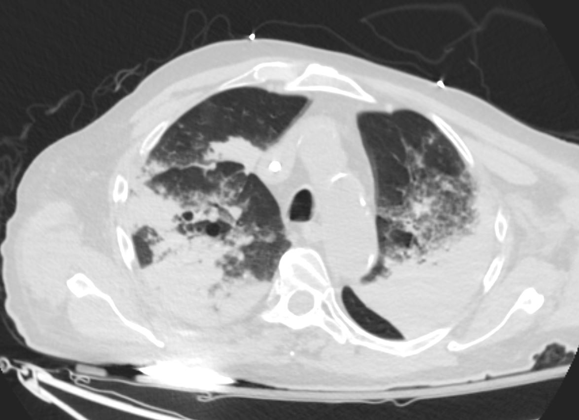
71 yo m w/ a hx of recently diagnosed ANCA-associated vasculitis (diagnosed by kidney biopsy), presents with acute respiratory failure. Bronchoscopic findings were consistent with diffuse alveolar hemorrhage associated with MSSA (Methicillin Sensitive Staph Aureus ) pneumonia/bacteremia. Note Consolidation surrounded by ground glass opacity, the latter likely reflecting a hemorrhagic component
CTscan shows multicentric consolidations likely a combination of alveolar hemorrhage and pneumonia
Ashley Davidoff TheCommonVein.net
Granulomatosis with polyangiitis (GPA),
aka Wegener’s Granulomatosis
Case 1
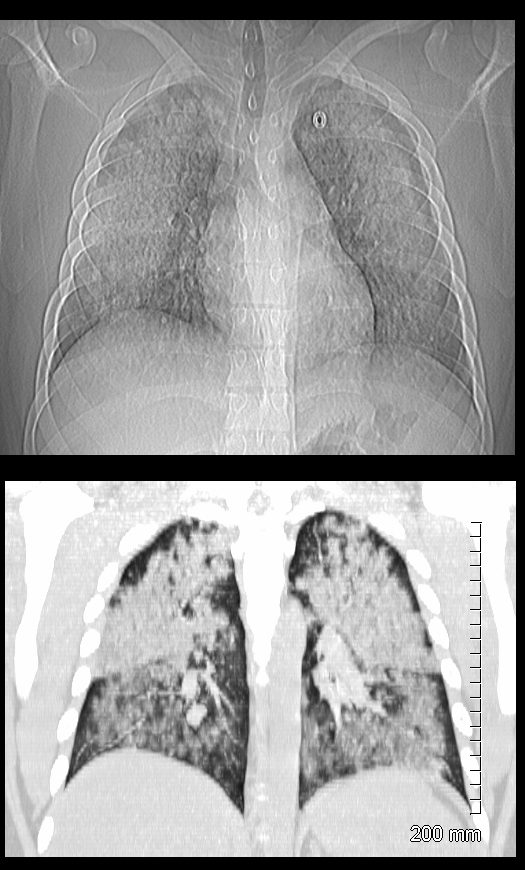
19 year old male previously well with history of hemoptysis, sweating, fevers, myalgias, arthritis over 3 weeks.
CT scan scout (above ) shows diffuse bilateral lobar infiltrates with subpleural sparing
Coronal CT shows bilateral symmetrical lobar nodular consolidations involving the upper and lower lobes. The upper lobes are more consolidative and the lower lobes have an acinar pattern. These finding are consistent with acute pulmonary hemorrhage
Lab shows ANCA positivity, acute renal failure (creatinine 6) and renal biopsy showing crescentic glomerulonephritis. Treated with cyclophosphamide
Ashley Davidoff MD TheCommonVein.net 139195c
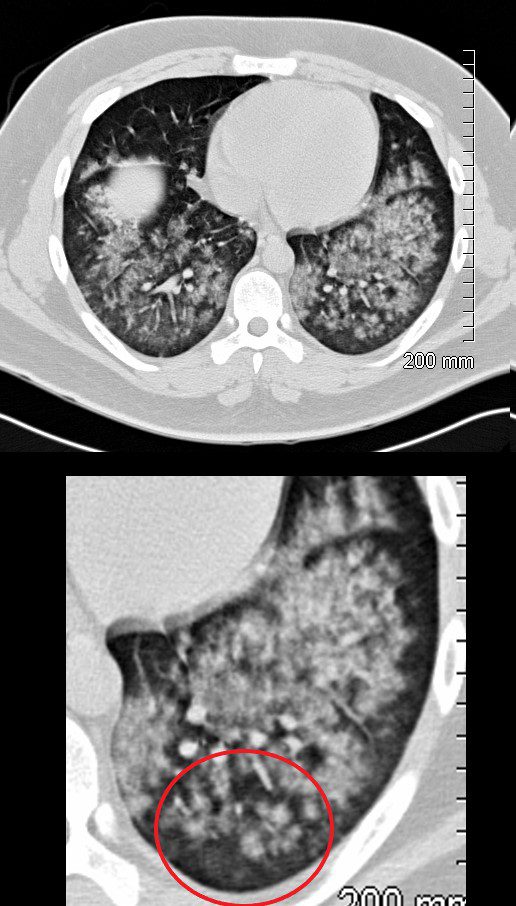
19 year old male previously well with history of hemoptysis, sweating, fevers, myalgias, arthritis over 3 weeks.
CT scan in the axial projection shows diffuse bilateral nodular consolidations (acinar pattern ringed in red) with subpleural sparing consistent with pulmonary hemorrhage
Lab shows ANCA positivity, acute renal failure (creatinine 6) and renal biopsy showing crescentic glomerulonephritis. These finding are consistent with a diagnosis of GPA. He was treated with cyclophosphamide
Ashley Davidoff MD TheCommonVein.net 139193cL
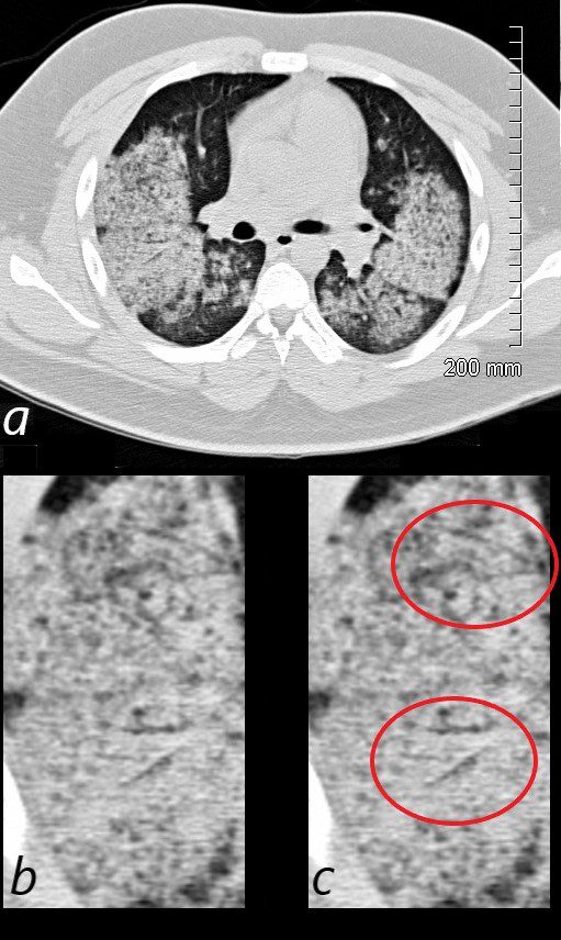
19 year old male previously well with history of hemoptysis, sweating, fevers, myalgias, arthritis over 3 weeks.
CT scan in the axial projection shows diffuse bilateral nodular consolidations with subpleural sparing consistent with pulmonary hemorrhage. Air bronchograms are noted in the posterior segment of the right upper lobe (b ringed in c) and the superior segment of the right lower lobe (b ringed in c)
Labs showed ANCA positivity, acute renal failure (creatinine 6) and renal biopsy showed crescentic glomerulonephritis. These finding are consistent with a diagnosis of GPA. He was treated with cyclophosphamide
Ashley Davidoff MD TheCommonVein.net 139185cL
Case 2 GPA
Pulmonary Hemorrhage
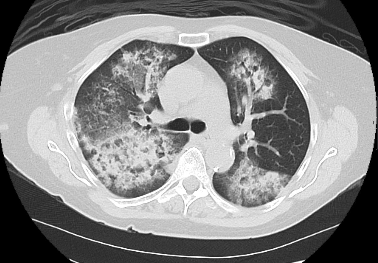
35 year old male in acute renal failure and respiratory distress with hemoptysis. CT shows multicentric segmental and subsegmental regions of consolidations and ground glass opacity in the upper and lower lobes with subpleural sparing consistent with pulmonary hemorrhage
Ashley Davidoff MD TheCommonVein.net 137273
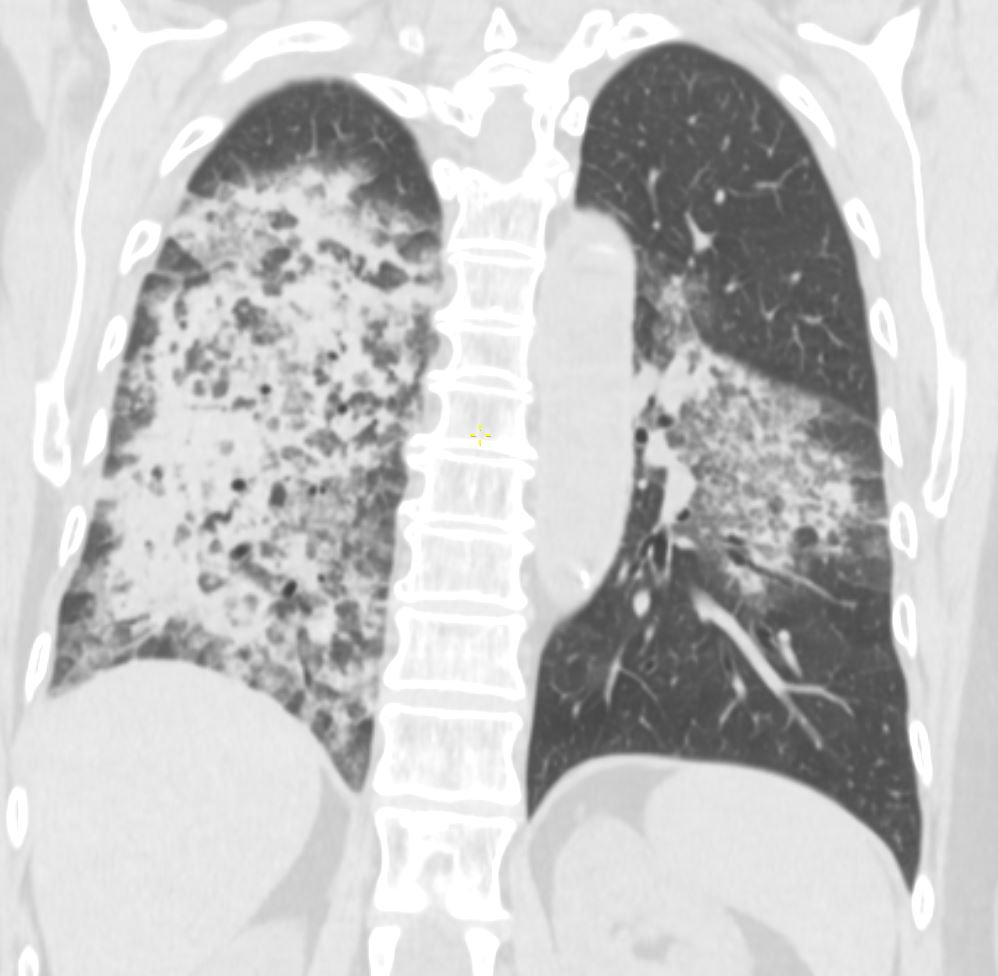
35 year old male in acute renal failure and respiratory distress with hemoptysis. CT shows multicentric segmental and subsegmental regions of consolidations and ground glass opacity in the upper and lower lobes with subpleural sparing consistent with pulmonary hemorrhage
Ashley Davidoff MD TheCommonVein.net 137275
Trauma Hemorrhage – Resuscitation Attempts
Acinar Shadows
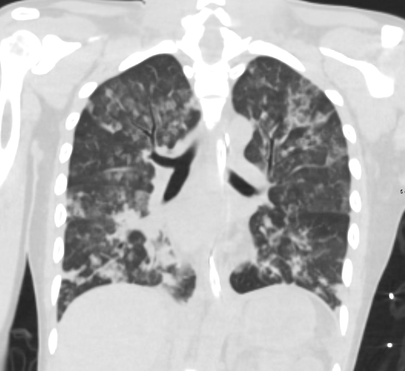
Coronal CT following trauma and resuscitative attempts in a 37 year old female shows 2-5mm solid and ground glass nodules in both the upper and lower lobes with confluence to form subsegmental foci of consolidation in the right lower lobe and right middle lobe. There is evidence of subpleural sparing with a more central distribution. These findings are consistent with hemorrhagic foci of acinar shadows or acinar nodules following trauma
Ashley Davidoff MD TheCommonVein.net 137270 key words .lungs GGO ground glass opacities acinar shadows hemorrhage contusion post resuscitation 37F 301Lu
Acinar Shadows Subpleural Sparing

Coronal CT following trauma and resuscitative attempts in a 37 year old female shows 2-5mm solid and ground glass nodules in both the upper and lower lobes with confluence to form subsegmental foci of consolidation in the right lower lobe and right middle lobe. There is evidence of subpleural sparing with a more central distribution. These findings are consistent with hemorrhagic foci of acinar shadows or acinar nodules following trauma
Ashley Davidoff MD TheCommonVein.net 137270 key words .lungs GGO ground glass opacities acinar shadows hemorrhage contusion post resuscitation 37F 301Lu
Crazy Paving
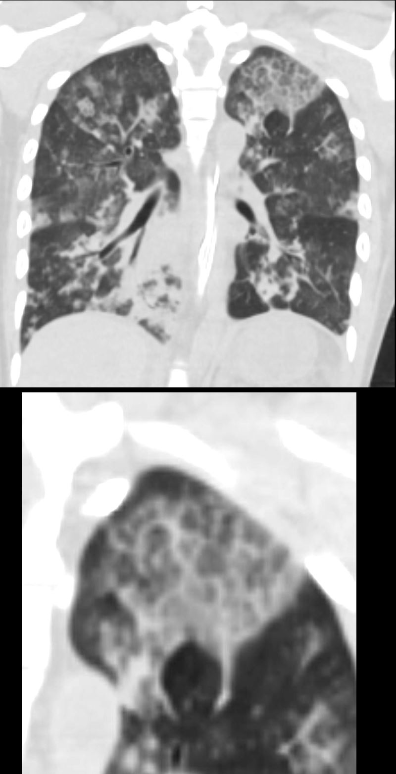
Coronal CT following trauma and resuscitative attempts in a 37 year old female shows a focus of crazy paving in the left upper lobe most likely secondary to contusion. In addition 2-5mm nodules in both the upper and lower lobes. In this clinical context. these findings are consistent with hemorrhagic foci of ground glass opacities acinar shadows and crazy paving following trauma.
Ashley Davidoff MD TheCommonVein.net 137272 301Lu
