Infection
Inflammation
Mounier Kuhn and Lady Windermere Syndrome

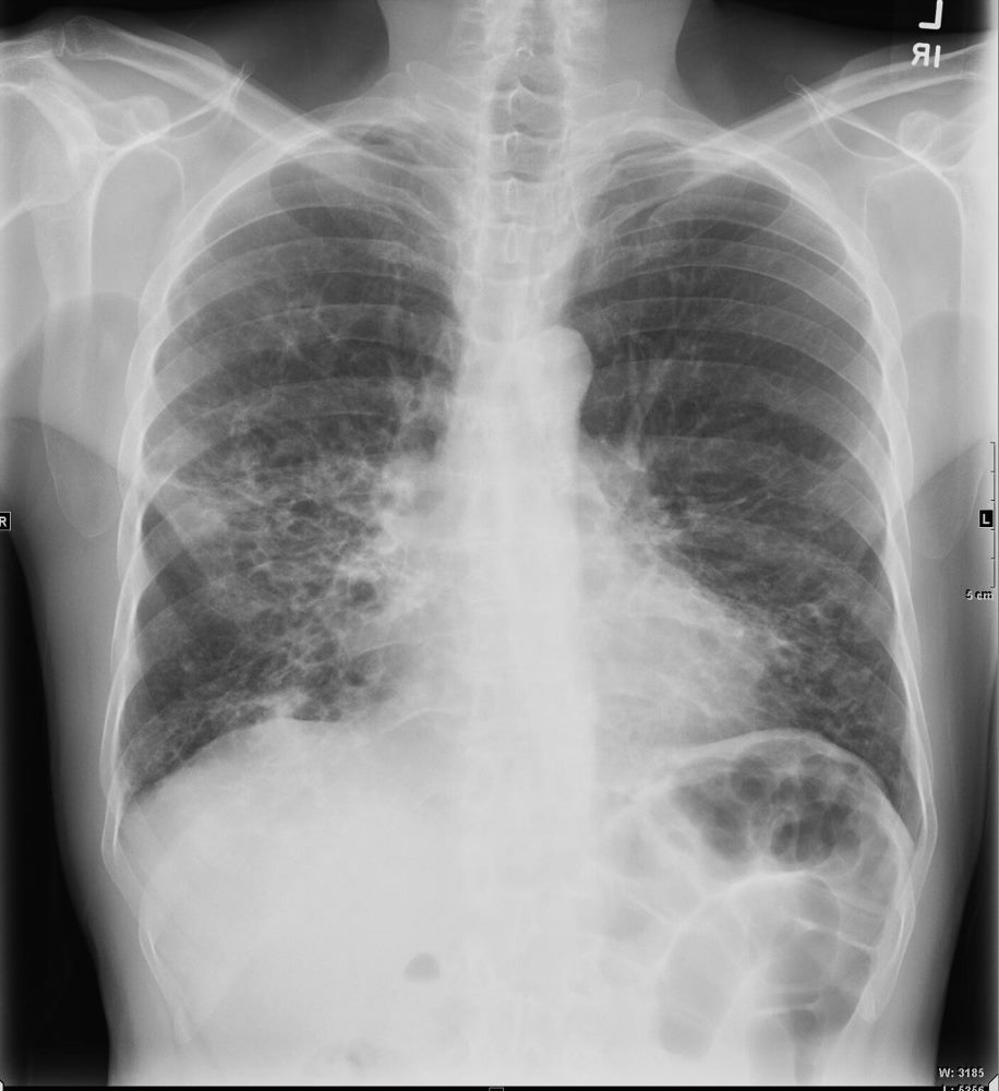
61 year old male with a history of treated mycobacterial infections and chronic cough
Frontal view shows shaggy heart borders with bibasilar cystic changes consistent with bronchiectasis in the middle lobe and lingula
Ashley Davidoff MD TheCommonVein.net 250Lu 135871
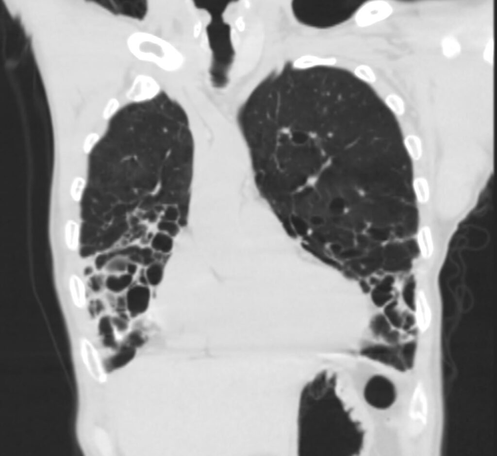
Correlative Coronal

61-year-old male with a history of treated mycobacterial infections including MAC and chronic cough.
Coronal CT at the level of the heart shows significant bronchiectasis to the middle lobe and lingula and as a result abut the right and left heart border accounting for the CXR findings of a “shaggy heart border”. There is a relative paucity of mucus in the ectatic airways. The history of MAC and the distribution of the bronchiectasis in the middle lobe and lingula are reminiscent of the diagnosis of Lady Windermere syndrome
Ashley Davidoff MD TheCommonVein.net 250Lu 135879
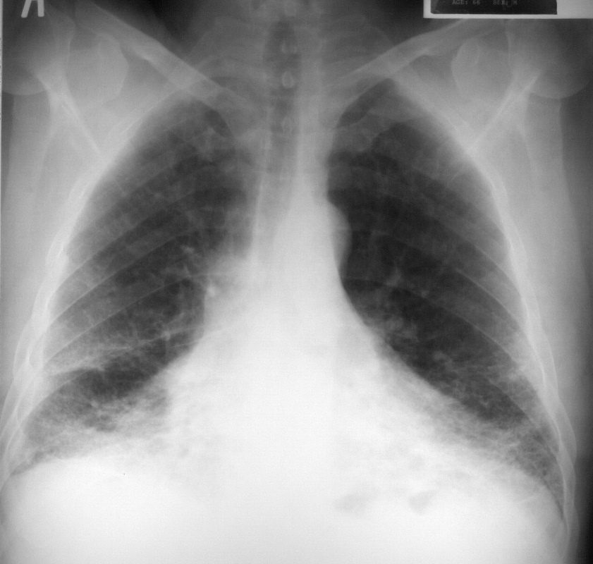
Asbetsosis

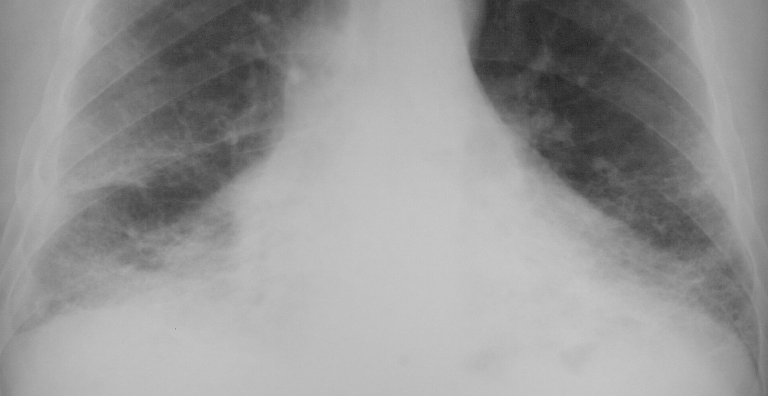
This is a frontal chest X-ray of a 66year old man who swept asbestos fibers off the floor for many years. The image characterises the “shaggy heart border” due to the interstitial process in the middle lobe and lingula.
Courtesy Ashley Davidoff MD TheCommonVein.net 32615.8

Courtesy Ashley Davidoff MD TheCommonVein.net 32617.8
Malignancy Mechanical/Atelectasis Trauma Metabolic Circulatory- Hemorrhage Immune Infiltrative Idiopathic Iatrogenic Idiopathic
