Tension Pneumothorax
Tension Pneumothorax
- Etymology:
- Derived from “pneuma” (air) and “thorax” (chest), combined with “tension,” reflecting the life-threatening accumulation of air in the pleural space under pressure.
- AKA:
- None specifically, but often described as a medical emergency.
- What is it?
- Tension pneumothorax refers to a life-threatening condition where air accumulates in the pleural space under pressure, causing compression and obstruction of low-pressure cardiovascular structures such as the pulmonary veins, SVC, and IVC. This reduces venous return and hence cardiac output, contributing to the hemodynamic instability seen in tension pneumothorax.
- Characterized by:
- Progressive accumulation of air in the pleural space with each breath.
- Collapse of the affected lung and compression of the contralateral lung.
- Mediastinal shift with compression of major vessels, reducing venous return and cardiac output.
- Anatomically affecting:
- Pleural space, lungs, mediastinum, and great vessels.
- Pathophysiology:
- Air enters the pleural space during inspiration but cannot escape during expiration due to a one-way valve mechanism.
- One-way valve mechanism occurs where air enters the pleural space during inspiration but cannot escape during expiration, causing increasing intrapleural pressure.
Progressive increase in intrapleural pressure compresses mediastinal structures, impairs venous return, and reduces cardiac output.
- How does it appear on each relevant imaging modality?
- CXR:
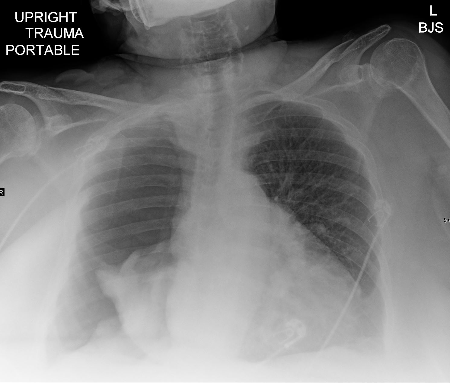
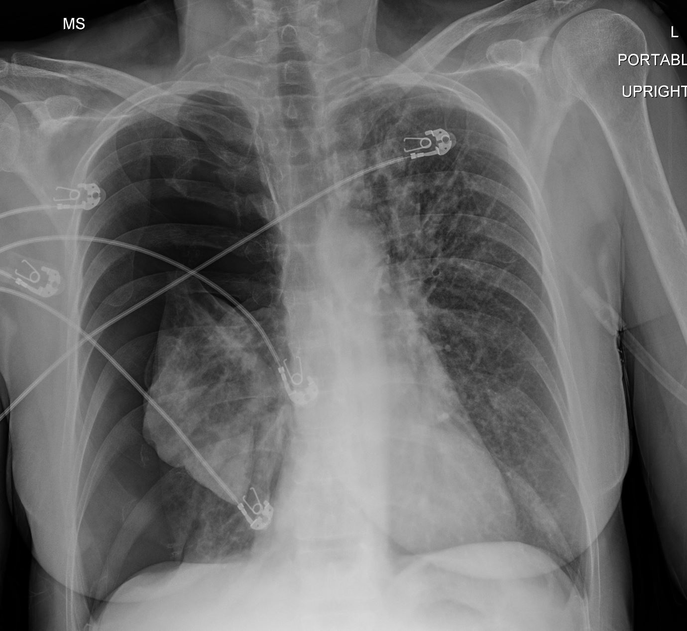
- Visible pleural line with absence of lung markings beyond it.
- Mediastinal shift away from the affected side.
- Flattening or inversion of the diaphragm on the affected side. Cardiovascular manifestations include venous congestion seen as engorgement of the SVC and IVC, with associated dilation of the azygos vein. Pulmonary veins may also appear engorged, contributing to vascular prominence on the expanded lung .
- Widening of the intercostal spaces.
- CT:
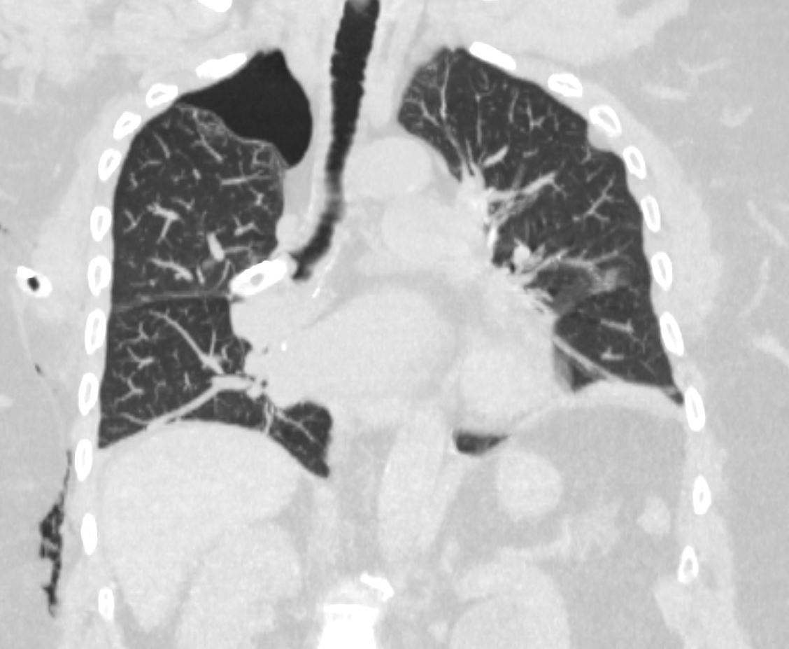
- Confirms presence of air in the pleural space and identifies associated findings such as collapsed lung, contralateral mediastinal shift, and compression of the great vessels. Specific manifestations include compression of the superior vena cava (SVC), inferior vena cava (IVC), and pulmonary veins, which appear as narrowing or flattening on imaging. Ipsilateral pulmonary veins may show increased compression due to direct intrapleural pressure, while contralateral pulmonary veins can appear stretched, displaced, and engorged due to mediastinal shift and increased venous pressure from obstructed pulmonary venous return.
- Ultrasound:
- Absence of lung sliding and barcode sign on M-mode.
- Presence of lung point confirming pneumothorax.
- Echocardiogram may reveal impaired right heart filling, septal bowing towards the left ventricle due to increased intrathoracic pressure, and diminished cardiac output.
- CXR:
- Differential Diagnosis:
- Massive hemothorax.
- Large pleural effusion.
- Diaphragmatic rupture.
- Recommendations:
- Immediate needle decompression in the second intercostal space at the midclavicular line.
- Followed by chest tube insertion to maintain decompression.
- Key Points and Pearls:
- Always suspect tension pneumothorax in cases of acute respiratory distress with hemodynamic compromise.
- Prompt recognition and intervention are critical to prevent cardiac arrest.
- Ultrasound is a rapid bedside tool for confirming the diagnosis in emergency settings.
- CT is useful in stable patients for detailed evaluation and identifying underlying causes.
30 year old female presents with tension pneumothorax 1 month prior to current admission

Ashley Davidoff MD TheCommonVein.net

Ashley Davidoff MD TheCommonVein.net

? Tension Pneumoythorax
Ashley Davidoff MD TheCommonVein.net
