- Respiratory Zone (Air Space)
Derived from the Latin word “respirare,” meaning “to breathe,” reflecting its primary function in gas exchange.
- Etymology
- AKA and Abbreviation
- Also known as the “Respiratory Zone.”
- What is it?
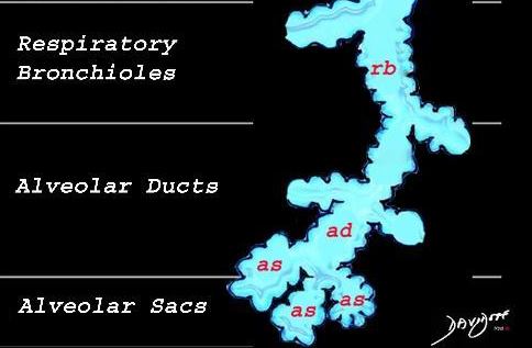
- The air space is a collective term for the distal portions of the lung, including the respiratory bronchioles, alveolar ducts, alveolar sacs, and alveoli. It represents the primary functional unit of the lungs where gas exchange occurs.
- Principles:
- Parts:
- Respiratory bronchioles.
- Alveolar ducts.
- Alveolar sacs.
- Alveoli (functional unit).
- Size:
- Respiratory bronchioles: Diameter ranges from 0.3 to 0.5 mm.
- Alveolar ducts: Diameter ranges from 0.2 to 0.3 mm.
- Alveolar sacs: Diameter ranges from 0.2 to 0.4 mm, depending on lung inflation.
- Alveoli: ~200 micrometers in diameter, varying with lung inflation.
- Total surface area: ~70 square meters in an adult lung, equivalent to the size of a tennis court.
- Shape:
- Respiratory bronchioles are tubular structures transitioning into alveolar ducts.
- Alveolar ducts are elongated corridors lined with alveoli.
- Alveolar sacs are clusters of alveoli arranged in a spherical configuration.
- Alveoli are polygonal in shape, arranged in a honeycomb-like network.
- Position:
- Found in the distal lung, branching off terminal bronchioles.
- Character:
- Thin walls lined with a single layer of epithelial cells (type I and type II pneumocytes).
- Richly vascularized by pulmonary capillaries.
- Time:
- Develops primarily during the late fetal and early postnatal periods, continuing to mature into early childhood.
- Parts:
- Blood supply:
- Provided by the pulmonary arteries, carrying deoxygenated blood to the capillaries surrounding the alveoli.
- Venous Drainage:
- Blood exits via the pulmonary veins, carrying oxygenated blood back to the left atrium of the heart.
- Lymphatic Drainage:
- Drains to peribronchial and subpleural lymphatics, eventually reaching the hilar and mediastinal lymph nodes.
- Nerve Supply:
- Innervated by autonomic fibers:
- Parasympathetic: Promotes bronchoconstriction and mucus secretion.
- Sympathetic: Induces bronchodilation.
- Innervated by autonomic fibers:
- Embryology:
- The air space originates from the distal end of the lung bud during the pseudoglandular stage of lung development (5-16 weeks gestation). Alveolarization begins late in the canalicular stage and continues postnatally.
- Histology:
- Respiratory bronchioles: Lined by a combination of ciliated cuboidal epithelium and non-ciliated Clara (club) cells, transitioning to alveolar epithelium distally.
- Alveolar ducts: Lined by simple squamous epithelium, with openings into alveoli along their walls.
- Alveolar sacs: Comprised entirely of alveoli, lined by type I and type II pneumocytes and surrounded by capillary networks.
- Alveoli:
- Type I pneumocytes: Thin, squamous cells facilitating gas exchange.
- Type II pneumocytes: Cuboidal cells producing surfactant.
- Alveolar macrophages: Immune cells clearing debris and pathogens.
- Tree-in-Bud Sign:
- The “tree-in-bud” sign is a radiological finding representing small airway inflammation or obstruction. It reflects impacted bronchioles and alveolar ducts filled with mucus, pus, or fluid.
- Relation to Anatomy: The sign appears as small, centrilobular nodules with branching opacities, mirroring the structure of respiratory bronchioles, ducts, and alveoli. It is typically associated with infectious processes, such as endobronchial tuberculosis or bronchiolitis.
- Physiology and Pathophysiology:
- Physiology:
- Facilitates oxygen and carbon dioxide exchange through diffusion across the alveolar-capillary membrane.
- Pathophysiology:
- Diseases affecting air spaces include:
- Infectious: Pneumonia.
- Inflammatory: Acute respiratory distress syndrome (ARDS).
- Obstructive: Emphysema leading to alveolar wall destruction.
- Fibrotic: Interstitial lung diseases (e.g., idiopathic pulmonary fibrosis).
- Diseases affecting air spaces include:
- Physiology:
- Applied Anatomy to Radiology:
- CXR:
- Consolidation: Air spaces filled with fluid, pus, or blood appear as increased opacities.
- Air bronchograms: Visible air-filled bronchi within consolidated lung.
- CT:
- Ground-glass opacities: Partial filling or thickening of air spaces.
- Consolidation: Dense opacification representing fully filled air spaces.
- Hyperlucency: Seen in emphysema due to alveolar destruction.
- Tree-in-bud sign: Visualized as small centrilobular nodules with a branching pattern, indicative of bronchiolar and alveolar duct obstruction or inflammation.
- PFTs:
- Pulmonary Function Tests (PFTs) assess the functionality of the respiratory zone by evaluating parameters such as:
- Diffusing capacity for carbon monoxide (DLCO), which reflects alveolar-capillary membrane efficiency.
- Residual volume (RV) and total lung capacity (TLC), indicative of air trapping or hyperinflation.
- Forced expiratory volume in 1 second (FEV1) and forced vital capacity (FVC), providing insights into obstructive or restrictive patterns.
- Pulmonary Function Tests (PFTs) assess the functionality of the respiratory zone by evaluating parameters such as:
- CXR:
- Pathological Implications:
- Infectious: Viral or bacterial pneumonia leading to air space opacities.
- Inflammatory: Pulmonary edema causing fluid-filled air spaces.
- Fibrotic: Scarring and collapse of air spaces reducing gas exchange efficiency.
- Tree-in-Bud Specific: Reflects bronchiolar obstruction or inflammation due to infections (e.g., tuberculosis, fungal infections) or inflammatory processes (e.g., cystic fibrosis).
- Key Points and Pearls:
- The air space is the primary site for gas exchange and a critical target in lung imaging.
- Differentiating air space diseases (e.g., infection vs. malignancy) often requires integration of clinical, imaging, and histological findings.
- The “tree-in-bud” sign provides crucial insights into small airway pathologies and their anatomical correlates.
- AKA and Abbreviation
Respiratory Zone = Structures that
Participate in Gas Exchange

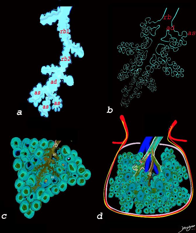
Airspace refers to the part of the lung that serves in gas exchange and therefore structurally refers to the acinus that serves as the unit that participates in air exchange including the respiratory bronchiole (rb) alveolar duct,(ad) alveoli sac (as) and the alveoli
The diagram shows the components of the air spaces in their branching forms (a and b) and then within the acinus with the alveoli (c) Image shows a secondary lobule consisting of 20-30 acini
Ashley Davidoff MD TheCommonVein.net 32645a09.8

Airspace refers to the part of the lung that serves in gas exchange and therefore structurally refers to the acinus that serves as the unit that participates in air exchange including the respiratory bronchiole (rb) alveolar duct,(ad) alveoli sac (as) and the alveoli
The diagram shows the components of the air spaces in their branching forms (a and b) and then within the acinus with the alveoli (c) Image shows a secondary lobule consisting of 20-30 acini
Ashley DavidoffMD TheCommonVein.net 32645a09.8

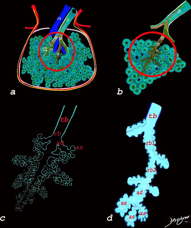
The diagram allows us to understand the the components and the position of the small airways starting in (a) which is a secondary lobule that is fed by a lobular bronchiole(lb) which enters into the secondary lobule and divides into terminal bronchioles (tb) which is the distal part of the conducting airways, and at a diameter of 2mm or less . It divides into the respiratory bronchiole (rb) a transitional airway which then advances into the alveolar ducts(ad) and alveolar sacs (as) Diseases isolated to the small airways do not affect the alveoli and hence there is peripheral sparing Ashley Davidoff MD TheCommonVein.net lungs-0749
A

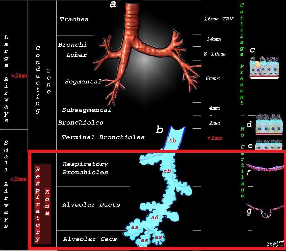
The respiratory zone or airspace refers to the alveolar region where gas exchange occurs, including the respiratory bronchioles, alveolar ducts, alveolar sacs, and alveoli.
Courtesy Ashley Davidoff MD TheCommonVein.net lungs-0740n

The respiratory zone or airspace outlined in red refers to the alveolar region where gas exchange occurs, including the respiratory bronchioles, alveolar ducts, alveolar sacs, and alveoli.
Courtesy Ashley Davidoff MD TheCommonVein.net
Fleischner Society
airspace
Anatomy.—An airspace is the gas-containing part of the lung, including the respiratory bronchioles but excluding purely conducting airways, such as terminal bronchioles.
Radiographs and CT scans.—This term is used in conjunction with consolidation, opacity, and nodules to designate the filling of airspaces with the products of disease (,14).
