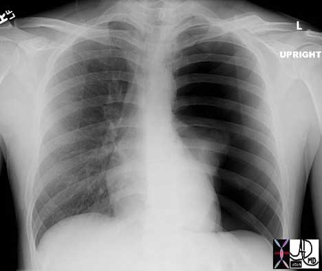
 This is the type of CXR that sends shivers down the spine. The overall blackness of the left chest cavity, in association with a nubbin of lung tissue in the ipsilateral hilum and rightward mediastinal shift is characteristic of a tension pneumothorax with total atelectasis of the left lung. Immediate and urgent decompression with a chest drain is indicated.
Courtesy of: Ashley Davidoff, M.D.
42525
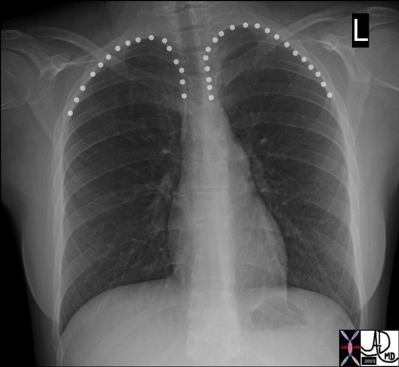
In the upright position air will rise to the apex (as long as there are no pleura adhesions.
In the apex it will assume a sickle cell shape since it can exert force on the lung but not onthe chest wall so it assumes the shape of the apex of the lung
Ashley Davidoff MD
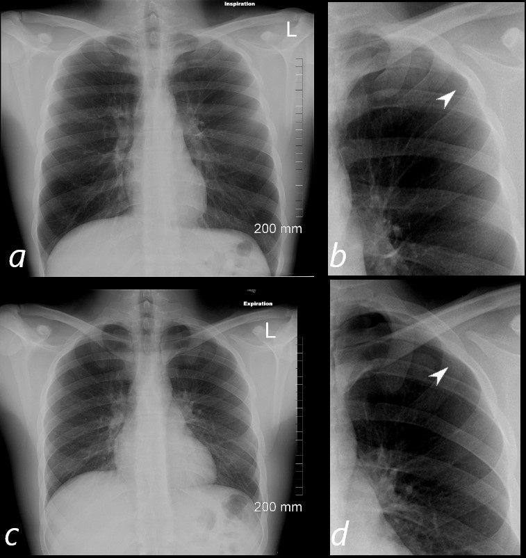
In an upright position a pneumothorax rises to the apex of the lung and assumes the shape of the apex because it exerts pressure on the lung apex which yields to the grater pressure. On the inspiration film (a) the pneumothorax is barely seen, even on the enhanced magnified view (b, white arrowhead) On the expiration film the pneumothorax is better seen (d white arrowhead) because it is larger and because the intraparenchymal pressure of the lungs is reduced thus allowing the pneumotorax to accumulate in the apical region and less spread out along the surface of the lung
Ashley Davidoff MD
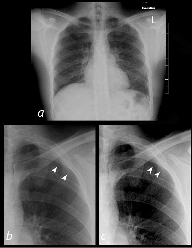
In an upright position a pneumothorax rises to the apex of the lung and assumes the shape of the apex because it exerts pressure on the lung apex which yield to the greater pressure. The expiration film accentuates the pneumothorax because it further reduces the pressure in the lungs and increases the pressure difference between the PTX and the intraparenchymal pressure.
The PTX is barely seen in (a) and is better seen in the magnified views (b and c) and with increasing contrast (c) the faint line of the the pleura becomes better visualized (white arrowheads).
Ashley Davidoff MD
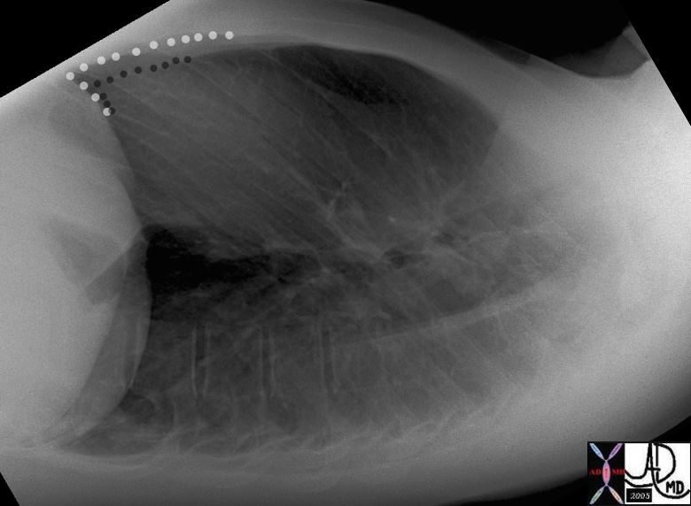
The highest point in the chest is anterior and inferior
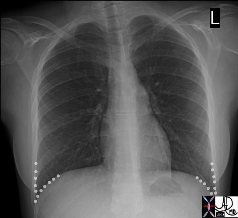
When the a patient with a pneumothorax is in the supine position air will accumulate in the sulcus and appear as a lucency in the sulcus. This is called the deep sulcus sign
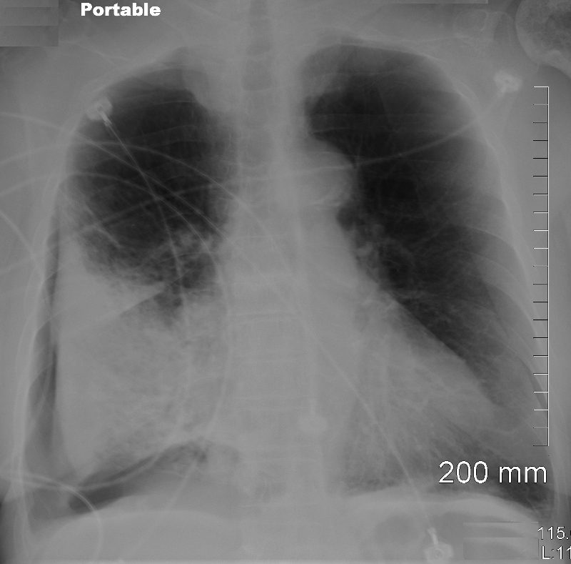
Portable supine examination in the ICU shows a pneumothorax in the right subpulmonic region
Ashley Davidoff MD
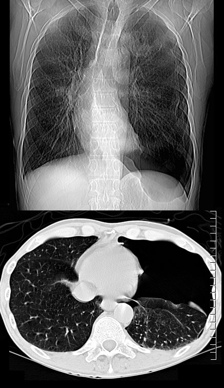
CT scout film (above) shows a lucency of air accumulation in the left deep sulcus that also extends to the lateral margin of the left lung. The axial CT image confirms the presence of a large pneumothorax, dominant at the highest point in the chest in the supine position Portable supine examination in the ICU shows a pneumothorax in the right subpulmonic region
Ashley Davidoff MD
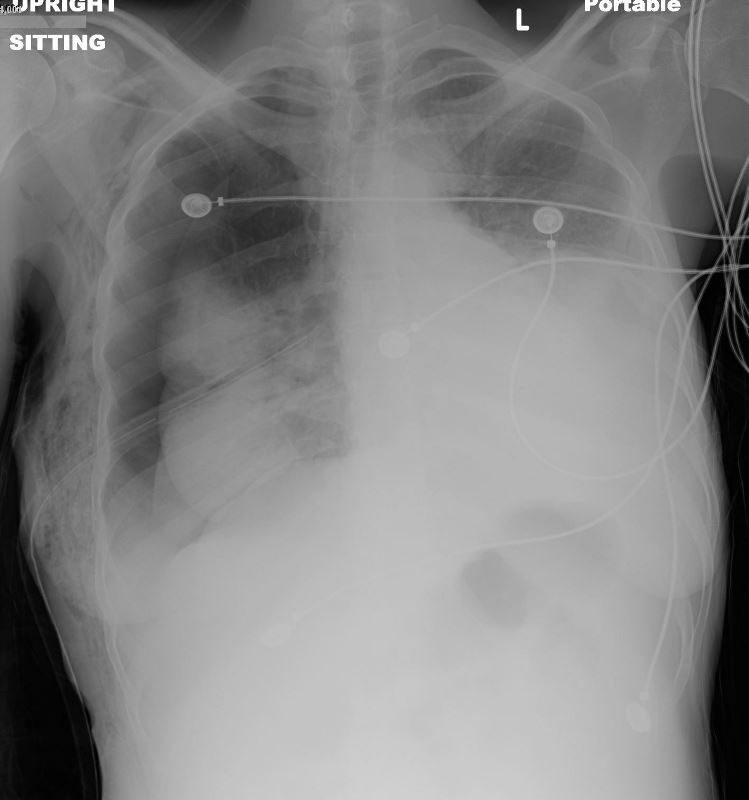
30 year old female presented in 2013 with acute pneumothorax with tamponade requiring placement of a chest tube
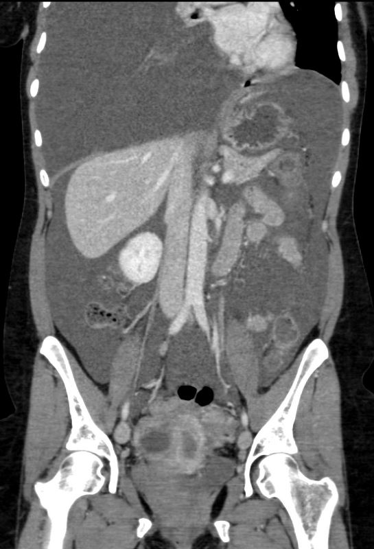
RECURRENT PLEURAL FLUID COLLECTION and LARGE VOLUME ASCITES
30 year old female
presented in 2013 with acute pneumothorax with tamponade requiring placement of a chest tube
The PTX and right hemothorax slowly resolved but a month later the large pleural effusion or hemothorax recurred
In December 2013 the right effusion was large enough to cause total collapse of the right lung. A small left effusion was also present
In February 2014 a large volume of ascites was present as well as a subcutaneous collection either from a wall defect or perhaps an endometrial deposit.
At the same endometriomas were again noted in the pelvis
Feb 2014
Ashley Davidoff MD
PCP Infection
- feature of PCP infection,
- incidence 35% in patients with cysts
- frequently bilateral
- often refractory to chest tube Rx,
- frequently need surgery eg pleurodesis
- associated higher mortality rate,
- especially in patients on ventilation.
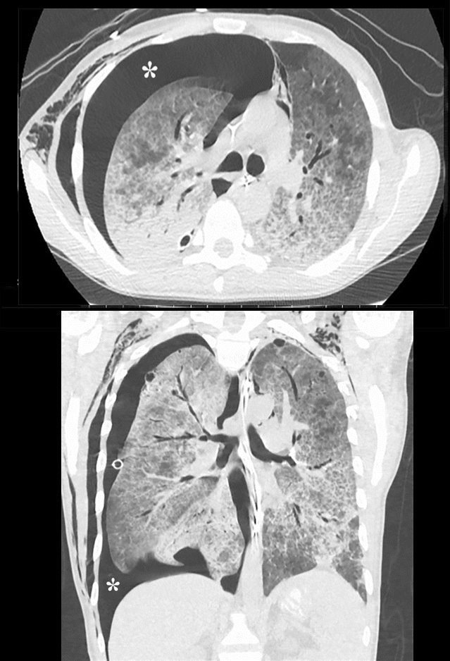
Parekh, M et al Review of the Chest CT Differential Diagnosis of Ground-Glass Opacities in the COVID Era Radiology Vol. 297, No. 3 July 2020
Ashley Davidoff MD TheCommonVein.net
77949
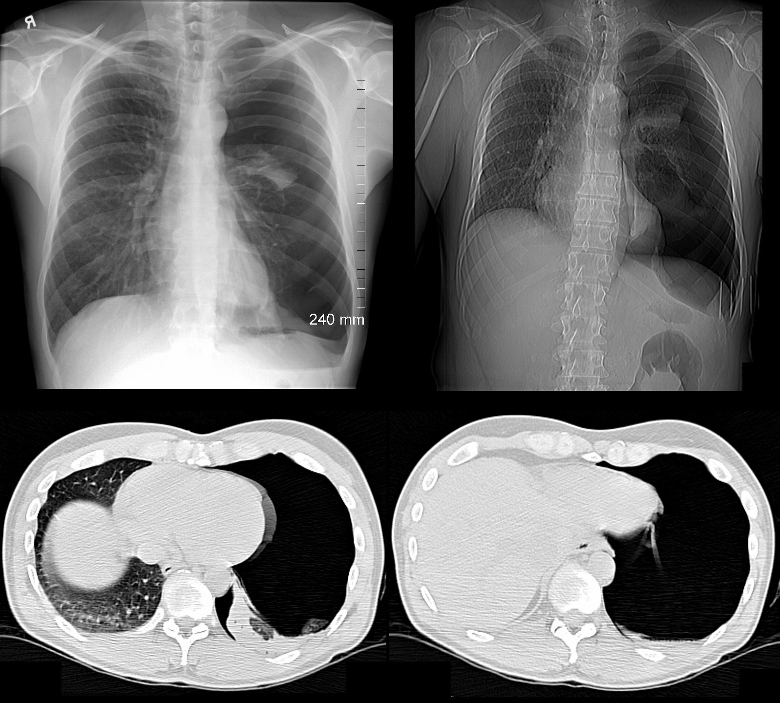
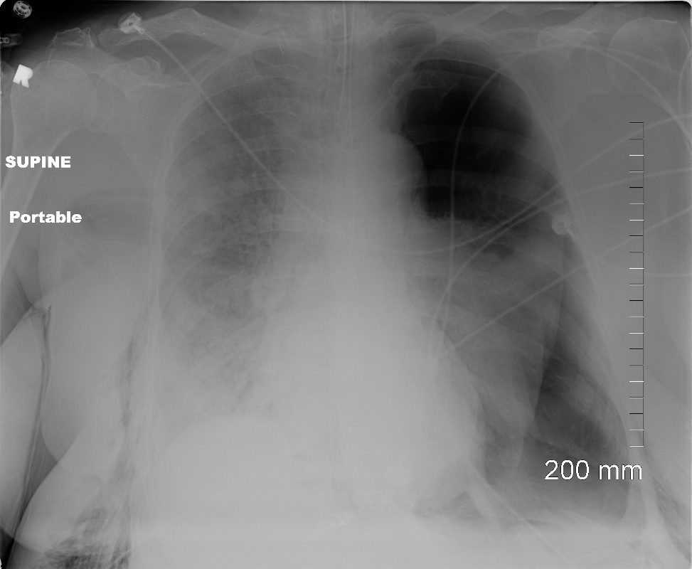
49 year old male with a cough presents for a Chest Xray which showed a tension pneumothorax. Chest tube was placed emergently in the radiology department.
Ashley Davidoff MD TheCommonVein.net
117300c
