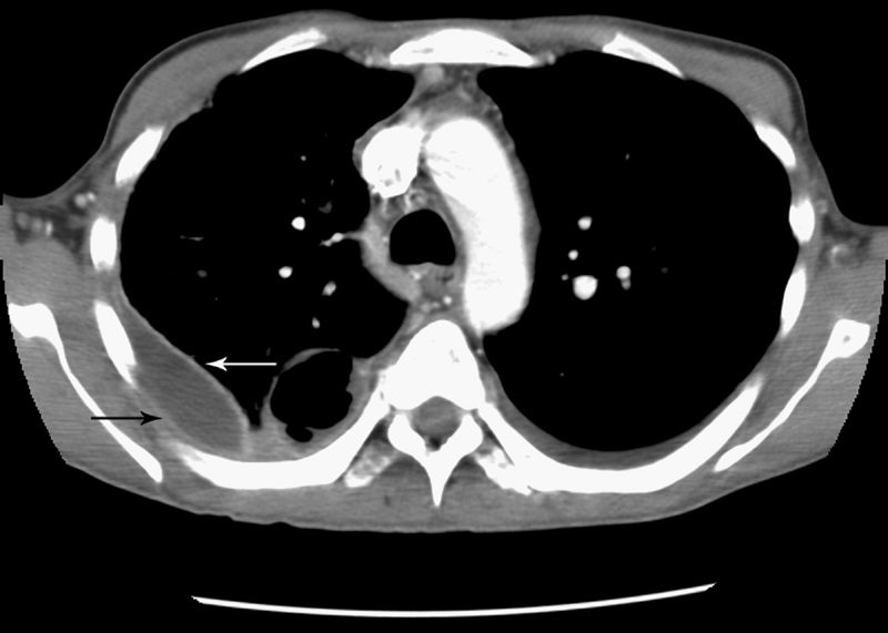
The split pleura sign is a radiological finding seen on contrast
enhanced CT scans of the chest, indicating the presence of a
pleural effusion, often related to empyema (infected pleural fluid). It
occurs when the visceral and parietal pleura become separated and
are visible as two distinct, thickened, enhancing layers, outlining
the fluid collection within the pleural cavity. The pathogenesis
involves inflammation or infection causing thickening and
enhancement of the pleura, which leads to the characteristic
appearance. The split pleura sign helps differentiate empyema from
other conditions like a simple pleural effusion or pleural abscess,
as it suggests an inflammatory process.

Source
Signs in Thoracic Imaging
Journal of Thoracic Imaging 21(1):76-90, March 2006.
Separation and enhancement of the visceral and parietal pleural layers on CT (Fig. 22) is considered strong evidence of empyema.3,61 Normally, individual pleural layers are not discernable as discrete structures.3 Empyemic fluid fills the pleural space, resulting in thickening and enhancement of the pleura with a denotable separation.3,61
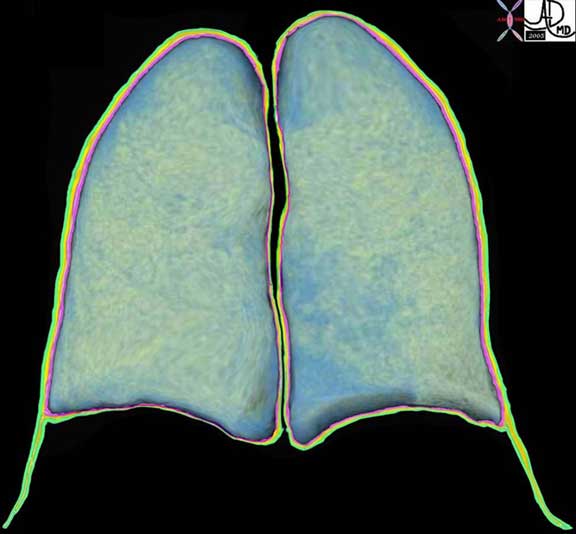
 The coronally reformatted image of the lung parenchyma has been outlined with the visceral pleura, (pink) the pleural fluid in the pleural space, (orange) and the parietal pleura (green). Note how at end expiration the parietal pleura in the costophrenic sulcus extends beyond the lung margin so that the visceral pleura is absent in the costophrenic sulcus and there are two layers of parietal pleura facing each other. During inspiration the lung expands into this space.
Courtesy of: Ashley Davidoff, M.D.
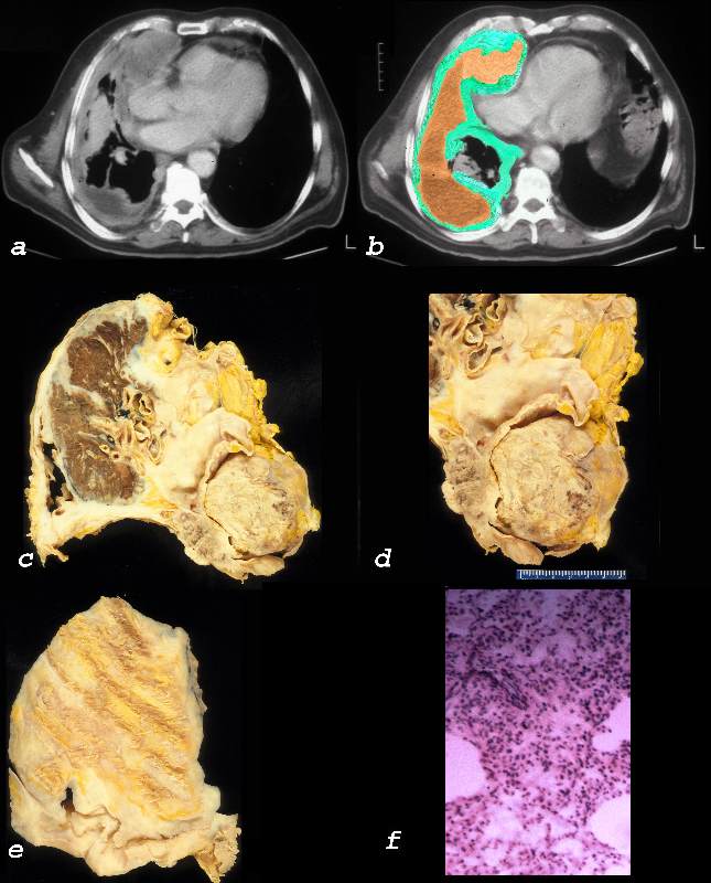
Mesothelioma

Ashley Davidoff MD TheCommonVein.net 32640c01L01.8

Ashley Davidoff MD TheCommonVein.net
pleura mesothelioma 0028c01
