- Active TB
- Inhomogeneous
- Affecting
- Upper lobe
- superior segment of the lower lobe
- lobar
- segmental
- Active TB
- Reactivation
- Post primary
- Characterised by
- consolidation
- cavitation
- miliary pattern
- lymphadenopathy
- pleural effusion
- Characterised by
- Healed TB
-
- Fibronodular scarring
- traction bronchiectasis
- calcified lymph nodes
- volume loss
-
- Affecting
- Position
- Inhomogeneous
- Reactivated tuberculosis (TB) can affect any part of the lungs, including the
- tends to affect the upper lobes of the lungs more commonly than the lower lobes.
-
- but can affect the
- lower lobes, or
- both.
- upper lobe pre-dominance because of
- greater oxygen concentration
- which promotes bacterial growth.
However, it’s important to note that TB can affect any part of the lungs and the location of TB disease in the lungs can vary depending on the individual’s immune status, the strain of TB bacteria involved, and the extent of disease.
-
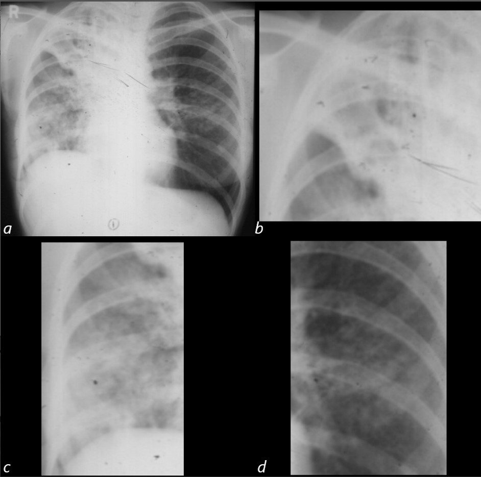
Reactivated TB
Multicentric findings showing volume loss and an infiltrate in the right upper lobe (a,b) diffuse infiltrate in the right lower lobe (a,c) in a background of chronic granulomatus nodules (a,b,c)
Historical Xray from 60 years ago from the teaching collection of Dr Lloyd Hawes
Ashley Davidoff
TheCommonVein.net68 female Reactivation Miliary TB
-

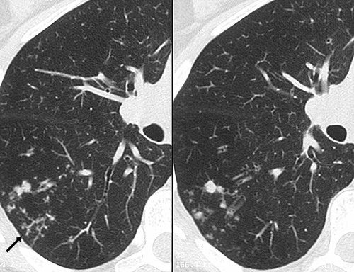
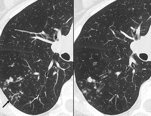
Post primary active tuberculosis in a 66-year-old woman with a chronic cough. High-resolution CT scans of the right lung show peripheral, poorly defined, small (2–4-mm-diameter) centrilobular nodules and branching linear opacities of similar caliber originating from a single stalk (the tree-in-bud pattern) in the lower lobe (arrow). These findings represent endobronchial spread of tuberculosis.
Rossi, SE et al Tree-in-Bud Pattern at Thin-Section CT of the Lungs: Radiologic-Pathologic Overview RadioGraphics Vol. 25, No. 3 2005
Postprimary active tuberculosis in a 66-year-old woman with a chronic cough. High-resolution CT scans of the right lung show peripheral, poorly defined, small (2–4-mm-diameter) centrilobular nodules and branching linear opacities of similar caliber originating from a single stalk (the tree-in-bud pattern) in the lower lobe (arrow). These findings represent endobronchial spread of tuberculosis.
Rossi, SE et al Tree-in-Bud Pattern at Thin-Section CT of the Lungs: Radiologic-Pathologic Overview RadioGraphics Vol. 25, No. 3 2005 -

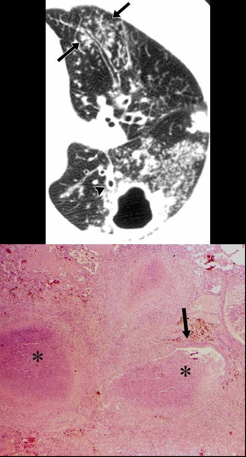
Postprimary active tuberculosis in a 34-year-old man with weight loss and a chronic cough. (a) High-resolution CT scan of the left lung shows a thick-walled cavity and multiple peripheral small nodules and branching linear structures (arrows). Note the thickening of the bronchial walls (arrowhead). (b) Photomicrograph (original magnification, ×400; hematoxylin-eosin stain) shows impacted caseous material (*) in small peripheral airways (arrow).
Rossi, SE et al Tree-in-Bud Pattern at Thin-Section CT of the Lungs: Radiologic-Pathologic Overview RadioGraphics Vol. 25, No. 3 2005 -

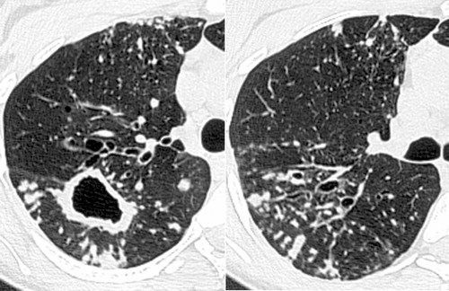
Infection with M avium-intracellulare complex in a 44-year-old woman with malaise and a chronic cough. High-resolution CT scans of the right lung show multiple peripheral small nodules connected to branching linear opacities and a thick-walled cavity in the superior segment of the lower lobe. Note the thickening of the bronchial walls, bronchial dilatation, and mucus impaction. The diagnosis was confirmed with bronchoalveolar lavage.
Rossi, SE et al Tree-in-Bud Pattern at Thin-Section CT of the Lungs: Radiologic-Pathologic Overview RadioGraphics Vol. 25, No. 3 2005
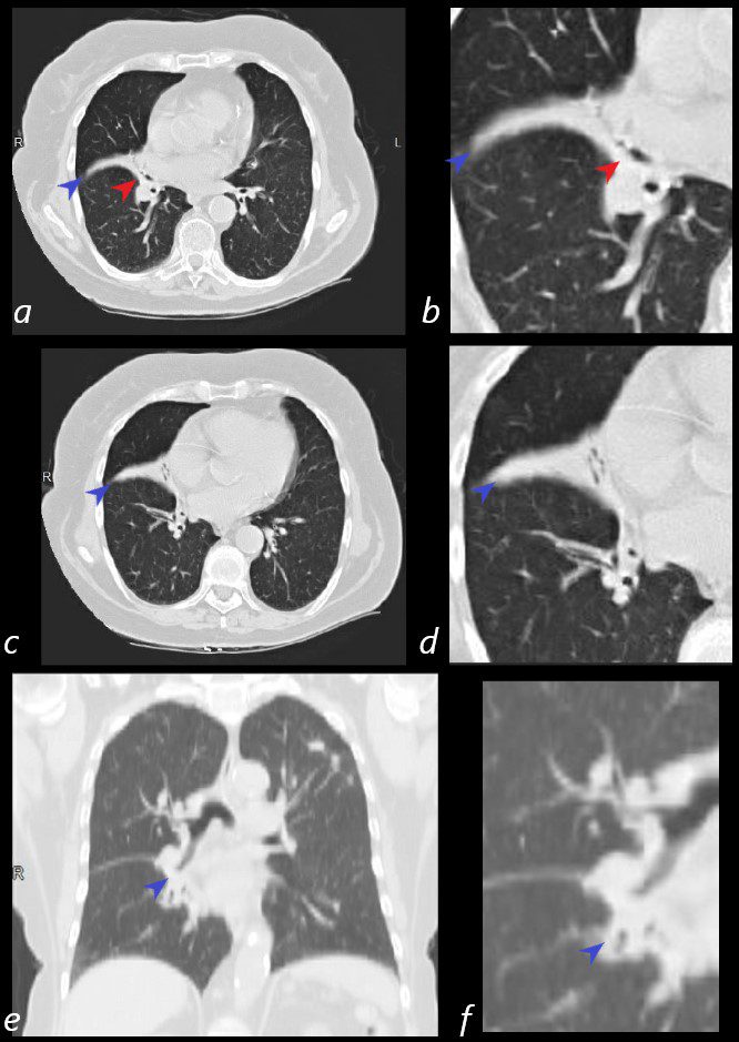
80-year-old Russian woman who initially presented with a cavitating LUL nodule that was biopsied and thought to represent sarcoidosis, nut subsequently shown to be TB. Axial CT’s shows thickening around the bronchovascular bundles of the middle lobe (red arrowhead – a, b) with post obstructive atelectasis of the lateral segment of the RML (blue arrowhead , a-f)
Ashley Davidoff MD Ashley Davidoff MD TheCommonVein.net
31645cL
-
TCV


