
-
- sarcoidosis (classic association)
- amyloidosis – nodular form
- lymphocytic interstitial pneumonia (LIP)
- lymphangitic carcinomatosis:
- silicosis
- coal worker’s pneumoconiosis:
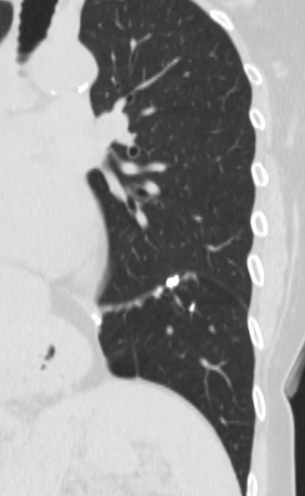
Sarcoidosis
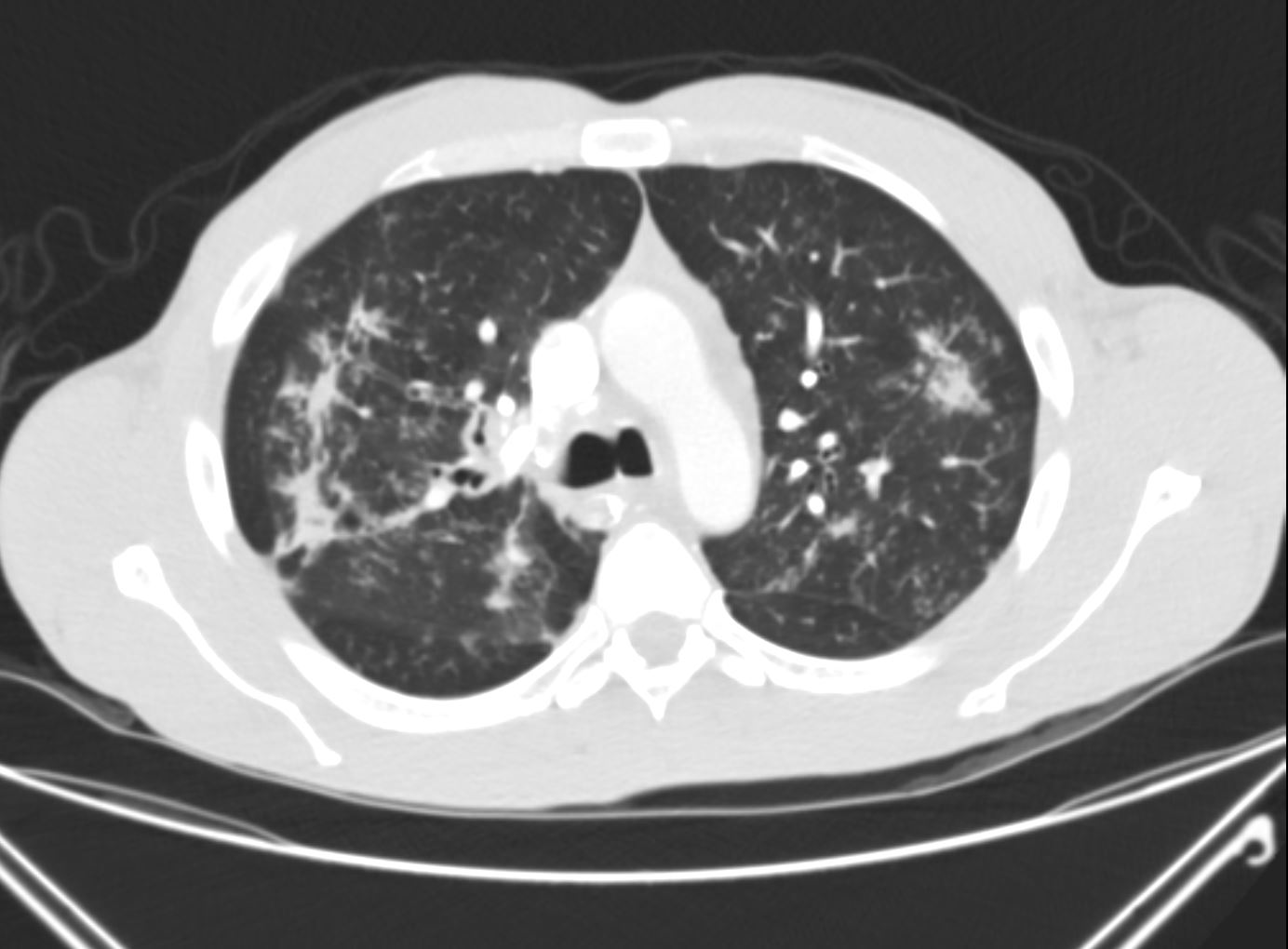
The axial CTscan shows thickening and irregularity of the major fissure, a band of fibrosis in the right upper lobe, thickening of a segmental bronchus in the right upper lobe and bronchocentric fibrosis in the left upper lobe and multiple micronodules
Ashley Davidoff MD TheCommonvein.net lungs sarcoid 002
Courtesy Paul Kohanteb MD
TheCommonVein.net
keywords lung pleura fissures and around the bronchi
key words lymphatics interstitium interstitial disease fx nodules dx sarcoidosis CTscan 446843
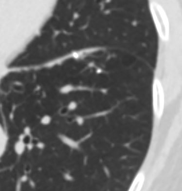
Ashley Davidoff MD TheCommonVein.net
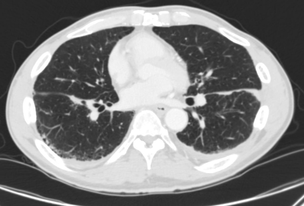
51-year-old male with history of sarcoidosis
Fissural based nodules Subpleural nodules Micronodules along the lymphovascular and
bronchovascular bundles of the secondary lobule
Calcified nodule some of which are surrounded by soft tissue of the granuloma
Ashley Davidoff MD TheCommonVein.net
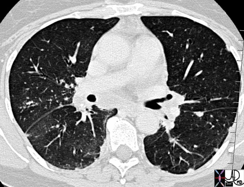
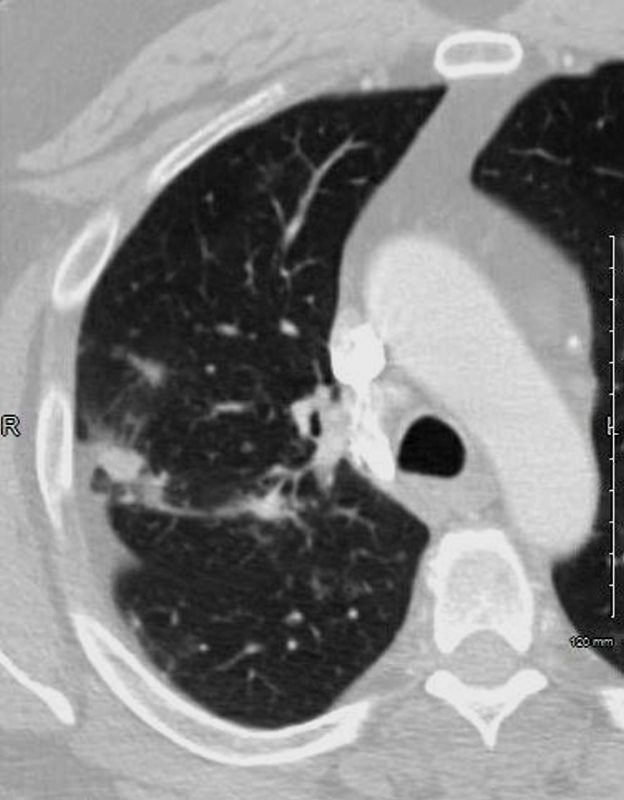
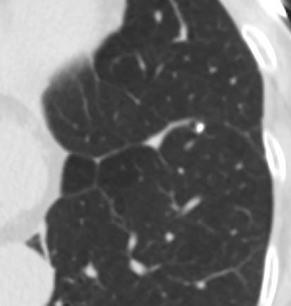
Amyloidosis
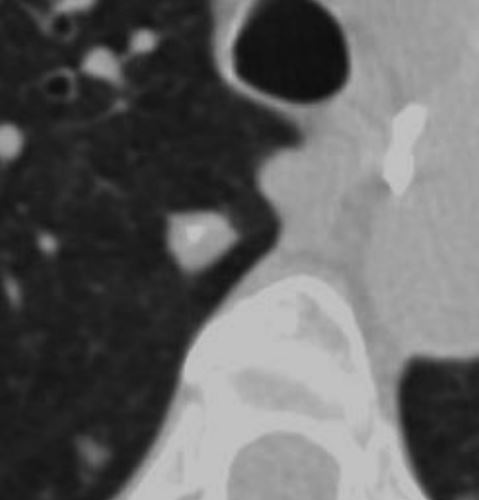
Axial CT images through the right upper lobe shows a solid amyloid nodule with central calcification abutting the major fissure. These features, although not pathognomonic are characteristic. Sarcoidosis would be a radiological consideration as well
Ashley Davidoff Boston Medical Center TheCommonvein.net LV-006
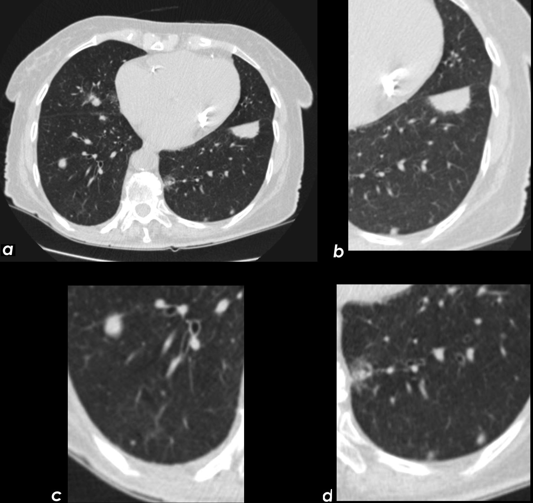
Axial CT images through the chest shows a Fissural based amyloid nodule along the left major fissure (a,b) Images c and d show posterior peripheral centrilobular nodules. In image c the nodules are associated with mosaic attenuation. The ground glass nodule in d could reflect alveolar septal disease withground glass changes surrounding a centrilobular nodule
Ashley Davidoff Boston Medical Center TheCommonvein.net LV-014c
TB
CT scan of TB with Calcified Graulomata Centered around Major fissure and Bronchioles with Atelectasis and Bronchiolectasis
Ashley Davidoff MD The CommonVein.net granulomata-along-fissures-010
CT scan of TB with Calcified Granulomata Centered around Major fissure and Bronchioles with Atelectasis and Bronchiolectasis
Ashley Davidoff MD The CommonVein.net granulomata-along-fissures-006
CT scan of TB with Calcified Graulomata Centered around Major fissure and Bronchioles with Atelectasis and Bronchiolectasis
Ashley Davidoff MD The CommonVein.net granulomata-along-fissures-007