53-year-old male with Head and Neck Cancer
3 Months Ago
Abnormal Secondary Lobules in the Right Upper Lobe
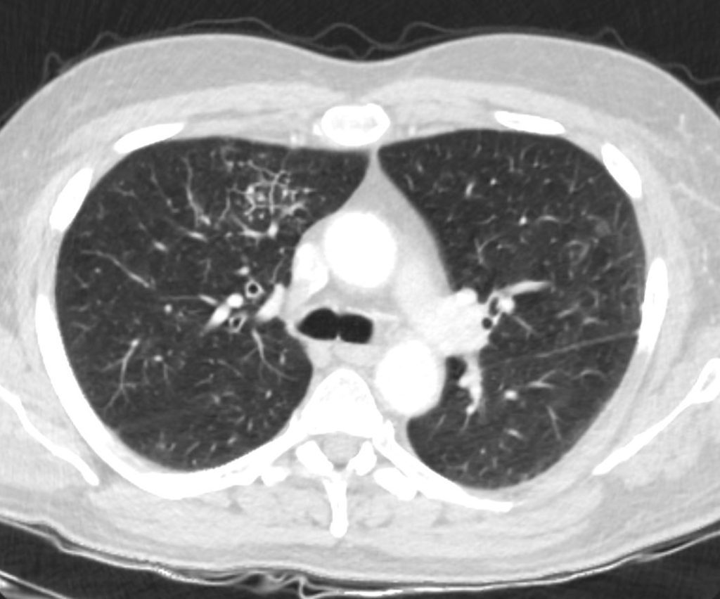
Abnormal Secondary Lobules –
CT of the chest in the axial plane of a 53 year old male with head and neck cancer. In the anterior segment of the right upper lobe are a few secondary lobules with prominent centrilobular nodules and irregularly thickened septa
Ashley Davidoff MD TheCommonVein.net 013Lu 136054
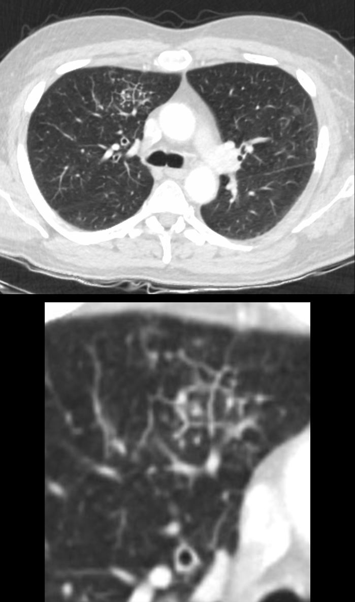
Abnormal Secondary Lobules –
CT of the chest in the axial plane of a 53year old male with head and neck cancer. In the anterior segment of the right upper lobe are a few secondary lobules with prominent centrilobular nodules and irregularly thickened septa
Ashley Davidoff MD TheCommonVein.net 013Lu 136054c
Findings in the Secondary Lobule in the Coronal Plane
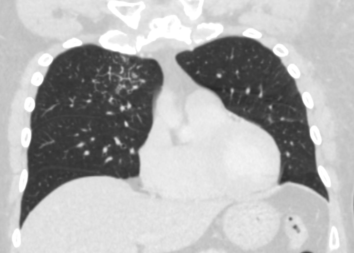
Abnormal Secondary Lobules –
CT of the chest in the coronal plane of a 53year old male with head and neck cancer. In the right upper lobe, there are a few secondary lobules with prominent centrilobular nodules and irregularly thickened septa
Ashley Davidoff MD TheCommonVein.net 013Lu 136057
Minimal Irregularity and Thickening of Secondary Lobules in the Right Lower Lobe
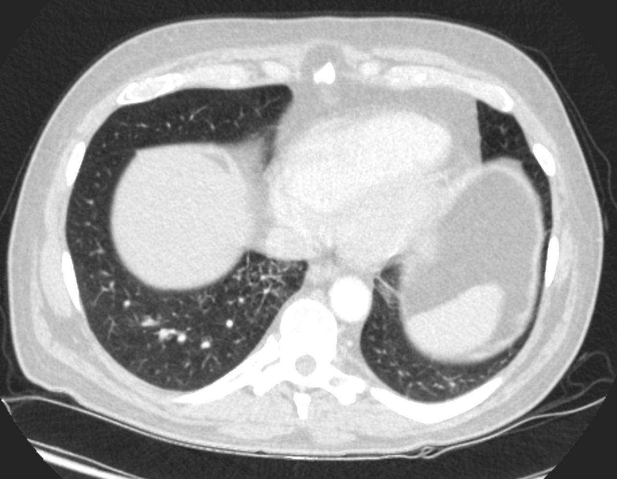
CT of the chest in the axial plane of a 53year old male with head and neck cancer. In the anterior segment of the right lower lobe are a few secondary lobules with prominent centrilobular nodules and irregularly thickened septa
Ashley Davidoff MD TheCommonVein.net 013Lu 136056
3 Month Later
CT Chest – Known Head and Neck Cancer Metastases
Development of Consolidative Masses
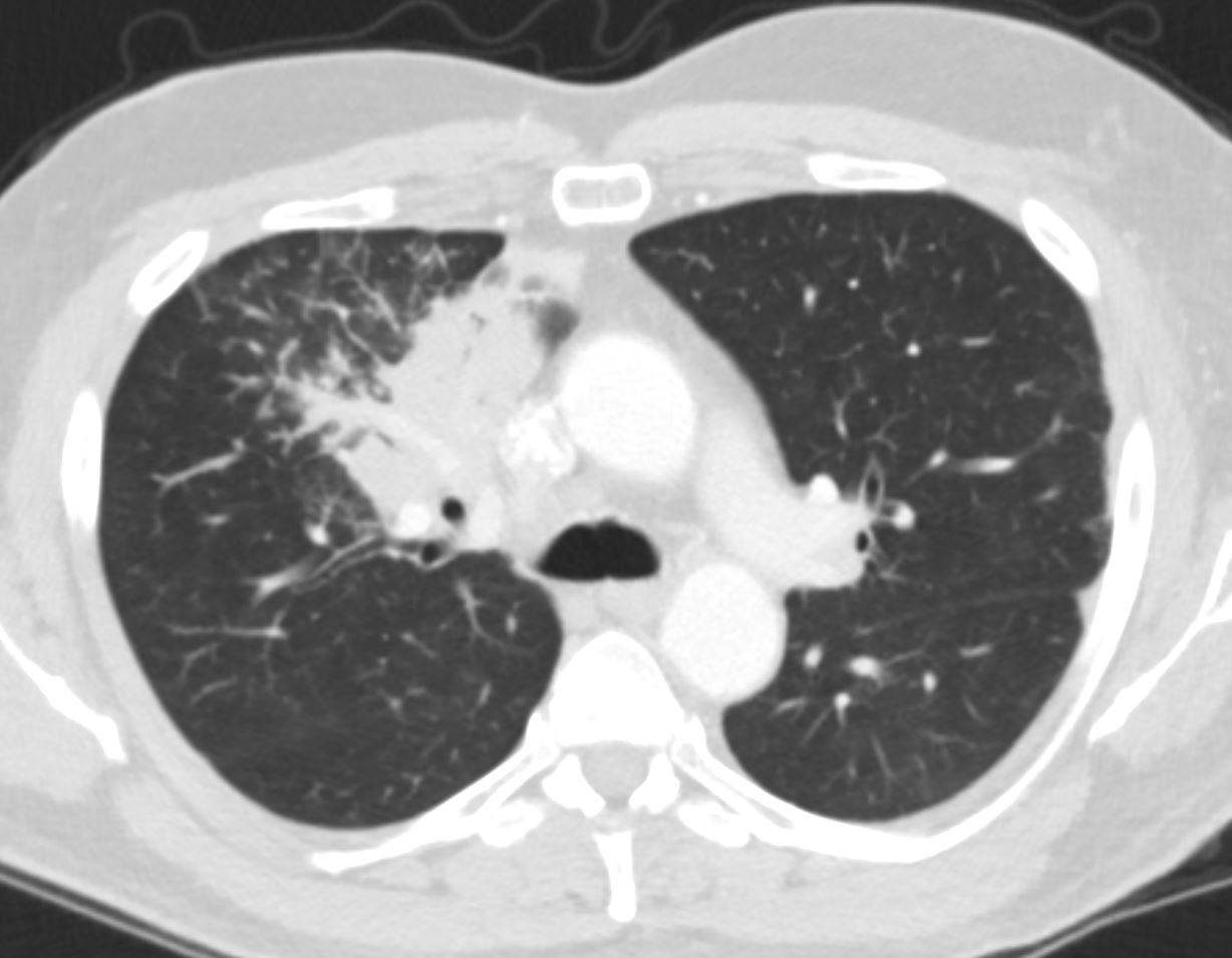
3 months later the patient present with chest pain and a cough. CT of the chest in the axial plane showed a right upper lobe mass abutting the mediastinum in the previous region of abnormality characterised by abnormal secondary lobules. The findings were suggestive of a rapidly developing metastasis in the right upper lobe.
Ashley Davidoff MD TheCommonVein.net 013Lu 136058
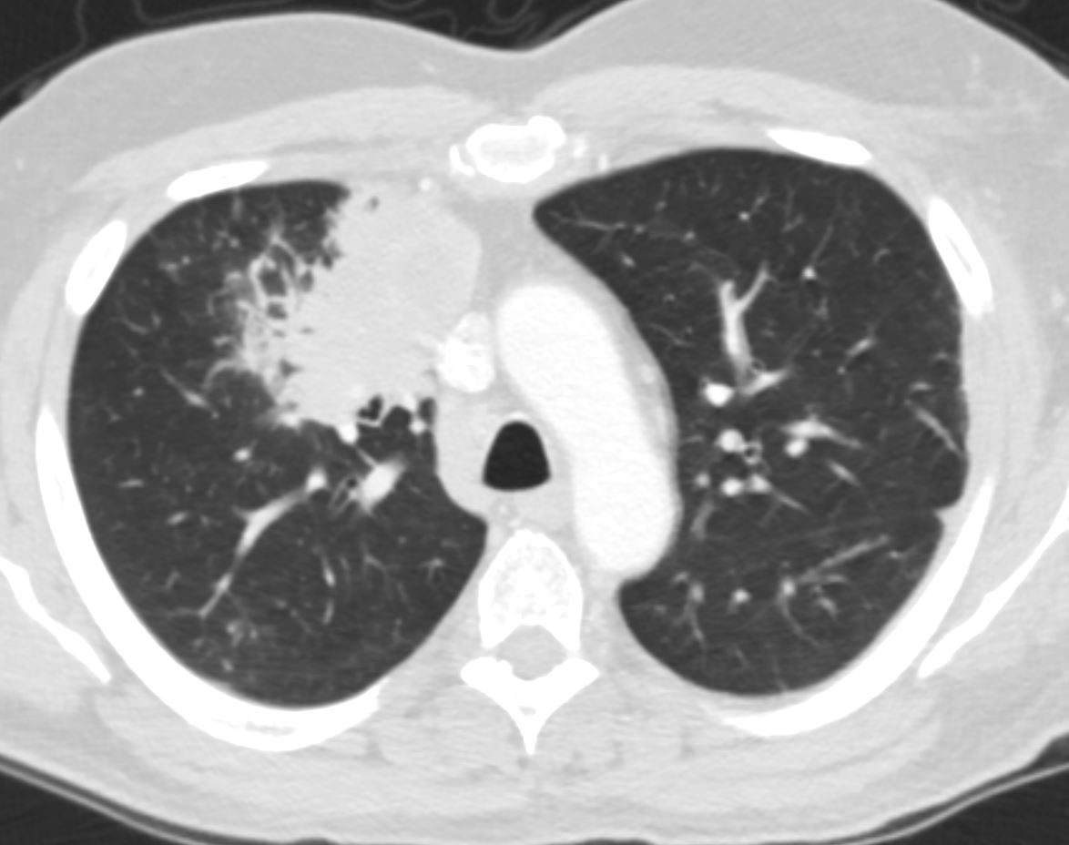
3months later the patient present with chest pain and a cough. CT of the chest in the axial plane showed a right upper lobe mass abutting the mediastinum in the previous region of abnormality characterised by abnormal secondary lobules. The findings were suggestive of a rapidly developing metastasis in the right upper lobe. The thickened septa likely reflect lymphangitis carcinomatosa
Ashley Davidoff MD TheCommonVein.net 013Lu 136060
The New Mass and Progressive Lymphangitis
In the Coronal Plane
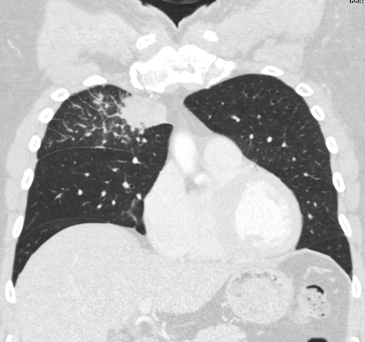
3months later the patient presented with chest pain and a cough. CT of the chest in the coronal plane showed a right upper lobe mass abutting the mediastinum in the previous region of abnormality characterised by abnormal secondary lobules. The findings are suggestive of a rapidly developing metastasis in the right upper lobe. The thickened septa likely reflect lymphangitis carcinomatosa
Ashley Davidoff MD TheCommonVein.net 013Lu 136064
Progressive Lymphangitis in the Right Upper Lobe
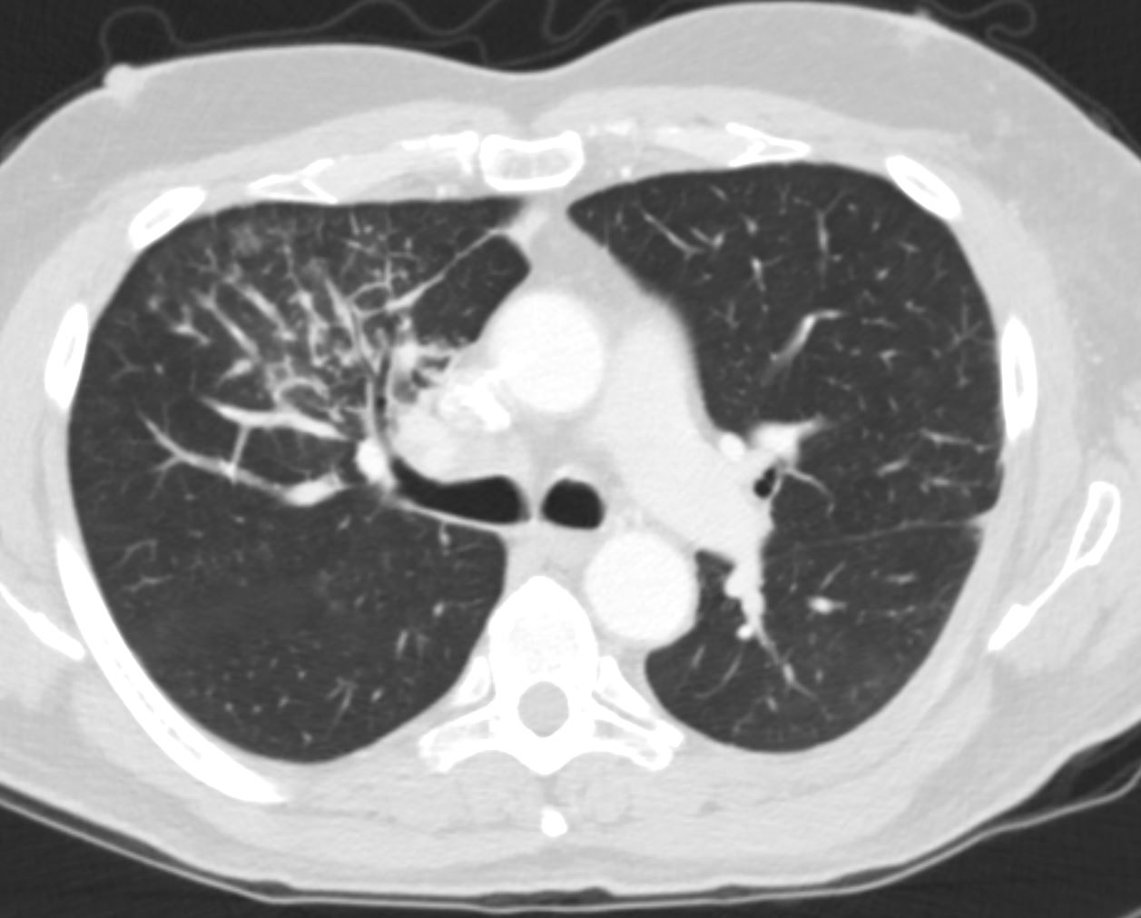
3 months later the patient presented with chest pain and a cough. CT of the chest in the axial plane showed a right upper lobe mass abutting the mass There is interlobular involvement of secondary lobules. The findings are suggestive of a rapidly developing metastasis in the right upper lobe with lymphangitis carcinomatosa
Ashley Davidoff MD TheCommonVein.net 013Lu 136061
Progressive Lymphangitis in the Lower Lobes Bilaterally
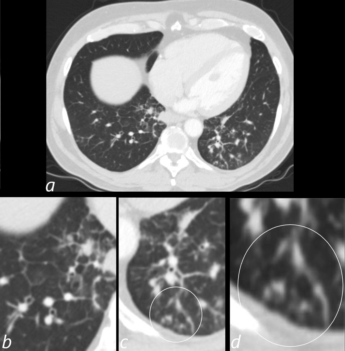
3 months later the patient presented with chest pain and a cough. CT of the chest in the axial plane shows new bilateral lower lobar regions of irregular interlobular septal thickening noted in the right lower lobe a, magnified in b). Ringed in image c and d are 2 side by side secondary lobules with irregular septal thickening centrilobular nodules and other intralobular nodules likely reflecting lymphatic involvement.
Given the changes in the right upper lobe these findings likely reflect lymphangitis carcinomatosa
Ashley Davidoff MD TheCommonVein.net 013Lu 136062cL
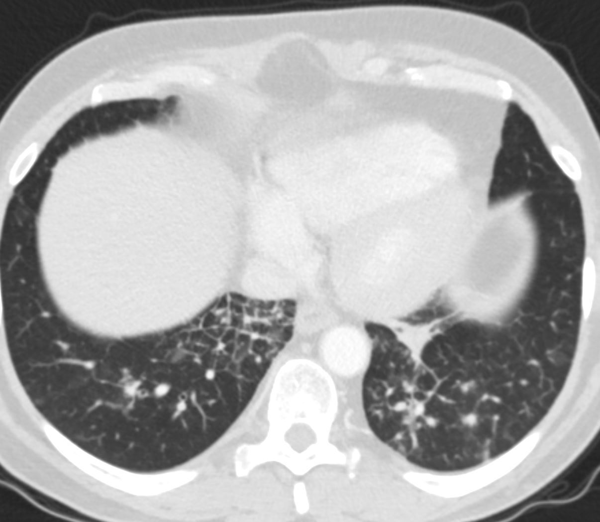
3 months later the patient presented with chest pain and a cough. CT of the chest in the axial plane shows new bilateral lower lobar regions of irregular interlobular septal thickening bilaterally more prominent on the right with nodular changes at the left base.
Given the changes in the right upper lobe these findings likely reflect lymphangitis carcinomatosa
Ashley Davidoff MD TheCommonVein.net 013Lu 136063
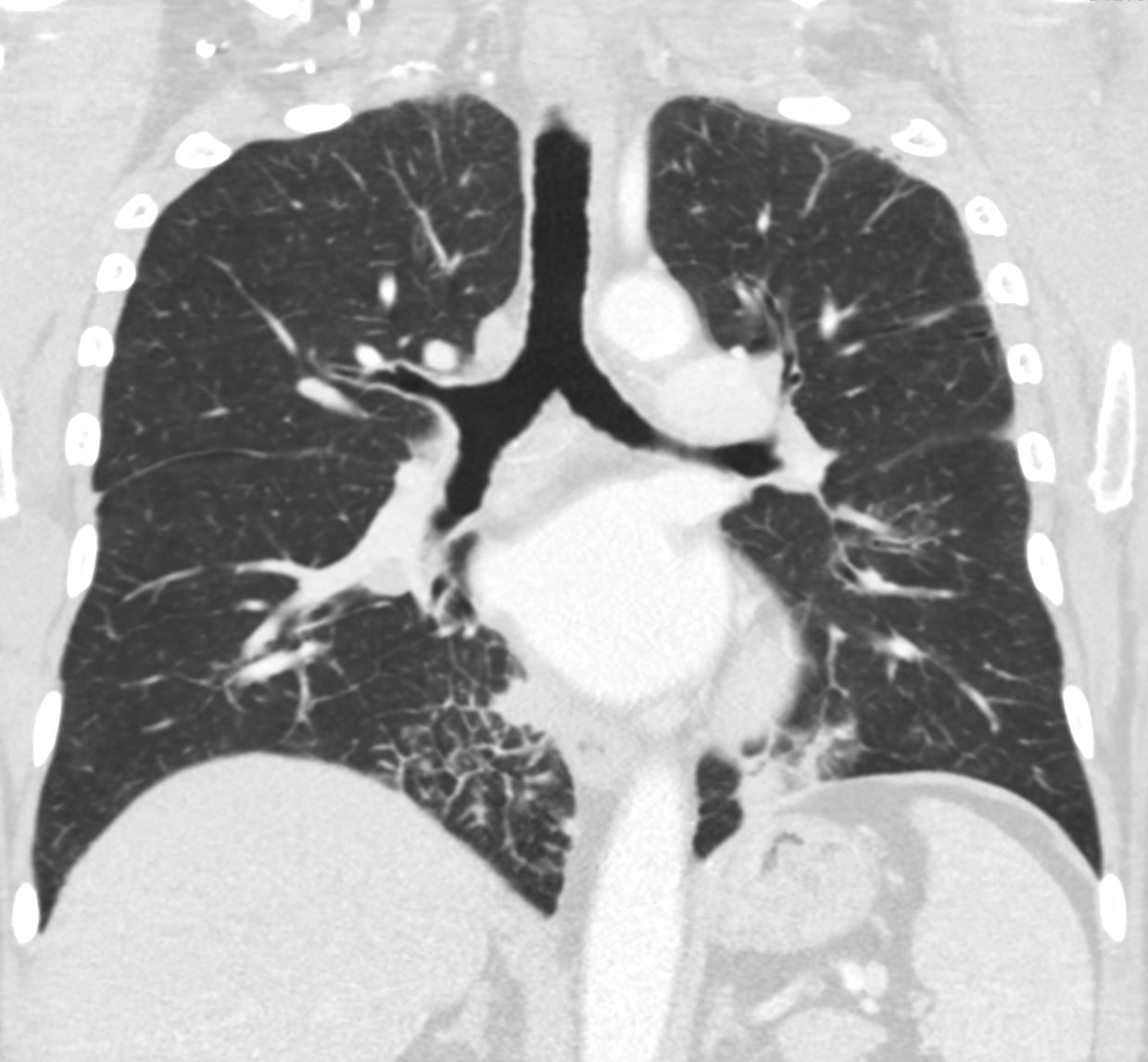
3 months later the patient presented with chest pain and a cough. CT of the chest in the coronal plane shows new bilateral lower lobar regions of irregular interlobular septal thickening noted in the right lower lobe and a subsegmental consolidation in the left lower lobe
Given the changes in the right upper lobe these findings likely reflect lymphangitis carcinomatosa
Ashley Davidoff MD TheCommonVein.net 013Lu 136067
Summary of the Events Over 3 Months
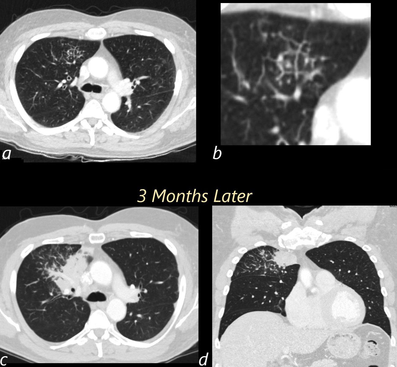
Images a and b show the initial presentation of disease characterised by right upper lobe changes in a few secondary lobules characterised by thickening of the septa and prominence of the centrilobular nodules. 3 months later (c and d) the patient presented with a cough A large right upper lobe consolidative mass and progressive involvement of more secondary lobules with thickening of interlobular septa is noted. These findings likely reflect lymphangitis carcinomatosa
Ashley Davidoff MD TheCommonVein.net 013Lu 13606cL
Search for
53M-H-N-ca-001-3-mths-prior
53M-H-


