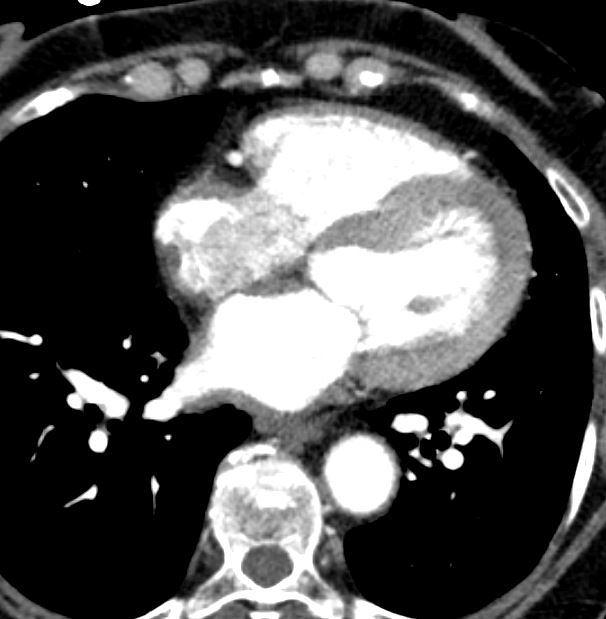
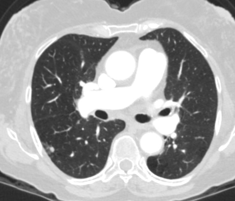
73-year-old female with history of ACL amyloidosis and cardiac involvement status post stem cell transplant
Ashley Davidoff
TheCommonVein.net
Ashley Davidoff
TheCommonVein.net
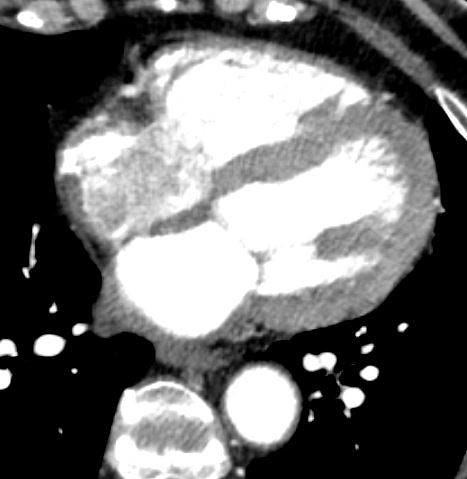
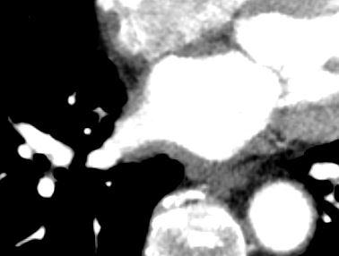
Left atrial wall thickening
Ashley Davidoff
TheCommonVein.net
Ashley Davidoff
TheCommonVein.net
Ashley Davidoff
TheCommonVein.net
Ashley Davidoff
TheCommonVein.net
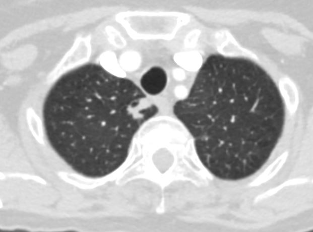
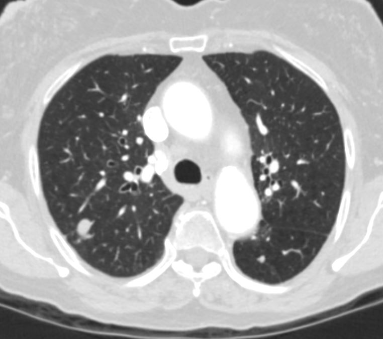
There are 3 similar appearing new nodules appearing in size from 7 mm to
11 mm within the right upper lobe right lower lobe and left lower lobe
characterized by a previously positioned dominant nodule associated with
the bronchovascular bundle, with some extension into the interlobular
septa.
PET CT
Abnormal uptake is seen in the right apical lung nodule. This is
worrisome for malignancy and biopsy may be attempted. Note that amyloid
nodule may also have uptake. Other lung nodules have less uptake and
remain within the benign range by PET imaging.
Echo
Normal LV cavity size with mildly to moderately increased LV wall thickness
(12-13 mm), and low normal global LV systolic function. Estimated LVEF is 50
55%.
Mild hypokinesis of basal LV wall segments.
