Definition
Squamous cell carcinoma (aka epidermoid carcinoma) is a malignant condition of the airway epithelium, usually arising from the central portions of the airways and has a strong causative association with smoking. It is the second most common lung cancer, making up 30% of cases.
Structurally squamous cell carcinoma is characterized by its central location and occurs more commonly in the upper lobes. It tends to spread locally to regional nodes and may cavitate which is a characteristic feature of squamous cell carcinomas in general.
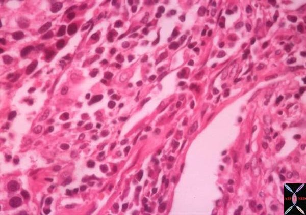
The histopathology is characterised by the proliferation of flat fish-scale like cells and may be associated and characterized by the production of keratin and intercellular bridges. Well differentiated SCC contains keratin pearls. The cells have large irregular nuclii, large nucleoli, and coarse chromatin. The cells are arranged in sheets.
Metaplasia, dysplasia, and carcinoma in situ are sometimes present in the tissue surrounding the carcinoma.
Metastatic disease does occur but compared to other lung cancers it is relatively uncommon occurring in <20% at presentation.
Clinically it often produces symptoms of cough dyspnea or atelectasis, wheezing or hemoptysis since it is more central in its location and affects larger airways. It is also the lung malignancy that is most often associated with hypercalcemia
From a diagnostic point of view, its central location and tendency to exfoliate enables cytological diagnosis from the sputum or bronchial washings.
Treatment is based on staging so that stages I, II, and IIIA have curative and surgical possibility, stage IIIb and more advanced are treated with combinations of radiotherapy and chemotherapy

Cell Block of aspirate biopsy. Note glassy cytoplasm, nucleoli, and inter-cytoplasmic spaces with bridges.
Courtesy tumorboard.com 54405
TheCommonVein.net
32501
Keywords
lungs pulmonary neoplasm primary malignant malignancy squamous cell carcinoma SCC histopathology
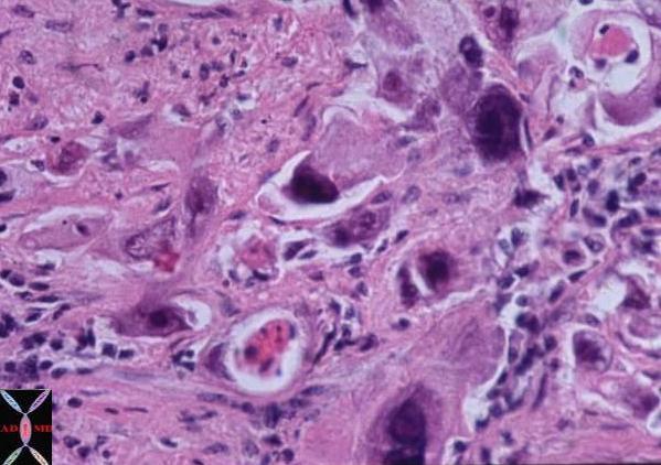
Note pleomorphic tumor cells and keratinous debris.
Courtesy Armando Fraire MD.
TheCommonVein.net
32814
Central Cavitating Nodule

Axial CTscan through the chest in 53 year old male shows a central 2.9cms cavitating lesion consistent with a squamous cell carcinoma of the lung.
Courtesy Ashley Davidoff MD
TheCommonVein.net
87739c.8s
Central Obstructing Lesion
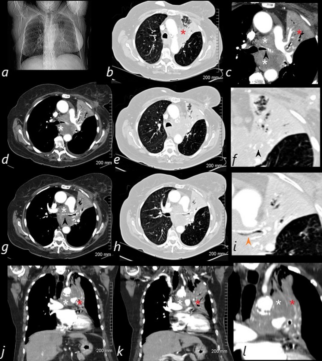
82-year-old female with dyspnea presents with an obstructing lesion of the left main stem bronchus and total atelectasis of the left lung caused by a central squamous cell carcinoma mass. The scout film (a) shows a vague area of increase density in the left upper lung field, with minimal elevation of the left mainstem bronchus. The central mass (white asterisk – is noted in c, d, g, j, k and l. Invasion and obstruction of the left main stem bronchus (black arrow – f, g) with invasion into the lumen without obstruction is notes in I – orange arrow. Post obstructive atelectasis of the left upper lobe is noted (red asterisk – b,c,j,k,l) with hyperinflated left lower lobe extending to the left apex.
Ashley Davidoff MD TheCommonVein.net
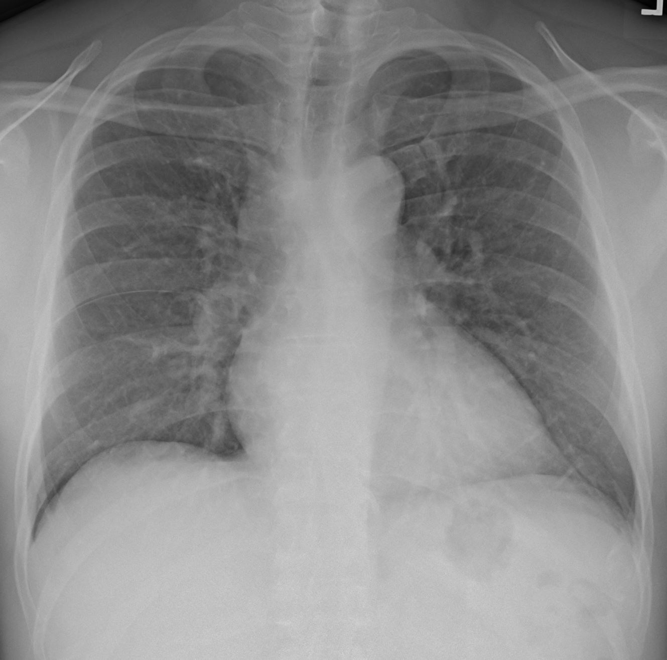
60M SCC arising from right mainstem with large mediastinal mass
Ashley Davidoff MD TheCommonVein.net
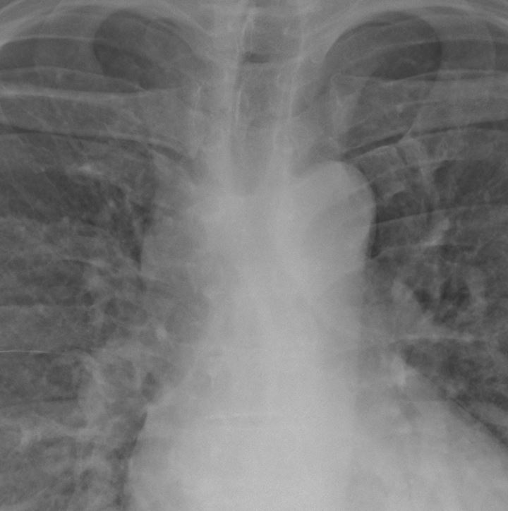
Ashley Davidoff MD TheCommonVein.net

Ashley Davidoff MD TheCommonVein.net
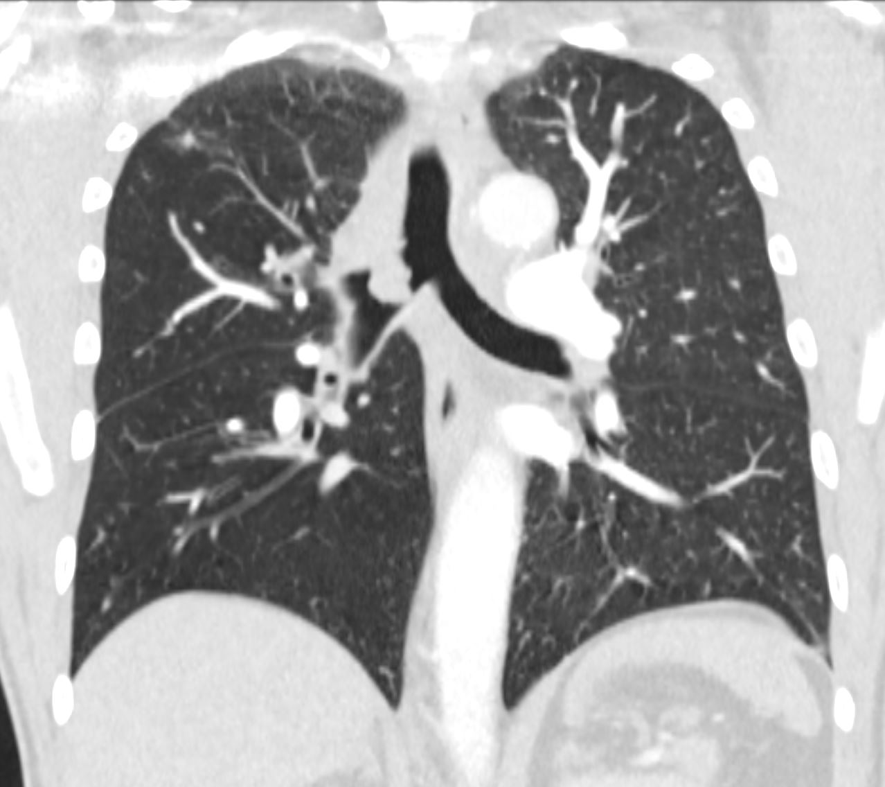
Ashley Davidoff MD TheCommonVein.net
S/P SVC Stent
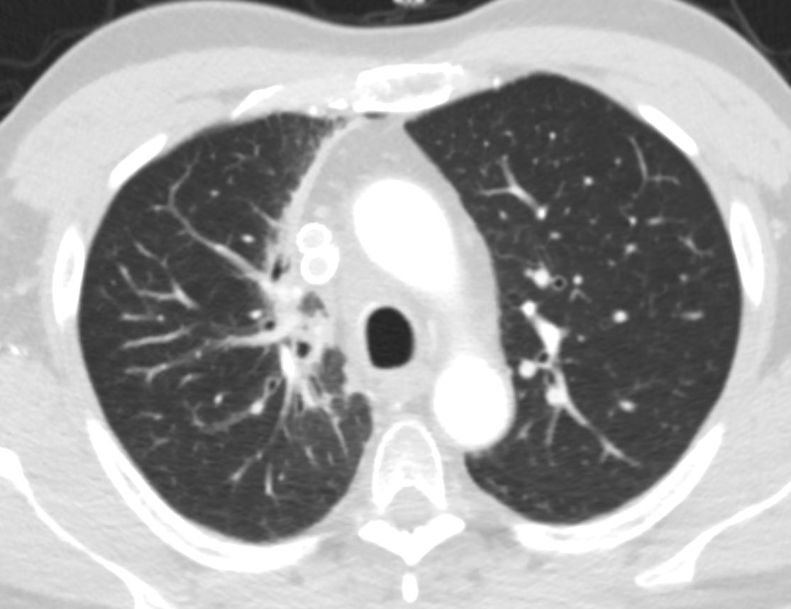
18mths later post XRT
Ashley Davidoff MD TheCommonVein.net
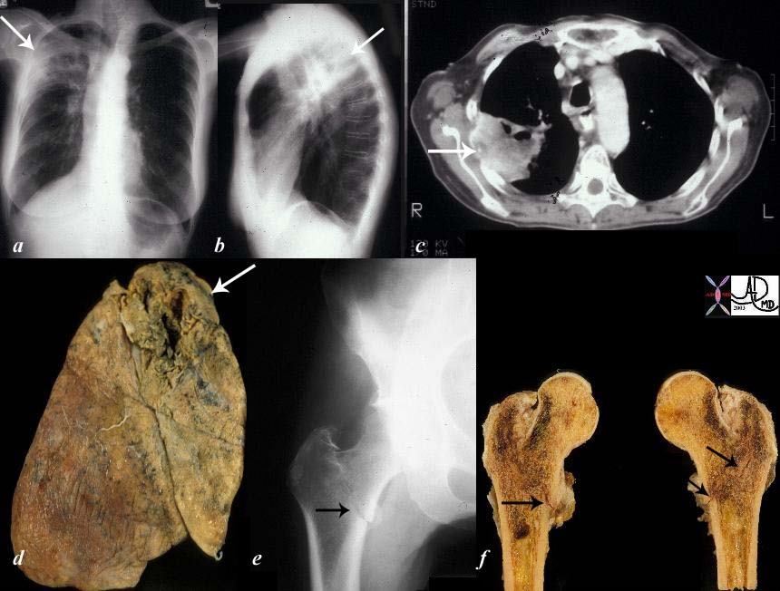
The collage of images reflect a patient with stage IV cavitating primary squamous carcinoma of the RUL (a,b,c,d – white arrows) with COPD. A metastatic lesion to the right femur was complicated by a pathological fracture. (e,f black arrows).
Courtesy Ashley Davidoff MD
TheCommonVein.net
32202c02
56 year Old male with Cavitating Squamous Cell Carcinoma in the Superior Segment of the Right Lower Lobe

Ashley Davidoff MD TheCommonVein.net squamous cell carcinoma cavitating 001

Ashley Davidoff MD TheCommonVein.net squamous cell carcinoma cavitating 002

Ashley Davidoff MD TheCommonVein.net squamous cell carcinoma cavitating 003

Ashley Davidoff MD TheCommonVein.net squamous cell carcinoma cavitating 004
Bubble Lucencies – Pseudocavitation
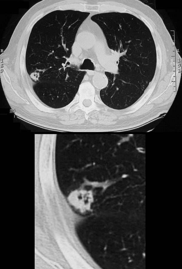
65 year male with peripheral lung nodule characterized by cavitation that was not present 2 years earlier . Pathology revealed squamous cell carcinoma
Ashley Davidoff
TheCommonVein.net
81 M SCC post XRT

Ashley Davidoff MD
TheCommonVein.net
Presenting Cavitating Mass Eroding 4th and 5th Rib
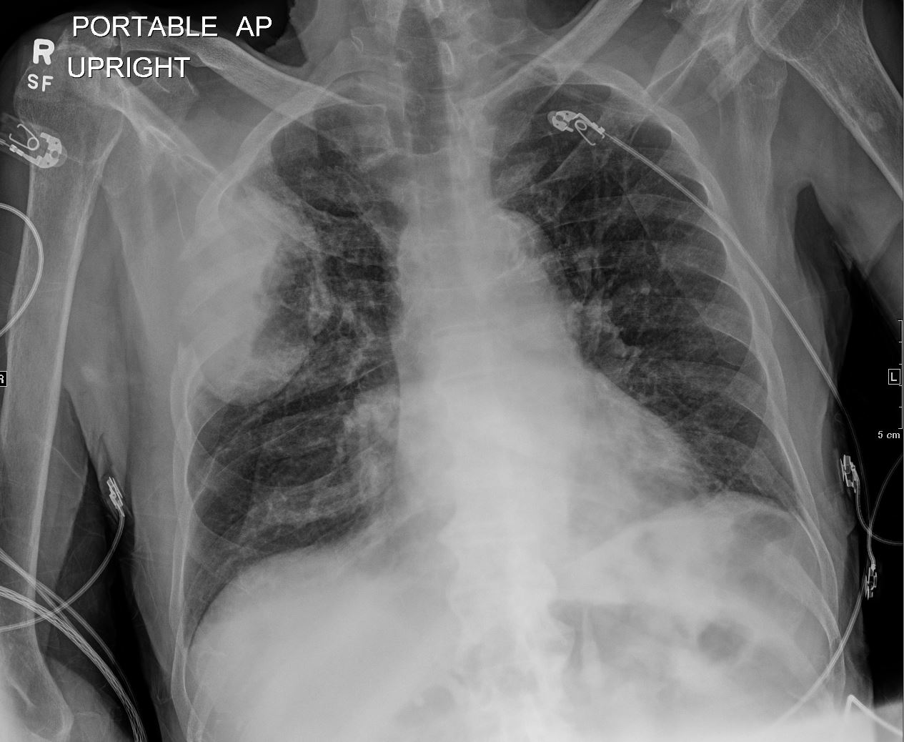
CXR
Ashley Davidoff MD
TheCommonVein.net
Presenting Cavitating Mass Eroding 4th and 5th Rib

CT Pre XRT
Ashley Davidoff MD
TheCommonVein.net
S/P XRT 4months Prior Showing Shrinkage of Mass and XRT Changes
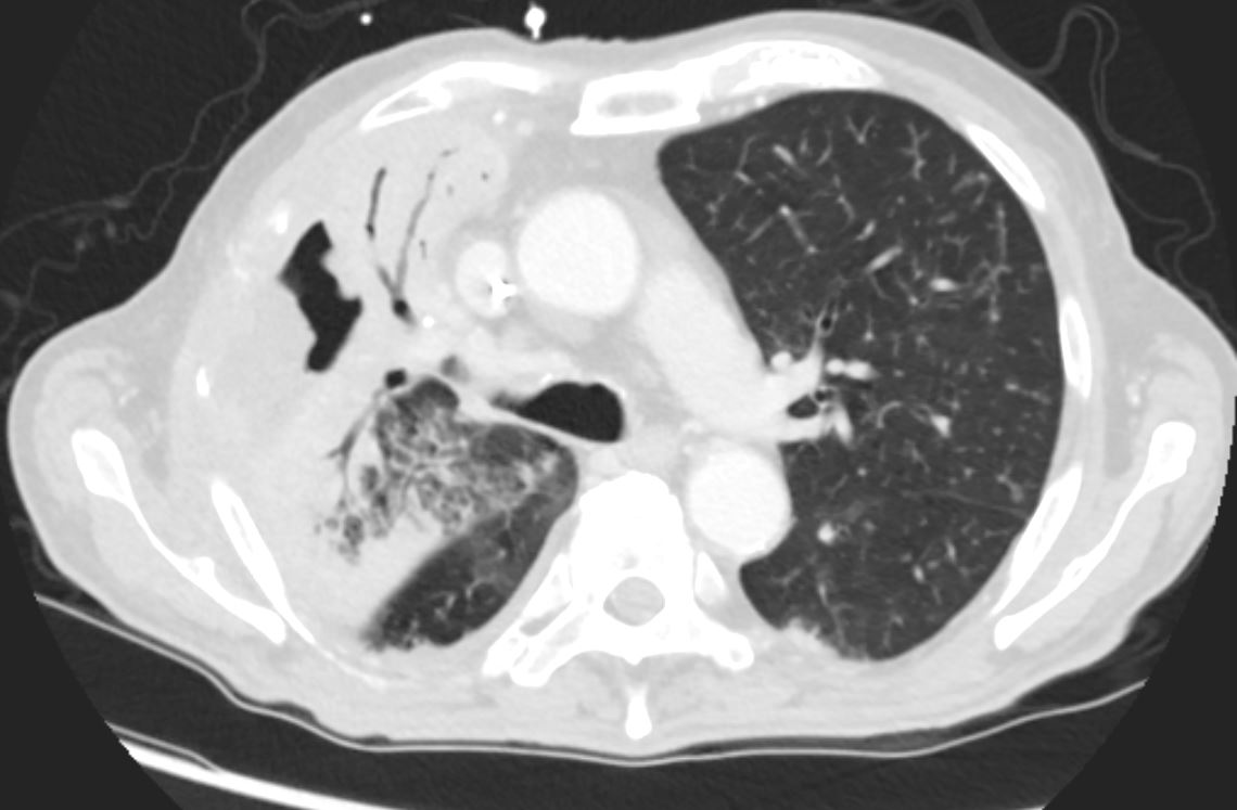
CT Post XRT
Ashley Davidoff MD
TheCommonVein.net
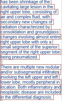
CT post XRT
Ashley Davidoff MD
TheCommonVein.net
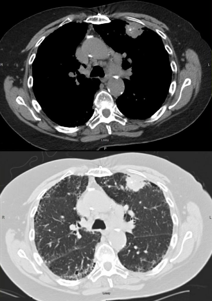
73 year old female with left mastectomy and IPF with new onset bilateral upper lobe masses. The LUL mass has scattered calcifications, and the right shows early cavitation.
Both lesions are PET positive
The LUL lesion was biopsied revealing squamous cell carcinoma with calcifications showing progressive growth and enlarging ipsilateral lymphadenopathy.
The right lesion shows progressive cavitation and enlargement likely a squamous cell carcinoma as well.
The ILD dominates in the periphery of the lower lobes but involves the upper lobes as well, reveals honeycombing in the right upper lobe, but shows no significant progression over the two years.
Ashley Davidoff MD TheCommonVein.net
