Cicatricial atelectasis refers to localized lung collapse caused by contraction or scarring of the lung tissue. It is a form of chronic atelectasis associated with fibrosis and architectural distortion, often secondary to inflammatory, infectious, or post-radiation processes. The term highlights the scarring (cicatrix) as the underlying cause of the volume loss.
Radiological Features
- Chest X-Ray (CXR):
- Findings:
- Localized volume loss in the affected lung region.
- Crowding of Bronchovascular Structures:
- Bronchi and vessels appear closer together due to volume contraction.
- Displacement of Adjacent Structures:
- Mediastinal structures, trachea, and fissures shift toward the collapsed lung.
- Irregular Opacities:
- Due to fibrotic scarring within the atelectatic region.
- Associated Findings:
- Rib crowding on the affected side.
- Compensatory hyperinflation in the contralateral lung.
- Findings:
- Computed Tomography (CT):
- Volume Loss:
- Reduced size of the affected lung or lobe with architectural distortion.
- Fibrotic Changes:
- Thickened interlobular septa and irregular opacities.
- Bronchiectasis:
- Traction bronchiectasis in areas of fibrosis and atelectasis.
- Pleural Abnormalities:
- Pleural thickening, particularly in post-tuberculous or post-radiation cases.
- Honeycombing:
- May be present in end-stage fibrotic diseases.
- Displacement of Fissures:
- Toward the affected region, indicating localized volume loss.
- Volume Loss:
- Magnetic Resonance Imaging (MRI):
- Rarely used but may show scarring and atelectatic regions with greater contrast resolution.
Causes and Associations
- Infectious Causes:
- Tuberculosis (TB):
- Common cause, especially in regions with high TB prevalence.
- Post-TB scarring leads to contraction atelectasis.
- Fungal Infections:
- Aspergillosis or histoplasmosis causing localized fibrosis and scarring.
- Tuberculosis (TB):
- Interstitial Lung Diseases:
- Idiopathic Pulmonary Fibrosis (IPF):
- Characterized by subpleural fibrosis, honeycombing, and associated atelectasis.
- Sarcoidosis:
- Granulomatous inflammation leads to scarring and atelectasis.
- Idiopathic Pulmonary Fibrosis (IPF):
- Post-Radiation Fibrosis:
- Scarring in the radiation field causes localized volume loss.
- Autoimmune Diseases:
- Rheumatoid Arthritis or Scleroderma:
- Chronic inflammation leads to fibrosis and cicatricial atelectasis.
- Rheumatoid Arthritis or Scleroderma:
- Pleural Diseases:
- Chronic pleural effusion or empyema resulting in pleural scarring and associated lung contraction.
Differential Diagnosis
Cicatricial atelectasis should be distinguished from:
- Obstructive Atelectasis:
- Results from airway obstruction; associated with central mass or mucus plug.
- Compression Atelectasis:
- Due to external compression (e.g., pleural effusion, pneumothorax).
- Lung Collapse due to Tumor:
- Volume loss associated with mass-like lesions or lymphadenopathy.
Clinical Relevance
- Diagnosis:
- Cicatricial atelectasis is a chronic process often identified on imaging during evaluation for fibrosis, infections, or inflammatory conditions.
- Management:
- Focuses on the underlying condition (e.g., tuberculosis treatment, autoimmune disease control).
- Complications:
- Progressive fibrosis can lead to respiratory impairment or secondary infections.
Atelectasis and Bronchiectasis

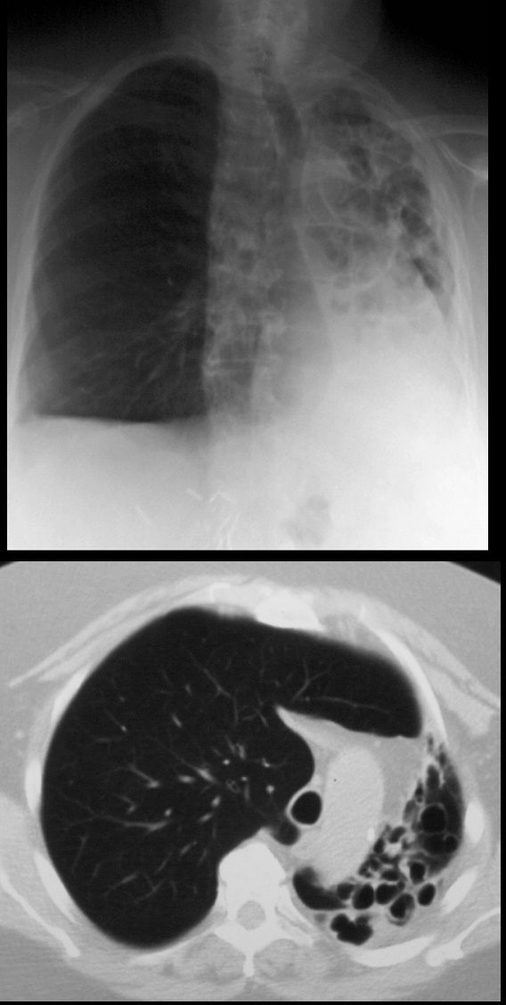
68year old male presents with chronic cough. Chest Xray reveals evidence of tram tracking and bronchiectasis in the left lung with volume loss of the entire right lung with secondary hyperinflation of the right lung, and mediastinal shift. CT scan shows severe volume loss and hyperinflation of the right lung.
Ashley Davidoff MD TheCommonVein.net

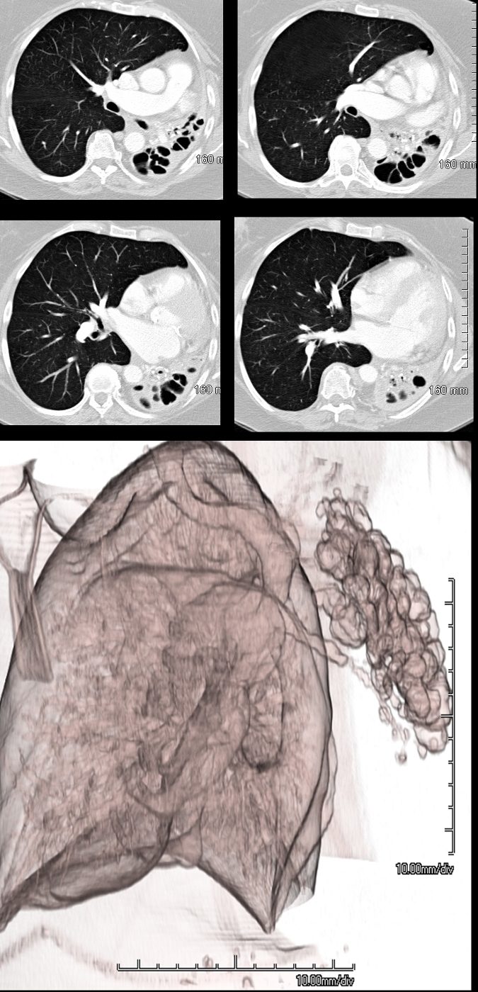
69 year female atrophied left lung secondary to chronic infection and resulting in cystic bronchiectasis (aka saccular bronchiectasis)
The axial images show a totally collapsed left lung with leftward mediastinal shift and hyperinflation of the right lung. The airways of the left lung are large and patent allowing for a 3Dreconstruction of the airways and confirming the diagnosis of saccular bronchiectasis. The atelectasis of the left lung is called cicatricial atelectasis secondary to chronic fibrosis
Ashley Davidoff MD TheCommonvein.net 117955c
Combination of Cicatrization and Obstruction

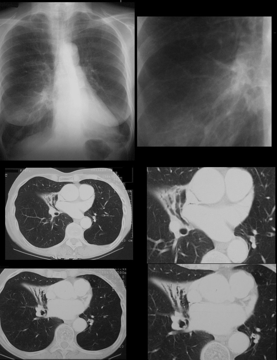
69year old female with varicose bronchiectasis atelectasis and mucus plugging. The CXR suggests a hilar process with an indistinct margin of the right heart border possibly relating to a middle lobe process. CT scans confirm atelectasis of the middle lobe, varicose bronchiectasis, and the presence of mucoid impaction best revealed in the left lower bronchi
Ashley Davidoff MD TheCommonVein.net

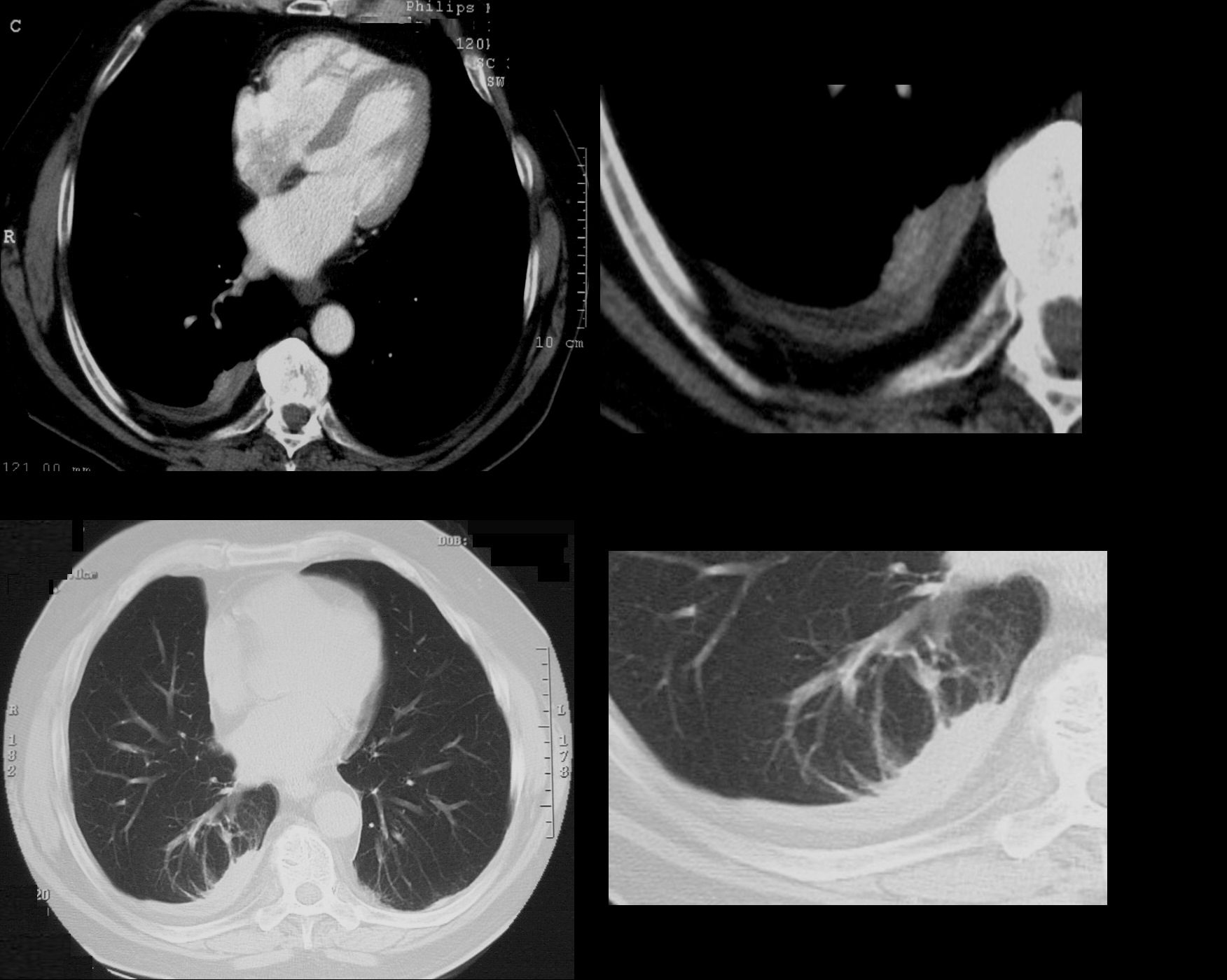
74 year old male with a cough.
CT shows split pleura sign with thickened visceral and parietal pleura with regions of early spiraling of an atelectatic process in the right lower lobe consistent with early rounded atelectasis
Ashley Davidoff MD TheCommonVein.net
31563c
