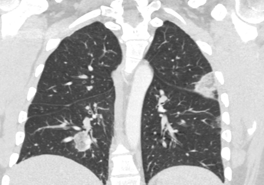
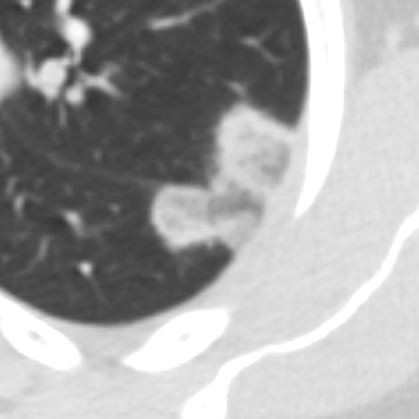
The reversed halo sign (also known as the atoll sign) is a radiological finding characterized by a central area of ground-glass opacity surrounded by a ring or crescent of denser consolidation. This pattern is most commonly observed on CT scans of the chest and indicates a variety of underlying pulmonary pathologies, typically involving inflammatory, infectious, or vascular processes.
Radiological Features
- Appearance on HRCT:
- Central Ground-Glass Opacity:
- Represents partial filling of alveoli with fluid, inflammatory cells, or fibrosis.
- Peripheral Ring of Consolidation:
- Denser opacity, typically indicating more complete filling of alveoli or fibrosis.
- The overall pattern resembles a halo or an atoll (a coral island formation).
- Ring thickness can vary and may be complete or incomplete.
- Central Ground-Glass Opacity:
- Location:
- May be solitary or multiple.
- Can occur in any region of the lungs, depending on the underlying pathology.
- Associated Findings:
- May coexist with other radiologic patterns such as nodules, interstitial thickening, or cavitations, depending on the underlying condition.
Common Causes and Associations
The reversed halo sign is a non-specific finding and can occur in several conditions:
- Infectious Diseases:
- Invasive Fungal Infections:
- Pulmonary mucormycosis and other invasive fungal infections, particularly in immunocompromised patients.
- Tuberculosis:
- Seen in tuberculous granulomas with central necrosis.
- Pneumocystis jirovecii Pneumonia (PJP):
- Rarely presents with this sign.
- Invasive Fungal Infections:
- Inflammatory/Immune-Mediated Diseases:
- Cryptogenic Organizing Pneumonia (COP):
- One of the most common causes.
- Peripheral consolidation represents organizing pneumonia, while the central ground-glass opacity reflects alveolar damage.
- Granulomatosis with Polyangiitis (GPA):
- May show reversed halo patterns in granulomatous inflammation.
- Cryptogenic Organizing Pneumonia (COP):
- Vascular Causes:
- Pulmonary Infarction:
- Central ground-glass opacity corresponds to alveolar hemorrhage, and the consolidation reflects infarcted lung tissue.
- Pulmonary Infarction:
- Other Causes:
- Sarcoidosis:
- Rare presentation of reversed halo due to granulomatous inflammation.
- Fibrotic Lung Diseases:
- May occur in subacute phases of organizing fibrosis.
- Sarcoidosis:
Differential Diagnosis
Conditions that may mimic the reversed halo sign:
- Classic Halo Sign:
- Opposite appearance: central consolidation surrounded by ground-glass opacity (e.g., in invasive aspergillosis).
- Cavitary Lesions:
- Central necrosis with thick-walled cavities, often due to infections or malignancy.
- Pulmonary Nodules with Ground-Glass Opacities:
- May appear similar but lack the organized peripheral consolidation.
Clinical Relevance
- The reversed halo sign is not specific to one disease, so clinical correlation and ancillary tests (e.g., serology, biopsy, or bronchoalveolar lavage) are essential.
- It helps radiologists and clinicians narrow the differential diagnosis based on patient history, immune status, and additional findings on imaging.

Reconstruction of an atoll which is an is a ring-shaped island, including a coral rim that encircles a lagoon. There may be coral islands on the rim. Atolls are located in warm tropical or subtropical parts of the oceans and seas where corals can develop. Most of the approximately 440 atolls in the world are in the Pacific Ocean.
Artistic rendering by ChatGPT
and Wikipedia
Ashley Davidoff MD TheCommonVein.net
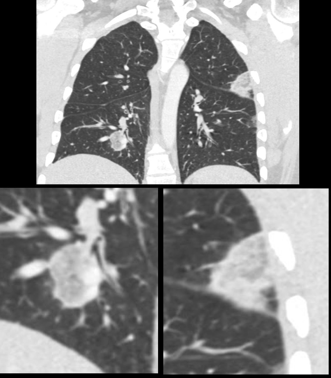
The nodule consists of a peripheral dense border surrounding a ground glass opacity, similar in appearance to an atoll
Ashley Davidoff MD TheCommonVein.net 139137c
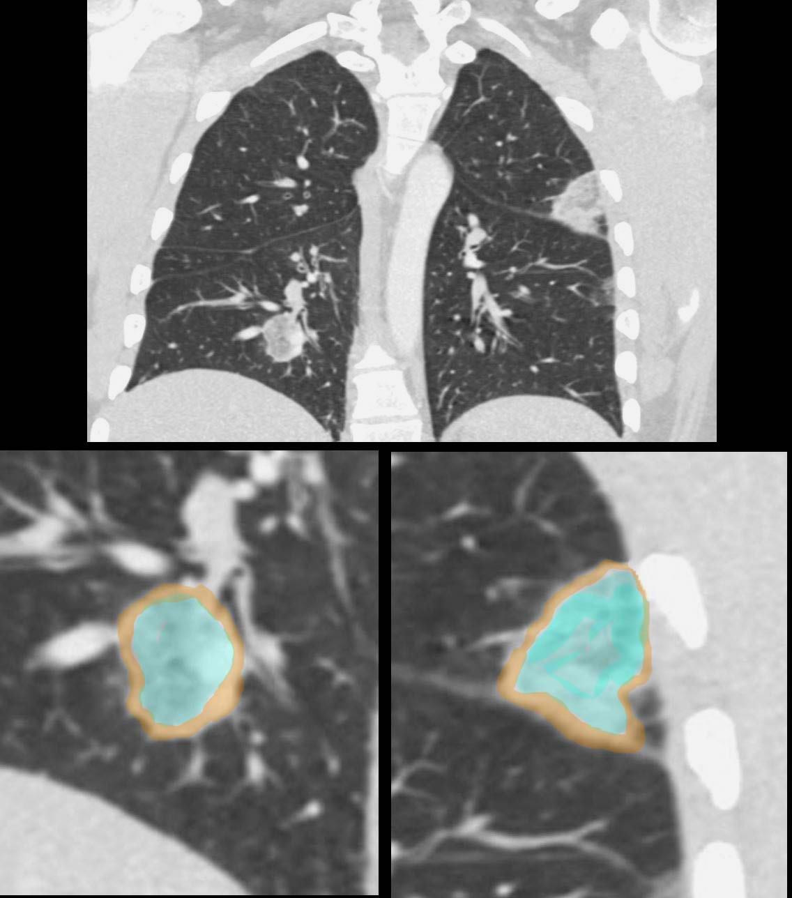
The nodule consists of a peripheral dense border (overlaid in brown) surrounding a ground glass opacity overlaid in turquoise)
similar in appearance to an atoll
Ashley Davidoff MD TheCommonVein.net 139137cL
It is associated with various lung conditions, most notably
organizing pneumonia (OP), although it can also be seen in other
diseases such as pulmonary infarction, granulomatosis with
polyangiitis, and fungal infections.
Lung biopsy may be carried out
to confirm the underlying cause.
Laboratory tests and cultures may
also be used if infection or autoimmune disease is suspected.
Hemorrhagic PE with with Reversed Halo Sign
32 year old male presents with acute PE and reversed halo sign indicative most likely of a hemorrhagic pulmonary infarction
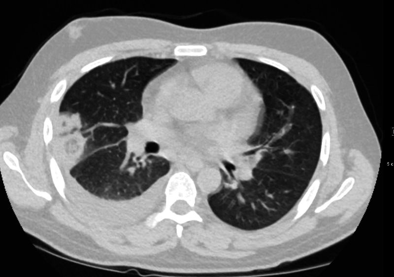
32 year old male presents with acute PE and reversed halo sign indicative most likely of a hemorrhagic pulmonary infarction
Ashley Davidoff MD
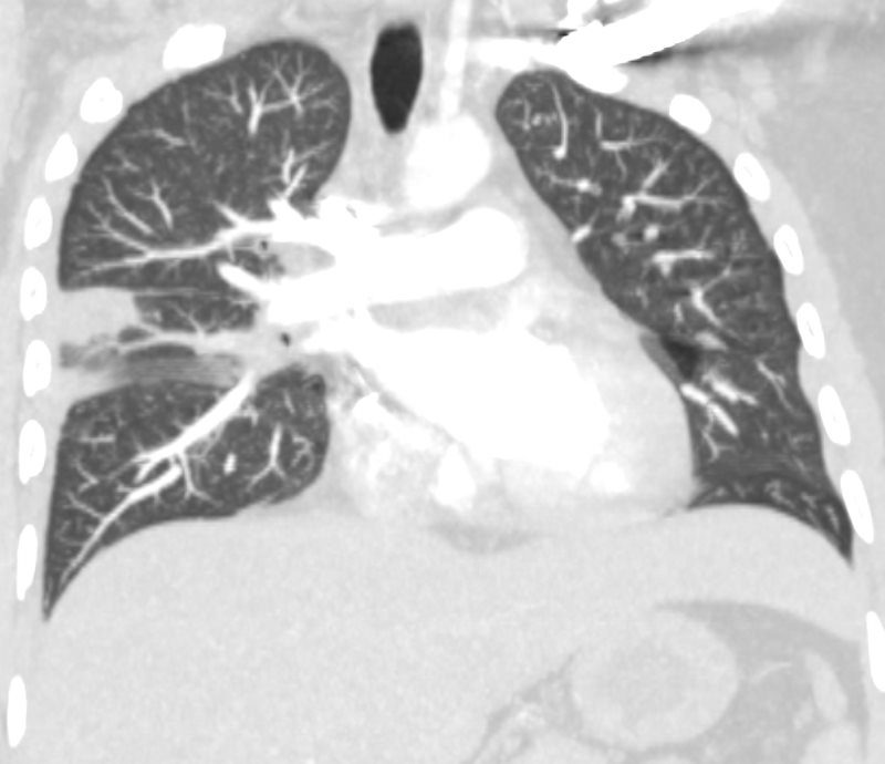
32 year old male presents with acute PE and reversed halo sign indicative most likely of a hemorrhagic pulmonary infarction
Ashley Davidoff MD
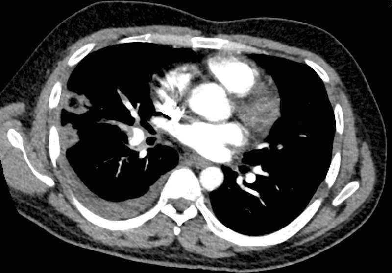
32 year old male presents with acute PE and reversed halo sign indicative most likely of a hemorrhagic pulmonary infarction
Ashley Davidoff MD
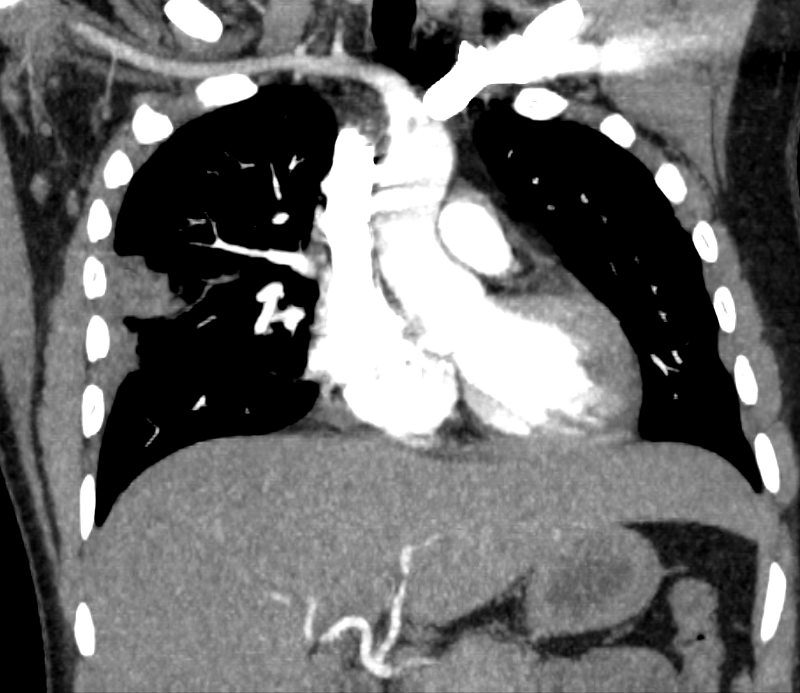
32 year old male presents with acute PE and reversed halo sign indicative most likely of a hemorrhagic pulmonary infarction
Ashley Davidoff MD
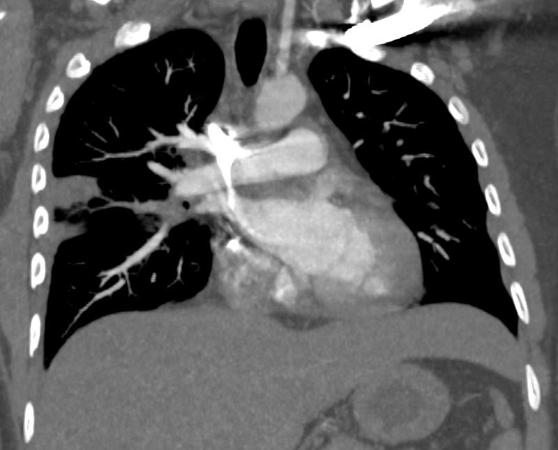
32 year old male presents with acute PE and reversed halo sign indicative most likely of a hemorrhagic pulmonary infarction
Ashley Davidoff MD
Reversed Halo Sign Related to Immunotherapy for Adenoca of the Lung
87-year-old female with stage IV adenocarcinoma s/p immunotherapy
In the initial scans there are prominent lesions demonstrating the reversed halo sign likely as a result of the therapy
Subsequent imaging (2 CT scans) shows progressive resolution of the multicentric changes
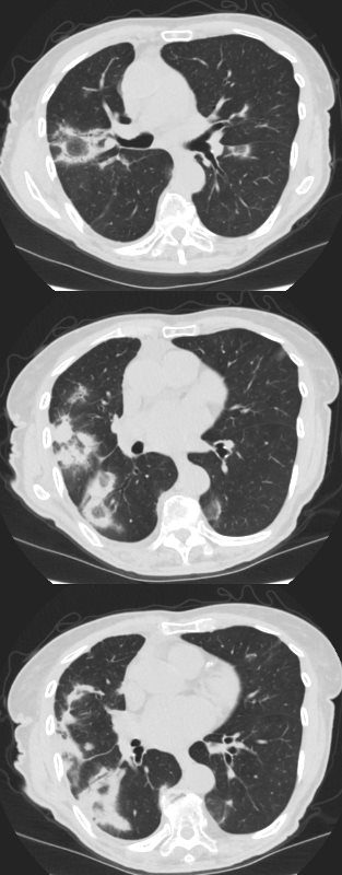
87-year-old female with stage IV adenocarcinoma s/p immunotherapy
In the initial scans there are prominent lesions demonstrating the reversed halo sign likely as a result of the therapy
Subsequent imaging (2 CT scans) shows progressive resolution of the multicentric changes
Ashley Davidoff MD 131478.8
Follow Up 2 Months Later
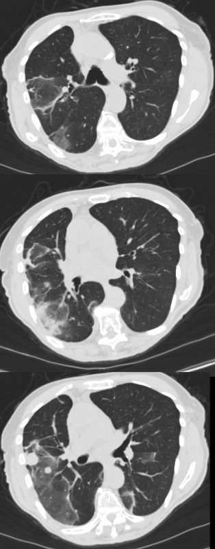
87-year-old female with stage IV adenocarcinoma s/p immunotherapy
In the initial scans there are prominent lesions demonstrating the reversed halo sign likely as a result of the therapy
Subsequent imaging (2 CT scans) shows progressive resolution of the multicentric changes
Ashley Davidoff MD
Follow Up 6 Months Later
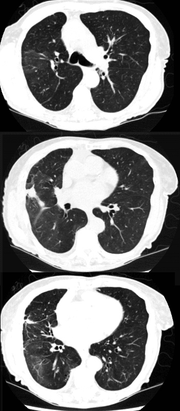
87-year-old female with stage IV adenocarcinoma s/p immunotherapy
In the initial scans there are prominent lesions demonstrating the reversed halo sign likely as a result of the therapy
Subsequent imaging (2 CT scans) shows progressive resolution of the multicentric changes
Ashley Davidoff MD
Young Man with Pleuritic Pain
Broad Differential Diagnosis

31 year old male with a history of DKA, brachial DVT and pleuritic pain presents for a CTPA to evaluate for pulmonary embolism. Findings show no acute PE’s, but he has multicentric, bilateral, peripheral based foci with reversed halo signs, (atoll sign). While chronic PE is on the differential diagnosis, the atoll sign is an unusual consequence of PE . Organizing pneumonia, fungal infections, – mucormycosis, aspergillosis, TB and chronic manifestations of a COVID infection are on the differential diagnosis
The nodule consists of a peripheral dense border surrounding a ground glass opacity, similar in appearance to an atoll
Ashley Davidoff MD TheCommonVein.net 139135

